Morpho 106 Copy
Maxillary Molars
- Maxillary molars are usually the first permanent teeth to erupt into the maxillary arch.
- Each maxillary molar usually has four major cusps, with two on the buccal portion of the occlusal table and two on the lingual.
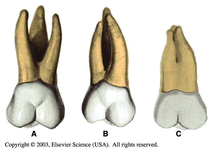
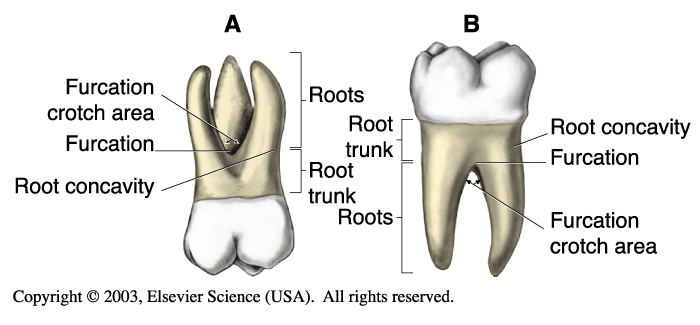
- Each maxillary molar has three well-separated and well-developed roots.
- A tooth with three roots is said to be trifurcated, which means “divided into thirds.”
Maxillary First Molars
- The maxillary first molars (No. 3 and No. 14) are the first permanent teeth to erupt into the maxillary arch.
- They erupt distal to the primary maxillary second molars and are therefore nonsuccedaneous.
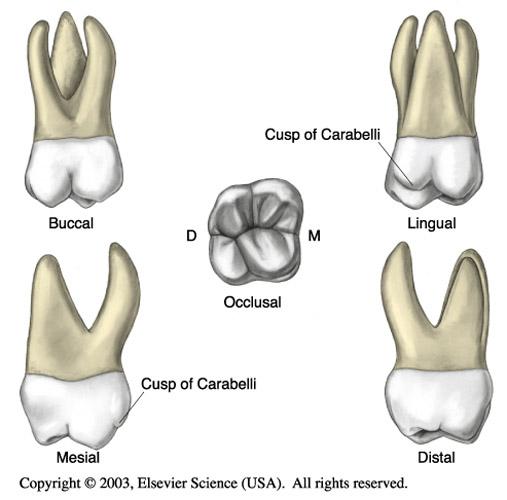
- The maxillary first molar is the largest tooth in the maxillary arch and also has the largest crown in the permanent dentition.
- This molar is composed of five developmental lobes, two buccal and three lingual.
- The fifth cusp is called the cusp of Carabelli.
Maxillary first molar. B, Mandibular first molar. (From Bath-Balogh MB, Fehrenbach MJ: Illustrated dental embryology, histology, and anatomy, ed 2, Philadelphia, 2005, Saunders.)
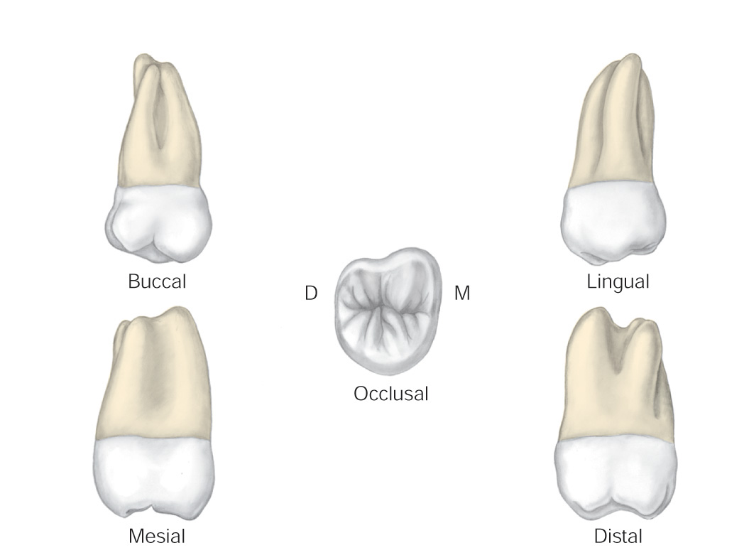
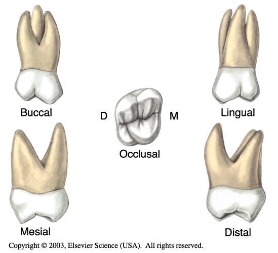
Various views of a permanent maxillary right first molar. (From Bath-Balogh MB, Fehrenbach MJ: Illustrated dental embryology, histology, and anatomy, ed 2, Philadelphia, 2005, Saunders.)
Maxillary Second Molars
- The crown of the maxillary second molar is somewhat shorter than that of the first molar, and it usually has four cusps.
- No fifth cusp is present.
- There are three roots.
- The roots of the secondary molars are smaller than those of the first molars. The lingual root is still the largest and longest.
- The buccal groove is located farther distally on the buccal surface of the second maxillary molar than on the first maxillary molar.
- The mesiobuccal cusp of the second maxillary molar is longer and has a more blunt cusp tip than the distobuccal cusp.
Various views of a permanent maxillary right second molar. (From Bath-Balogh MB, Fehrenbach MJ: Illustrated dental embryology, histology, and anatomy, ed 2, Philadelphia, 2005, Saunders.)
Maxillary Third Molars
- The maxillary third molars (No. 1 and No. 16) differ considerably in size and contour.
- The crown of the maxillary third molar is smaller and the roots are usually shorter.
- The roots of the maxillary third molar tend to fuse, and the result is a single tapered root.
- People sometimes refer to the maxillary third molars as the “wisdom” teeth because they erupt last.
Clinical Considerations with Maxillary Molars
- The roots of the maxillary molars may penetrate the maxillary sinus as a result of accidental trauma or during an extraction.
- The roots of the maxillary molars are close to the sinuses. Some patients confuse the pain caused by a sinus infection with pain related to their maxillary teeth, and vice versa.
- The permanent maxillary third molars may fail to erupt and may remain impacted within the alveolar bone.
- If the maxillary first molar is lost, the second molar can tip and drift into the open space, causing difficulty in chewing and furthering periodontal disease.