HRV - INGENIOUSLY SIMPLE!

More than software - a strong relationship with the patient.

Physiological basics

Complex control systems

Along with the central nervous system (CNS), the autonomic nervous system (VNS) is the most important neuronal control unit of the organism. The main function is to adapt the internal environment of the body to external and internal stresses (stimuli) and to maintain a constant function of the organism.

The peripheral VNS is involved in a complex system that is connected to the hypothalamus and other structures located in the CNS, in addition to connections to the brainstem.

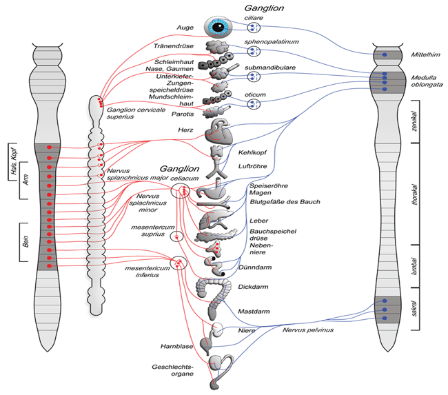
The autonomic nervous system (VNS) with sympathetic and parasympathetic nervous system

The peripheral part of the VNS consists essentially of the sympathetic and parasympathetic nervous systems. The sympathetic nervous system originates from the thoracic medulla and the upper three segments of the lumbar medulla and is therefore also called the thoracolumbar system. The parasympathetic nervous system originates from the brainstem and sacral medulla and is therefore also called the craniosacral system.

Control variables of heart rate variability (HRV)

The sinus node is located on the inner side of the posterior wall of the right atrium. The excitation originating there is transmitted via the muscles to the atrioventricular node (AV node), and from there to the ventricles. The sinus node is the fastest and therefore superior pacemaker of the heart with an intrinsic natural frequency of 80-120 beats per minute.

Modulation of heart rate by sympathetic and parasympathetic nervous system

The heart muscle is innervated by both sympathetic and parasympathetic parts. The sympathetic nervous system has a frequency-increasing (positive chronotropic) effect and the parasympathetic nervous system has a frequency-decreasing (negative chronotropic) effect. If the heart rate is lower than the intrinsic excitation frequency of the sinus node (80-120) the parasymapthic is dominant and if it is higher the sympathetic is dominant.

Baroreflex integration

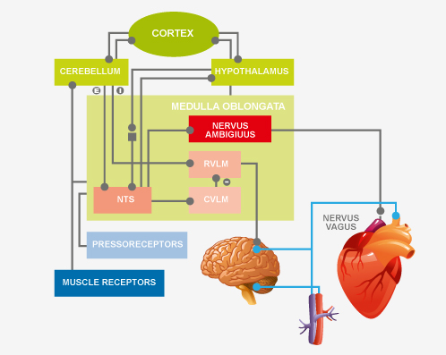
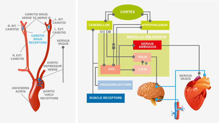
Baroreceptor signals reach the nucleus tractus solitarii (NTS) in the brainstem. From there, signals travel to the nucleus ambiguus and the rostroventrolateral and caudoventrolateral medulla oblongata from which, respectively, excitatory and inhibitory impulses are sent to the heart and vessels to control arterial blood pressure.

Baroreflex / Baroreceptors - Effects on HRV

The central mechanisms influencing HRV are baroreflex activity and respiratory sinus arrhythmia (RSA). Baroreflex control is used to permanently maintain adequate (mean) arterial blood pressure to supply all organ systems. The receptors located in the aortic arch and carotissinus have very high sensitivity and respond to minimal changes in pressure. During physical exertion or sport, there is a shift in the sensitivity threshold to meet the higher oxygen demand. An altered threshold due to permanent sympathetic stimulation plays a significant role in the development of hypertension.

Baroreflex / Baroreceptors - Effects on HRV

The dependence of heart rate on respiration is called respiratory sinus arrhythmia (RSA):

  • During inspiration, an increase in heart rate
  • During expiration a decrease inheart rate

RSA is mediated primarily by the alternating activity of the vagus nerve.

Influences on respiration-dependent heart rate variability:

  • Pulmonary, vascular and cardinal stretch receptors.
  • respiratory centers in the brainstem
  • different baroreflex sensitivity in the respective phases of the respiratory cycle

Due to inspiratory vagal inhibition, fluctuations in heart rate occur at the same frequency as respiration. Inspiratory inhibition is primarily caused by the influence of the medullary respiratory center on the medullary cardiovascular center. In addition, peripheral reflexes due to hemodynamic changes and thoracic stretch receptors are responsible.

Baroreflex / Baroreceptors - Effects on HRV

The resonance phenomenon describes the superposition of, in this case, biological oscillations. Physiological respiratory sinus arrhythmia describes a frequency range of about 0.3Hz.

Through timed breathing with a frequency of 6 breaths/min. this frequency shifts into the range of about 0.1Hz, where e.g. the fundamental frequency of baroreflex integration is located. This results in a change in HRV, which can be immediately displayed in the analysis.

Fig. left Clearly visible in the rhythmogram: left HRV with “normal” breathing until middle of rhythmogram then HRV with beat breathing.

In the longer term, this respiratory modulation leads to a strengthening of baroreflex control and improved parasympathetic nervous system activity.

X