DE69814629T2 - METHOD AND CONNECTIONS FOR ANALYZING NUCLEIC ACIDS BY MASS SPECTROMETRY - Google Patents
METHOD AND CONNECTIONS FOR ANALYZING NUCLEIC ACIDS BY MASS SPECTROMETRY Download PDFInfo
- Publication number
- DE69814629T2 DE69814629T2 DE69814629T DE69814629T DE69814629T2 DE 69814629 T2 DE69814629 T2 DE 69814629T2 DE 69814629 T DE69814629 T DE 69814629T DE 69814629 T DE69814629 T DE 69814629T DE 69814629 T2 DE69814629 T2 DE 69814629T2
- Authority
- DE
- Germany
- Prior art keywords
- acid
- nucleic acid
- group
- alkyl
- substituted
- Prior art date
- Legal status (The legal status is an assumption and is not a legal conclusion. Google has not performed a legal analysis and makes no representation as to the accuracy of the status listed.)
- Expired - Lifetime
Links
- 0 C*C1(C)CN(*)CCCC1 Chemical compound C*C1(C)CN(*)CCCC1 0.000 description 3
- USNCYWOZRFYMOR-UHFFFAOYSA-N CC(NC1C(CC2)CCN2C1)=O Chemical compound CC(NC1C(CC2)CCN2C1)=O USNCYWOZRFYMOR-UHFFFAOYSA-N 0.000 description 1
- QABAVCQYDZSKHP-UHFFFAOYSA-N CNC(c(cc1ccc2ccc3)cc4c1c2c3cn4)=O Chemical compound CNC(c(cc1ccc2ccc3)cc4c1c2c3cn4)=O QABAVCQYDZSKHP-UHFFFAOYSA-N 0.000 description 1
- PKXCBFHBLSOPSA-LBWKMTJJSA-N C[C@@H](C1(C)CC2(C)OC)C=Cc3c1c2c(C=C)nc3 Chemical compound C[C@@H](C1(C)CC2(C)OC)C=Cc3c1c2c(C=C)nc3 PKXCBFHBLSOPSA-LBWKMTJJSA-N 0.000 description 1
- UXRYHANGIUMFDO-UHFFFAOYSA-N Cc1cc(nc2CC=C3)c4c5c2c3ccc5cc2c4c1C=CC2 Chemical compound Cc1cc(nc2CC=C3)c4c5c2c3ccc5cc2c4c1C=CC2 UXRYHANGIUMFDO-UHFFFAOYSA-N 0.000 description 1
Classifications
-
- C—CHEMISTRY; METALLURGY
- C40—COMBINATORIAL TECHNOLOGY
- C40B—COMBINATORIAL CHEMISTRY; LIBRARIES, e.g. CHEMICAL LIBRARIES
- C40B70/00—Tags or labels specially adapted for combinatorial chemistry or libraries, e.g. fluorescent tags or bar codes
-
- C—CHEMISTRY; METALLURGY
- C07—ORGANIC CHEMISTRY
- C07F—ACYCLIC, CARBOCYCLIC OR HETEROCYCLIC COMPOUNDS CONTAINING ELEMENTS OTHER THAN CARBON, HYDROGEN, HALOGEN, OXYGEN, NITROGEN, SULFUR, SELENIUM OR TELLURIUM
- C07F9/00—Compounds containing elements of Groups 5 or 15 of the Periodic System
- C07F9/02—Phosphorus compounds
- C07F9/547—Heterocyclic compounds, e.g. containing phosphorus as a ring hetero atom
- C07F9/553—Heterocyclic compounds, e.g. containing phosphorus as a ring hetero atom having one nitrogen atom as the only ring hetero atom
- C07F9/576—Six-membered rings
- C07F9/59—Hydrogenated pyridine rings
-
- C—CHEMISTRY; METALLURGY
- C07—ORGANIC CHEMISTRY
- C07F—ACYCLIC, CARBOCYCLIC OR HETEROCYCLIC COMPOUNDS CONTAINING ELEMENTS OTHER THAN CARBON, HYDROGEN, HALOGEN, OXYGEN, NITROGEN, SULFUR, SELENIUM OR TELLURIUM
- C07F9/00—Compounds containing elements of Groups 5 or 15 of the Periodic System
- C07F9/02—Phosphorus compounds
- C07F9/547—Heterocyclic compounds, e.g. containing phosphorus as a ring hetero atom
- C07F9/6558—Heterocyclic compounds, e.g. containing phosphorus as a ring hetero atom containing at least two different or differently substituted hetero rings neither condensed among themselves nor condensed with a common carbocyclic ring or ring system
- C07F9/65586—Heterocyclic compounds, e.g. containing phosphorus as a ring hetero atom containing at least two different or differently substituted hetero rings neither condensed among themselves nor condensed with a common carbocyclic ring or ring system at least one of the hetero rings does not contain nitrogen as ring hetero atom
-
- C—CHEMISTRY; METALLURGY
- C07—ORGANIC CHEMISTRY
- C07F—ACYCLIC, CARBOCYCLIC OR HETEROCYCLIC COMPOUNDS CONTAINING ELEMENTS OTHER THAN CARBON, HYDROGEN, HALOGEN, OXYGEN, NITROGEN, SULFUR, SELENIUM OR TELLURIUM
- C07F9/00—Compounds containing elements of Groups 5 or 15 of the Periodic System
- C07F9/02—Phosphorus compounds
- C07F9/547—Heterocyclic compounds, e.g. containing phosphorus as a ring hetero atom
- C07F9/6561—Heterocyclic compounds, e.g. containing phosphorus as a ring hetero atom containing systems of two or more relevant hetero rings condensed among themselves or condensed with a common carbocyclic ring or ring system, with or without other non-condensed hetero rings
-
- C—CHEMISTRY; METALLURGY
- C40—COMBINATORIAL TECHNOLOGY
- C40B—COMBINATORIAL CHEMISTRY; LIBRARIES, e.g. CHEMICAL LIBRARIES
- C40B40/00—Libraries per se, e.g. arrays, mixtures
- C40B40/04—Libraries containing only organic compounds
-
- C—CHEMISTRY; METALLURGY
- C40—COMBINATORIAL TECHNOLOGY
- C40B—COMBINATORIAL CHEMISTRY; LIBRARIES, e.g. CHEMICAL LIBRARIES
- C40B50/00—Methods of creating libraries, e.g. combinatorial synthesis
- C40B50/14—Solid phase synthesis, i.e. wherein one or more library building blocks are bound to a solid support during library creation; Particular methods of cleavage from the solid support
- C40B50/16—Solid phase synthesis, i.e. wherein one or more library building blocks are bound to a solid support during library creation; Particular methods of cleavage from the solid support involving encoding steps
-
- C—CHEMISTRY; METALLURGY
- C40—COMBINATORIAL TECHNOLOGY
- C40B—COMBINATORIAL CHEMISTRY; LIBRARIES, e.g. CHEMICAL LIBRARIES
- C40B80/00—Linkers or spacers specially adapted for combinatorial chemistry or libraries, e.g. traceless linkers or safety-catch linkers
-
- B—PERFORMING OPERATIONS; TRANSPORTING
- B01—PHYSICAL OR CHEMICAL PROCESSES OR APPARATUS IN GENERAL
- B01J—CHEMICAL OR PHYSICAL PROCESSES, e.g. CATALYSIS OR COLLOID CHEMISTRY; THEIR RELEVANT APPARATUS
- B01J2219/00—Chemical, physical or physico-chemical processes in general; Their relevant apparatus
- B01J2219/00274—Sequential or parallel reactions; Apparatus and devices for combinatorial chemistry or for making arrays; Chemical library technology
- B01J2219/00277—Apparatus
- B01J2219/0054—Means for coding or tagging the apparatus or the reagents
- B01J2219/00572—Chemical means
- B01J2219/00581—Mass
-
- B—PERFORMING OPERATIONS; TRANSPORTING
- B01—PHYSICAL OR CHEMICAL PROCESSES OR APPARATUS IN GENERAL
- B01J—CHEMICAL OR PHYSICAL PROCESSES, e.g. CATALYSIS OR COLLOID CHEMISTRY; THEIR RELEVANT APPARATUS
- B01J2219/00—Chemical, physical or physico-chemical processes in general; Their relevant apparatus
- B01J2219/00274—Sequential or parallel reactions; Apparatus and devices for combinatorial chemistry or for making arrays; Chemical library technology
- B01J2219/00583—Features relative to the processes being carried out
- B01J2219/00603—Making arrays on substantially continuous surfaces
- B01J2219/00605—Making arrays on substantially continuous surfaces the compounds being directly bound or immobilised to solid supports
- B01J2219/00608—DNA chips
-
- B—PERFORMING OPERATIONS; TRANSPORTING
- B01—PHYSICAL OR CHEMICAL PROCESSES OR APPARATUS IN GENERAL
- B01J—CHEMICAL OR PHYSICAL PROCESSES, e.g. CATALYSIS OR COLLOID CHEMISTRY; THEIR RELEVANT APPARATUS
- B01J2219/00—Chemical, physical or physico-chemical processes in general; Their relevant apparatus
- B01J2219/00274—Sequential or parallel reactions; Apparatus and devices for combinatorial chemistry or for making arrays; Chemical library technology
- B01J2219/00583—Features relative to the processes being carried out
- B01J2219/00603—Making arrays on substantially continuous surfaces
- B01J2219/00605—Making arrays on substantially continuous surfaces the compounds being directly bound or immobilised to solid supports
- B01J2219/00623—Immobilisation or binding
- B01J2219/00626—Covalent
-
- B—PERFORMING OPERATIONS; TRANSPORTING
- B01—PHYSICAL OR CHEMICAL PROCESSES OR APPARATUS IN GENERAL
- B01J—CHEMICAL OR PHYSICAL PROCESSES, e.g. CATALYSIS OR COLLOID CHEMISTRY; THEIR RELEVANT APPARATUS
- B01J2219/00—Chemical, physical or physico-chemical processes in general; Their relevant apparatus
- B01J2219/00274—Sequential or parallel reactions; Apparatus and devices for combinatorial chemistry or for making arrays; Chemical library technology
- B01J2219/00583—Features relative to the processes being carried out
- B01J2219/00603—Making arrays on substantially continuous surfaces
- B01J2219/00605—Making arrays on substantially continuous surfaces the compounds being directly bound or immobilised to solid supports
- B01J2219/00632—Introduction of reactive groups to the surface
- B01J2219/00637—Introduction of reactive groups to the surface by coating it with another layer
-
- B—PERFORMING OPERATIONS; TRANSPORTING
- B01—PHYSICAL OR CHEMICAL PROCESSES OR APPARATUS IN GENERAL
- B01J—CHEMICAL OR PHYSICAL PROCESSES, e.g. CATALYSIS OR COLLOID CHEMISTRY; THEIR RELEVANT APPARATUS
- B01J2219/00—Chemical, physical or physico-chemical processes in general; Their relevant apparatus
- B01J2219/00274—Sequential or parallel reactions; Apparatus and devices for combinatorial chemistry or for making arrays; Chemical library technology
- B01J2219/00718—Type of compounds synthesised
- B01J2219/0072—Organic compounds
- B01J2219/00722—Nucleotides
-
- C—CHEMISTRY; METALLURGY
- C07—ORGANIC CHEMISTRY
- C07B—GENERAL METHODS OF ORGANIC CHEMISTRY; APPARATUS THEREFOR
- C07B2200/00—Indexing scheme relating to specific properties of organic compounds
- C07B2200/11—Compounds covalently bound to a solid support
Abstract
Description
TECHNISCHES GEBIETTECHNICAL TERRITORY
Die vorliegende Erfindung betrifft allgemein Verfahren und Zusammensetzungen zum Analysieren von Nukleinsäuremolekülen und, genauer gesagt, Markierungen, die in einer breiten Vielfalt von Nukleinsäurereaktionen eingesetzt werden können, bei denen die Abtrennung von Nukleinsäuremolekülen auf der Grundlage ihrer Größe erforderlich ist.The present invention relates to generally methods and compositions for analyzing nucleic acid molecules and, more specifically, markings that come in a wide variety of nucleic acid reactions can be used where the separation of nucleic acid molecules based on their Size required is.
HINTERGRUND DER ERFINDUNGBACKGROUND THE INVENTION
Nachweis und Analyse von Nukleinsäuremolekülen sind mit die wichtigsten Techniken in der Biologie. Solche Techniken sind wesentlich in der Molekularbiologie und spielen eine sich schnell erweiternde Rolle im Rest der Biologie.Detection and analysis of nucleic acid molecules are with the most important techniques in biology. Such techniques are essential in molecular biology and play themselves quickly expanding role in the rest of biology.
Im allgemeinen umfaßt eine Art von Analyse von Nukleinsäurereaktionen die Abtrennung von Nukleinsäuremolekülen auf der Grundlage ihrer Länge. Zum Beispiel ist eine im breiten Umfang verwendete Technik, die Polymerasekettenreaktion (PCR) (siehe U.S.-Patente Nrn. 4,683,195, 4,683,202 und 4,800,159), eine im breiten Umfang eingesetzte Technik geworden, um sowohl in einer Probe vorliegende Sequenzen zu identifizieren als auch DNA-Moleküle für weitere Manipulation zu synthetisieren.Generally includes one Kind of analysis of nucleic acid reactions the separation of nucleic acid molecules based on their length. For example, one technique widely used is that Polymerase chain reaction (PCR) (see U.S. Patent Nos. 4,683,195, 4,683,202 and 4,800,159), a widely used technique to both identify sequences present in a sample as well as DNA molecules for further To synthesize manipulation.
Kurz gesagt, werden bei der PCR DNA-Sequenzen durch eine enzymatische Reaktion amplifiziert, die neue DNA-Stränge in entweder einer geometrischen oder linearen Art und Weise synthetisiert. Im Anschluß an die Amplifikation müssen die DNA-Sequenzen nachgewiesen und identifiziert werden. Wegen nicht-spezifischer Amplifikationen, die ansonsten die Analyse durcheinanderbringen würden, oder einer Reinheitsanforderung werden die PCR-Reaktionsprodukte im allgemeinen vor dem Nachweis einer Trennung unterzogen. Die Trennung auf der Grundlage der Größe (d. h. Länge) der Produkte liefert die nützlichste Information. Das Verfahren, das die höchste Auflösung von Nukleinsäuremolekülen ergibt, ist die elektrophoretische Trennung. Bei diesem Verfahren wird jede einzelne PCR-Reaktion auf ein geeignetes Gel aufgebracht und einem Spannungspotential ausgesetzt. Die Anzahl von Proben, die verarbeitet werden können, ist durch die Anzahl von Näpfen im Gel beschränkt. Auf den meisten Gelvorrichtungen können ungefähr 10 bis 64 Proben in einem einzigen Gel getrennt werden. Somit ist die Verarbeitung einer großen Anzahl von Proben sowohl arbeits- als auch materialintensiv.In short, DNA sequences are used in PCR amplified by an enzymatic reaction, the new DNA strands in either synthesized in a geometric or linear manner. in the Connection to need the amplification the DNA sequences are detected and identified. Because of non-specific Amplifications that otherwise mess up the analysis would or a purity requirement, the PCR reaction products generally subjected to separation before detection. The separation based on size (i.e. Length) of the products delivers the most useful Information. The process that gives the highest resolution of nucleic acid molecules is electrophoretic separation. With this procedure, everyone single PCR reaction applied to a suitable gel and one Voltage potential exposed. The number of samples processed can be is by the number of bowls limited in the gel. Most gel devices can hold approximately 10 to 64 samples in one single gel can be separated. So the processing is a large number of samples both labor and material intensive.
Elektrophoretische Trennung muß mit irgendeinem Nachweissystem gekoppelt werden, um Daten zu erhalten. Nachweissysteme für Nukleinsäuren verwenden im allgemeinen, und fast ausschließlich, einen Einlagerungsfarbstoff oder eine radioaktive Markierung und weniger häufig eine nicht-radioaktive Markierung. Einlagerungsfarbstoffe, wie etwa Ethidiumbromid, sind einfach zu verwenden. Der Farbstoff wird während der Elektrophorese in die Gelmatrix einbezogen oder das Gel wird, im Anschluß an die Elektrophorese, in einer farbstoffhaltigen Lösung getränkt. Der Farbstoff kann in einigen Fällen direkt sichtbar gemacht werden, wird aber häufiger, und insbesondere für Ethidiumbromid, mit Licht (z. B. UV) angeregt, um zu fluoreszieren. Trotz dieser offensichtlich einfachen Anwendung haben solche Farbstoffe einige merkbare Nachteile. Erstens sind die Farbstoffe unempfindlich und es muß eine große Menge an Nukleinsäuremolekülen vorliegen, um die Produkte sichtbar zu machen. Zweitens sind die Farbstoffe typischerweise mutagen oder karzinogen.Electrophoretic separation must be with any Detection system are coupled to receive data. detection systems use for nucleic acids in general, and almost exclusively, an intercalation dye or a radioactive label and less often a non-radioactive one Mark. Intercalation dyes such as ethidium bromide easy to use. The dye is in during electrophoresis the gel matrix is included or the gel is following the Electrophoresis, soaked in a dye-containing solution. The dye can be in some cases directly visible, but is becoming more common, and especially for ethidium bromide, excited with light (e.g. UV) to fluoresce. Despite this some dyes have obviously easy application noticeable disadvantages. First, the dyes are insensitive and there must be one size Amount of nucleic acid molecules present, to make the products visible. Second are the dyes typically mutagenic or carcinogenic.
Eine empfindlichere Nachweistechnik als Farbstoffe verwendet eine radioaktive (oder nicht-radioaktive) Markierung. Typischerweise wird entweder ein radioaktiv markiertes Nukleotid oder ein radioaktiv markierter Primer in die PCR-Reaktion einbezogen. Im Anschluß an die Trennung wird die radioaktive Markierung durch Autoradiographie "sichtbar gemacht". Obgleich empfindlicher leidet der Nachweis an Filmbeschränkungen, wie etwa Abweichung vom Reziprozitätsgesetz und Nicht-Linearität. Diese Beschränkungen können überwunden werden, indem die Markierung durch Phosphorbildanalyse nachgewiesen wird. Radioaktive Markierungen haben jedoch hohe Anforderungen an die Sicherheit, was die Ausnutzung von Resourcen erhöht und spezialisierte Ausrüstung und Personaltraining erfordert. Aus solchen Gründen hat die Verwendung von nicht-radioaktiven Markierungen zunehmend an Popularität gewonnen. In solchen Systemen enthalten Nukleotide eine Markierung, wie etwa ein Fluorphor, Biotin oder Digoxin, die durch einen Antikörper oder ein anderes Molekül (z. B. anderes Teil eines Ligandenpaares) nachgewiesen werden kann, der (das) mit einem Enzym markiert ist, das mit einem chromogenen Substrat reaktiv ist. Diese Systeme haben nicht die Sicherheitsbedenken, wie oben beschrieben, verwenden aber Komponenten, die oft labil sind und nicht-spezifische Reaktionen liefern können, was zu einem hohen Hintergrund führt (d. h. niedrigem Signal-Rausch-Verhältnis).A more sensitive detection technique uses a radioactive (or non-radioactive) label as dyes. Typically, either a radioactively labeled nucleotide is used or a radioactively labeled primer was included in the PCR reaction. In connection to the separation, the radioactive labeling is "made visible" by autoradiography. Although more sensitive the evidence suffers from film restrictions, such as deviation from the reciprocity law and non-linearity. These restrictions can overcome are detected by the label by phosphor image analysis becomes. Radioactive markings, however, have high requirements security, which increases the use of resources and specializes equipment and personal training requires. For such reasons, the use of non-radioactive Markings have become increasingly popular. In such systems nucleotides contain a label, such as a fluorophore, biotin or digoxin, which is caused by an antibody or another molecule (e.g. other part of a ligand pair) that can be detected (which) is labeled with an enzyme that with a chromogenic substrate is reactive. These systems don't have security concerns as described above, but use components that are often unstable are and can deliver non-specific reactions, leading to a high background leads (i.e. H. low signal-to-noise ratio).
US-A-5118605 betrifft Verfahren zum Testen eines Nukleinsäureanalyten. WO-A-9504160 betrifft ein Markierungsreagenz und ein Testverfahren.US-A-5118605 relates to methods of Testing a nucleic acid analyte. WO-A-9504160 relates to a labeling reagent and a test method.
ZUSAMMENFASSUNG DER ERFINDUNGSUMMARY THE INVENTION
Kurz gesagt stellt die vorliegende Erfindung Zusammensetzungen und Verfahren zur Verfügung, die in einer breiten Vielfalt von Ligandenpaarreaktionen eingesetzt werden können, bei denen die Trennung von interessierenden Molekülen, wie etwa Nukleinsäuremolekülen; auf der Grundlage ihrer Größe erforderlich ist. Repräsentative Beispiele für Verfahren, die mit dem Hintergrund der hierin zur Verfügung gestellten Offenbarung verbessert werden können, schließen PCR, Differential Display, RNA-Fingerprinting, PCR-SSCP, Oligo-Ligationsassays, Nukleaseverdauungsverfahren (z. B. Assays auf der Basis von Exo- und Endonuklease) und Didesoxy-Fingerprinting ein. Die hierin beschriebenen Verfahren können in einem weiten Bereich von Feldern eingesetzt werden, einschließlich z. B. bei der Entwicklung von Diagnostika für klinische Anwendung oder Forschung, der Bestimmung von Polymorphismen und der Entwicklung genetischer Karten.In short, the present one Invention compositions and methods available in a wide variety of ligand pair reactions can be used can, where the separation of molecules of interest, such as about nucleic acid molecules; on based on their size required is. Representative examples for Procedures with the background of those provided herein Revelation can be improved conclude PCR, differential display, RNA fingerprinting, PCR-SSCP, oligo ligation assays, nuclease digestion methods (e.g. B. assays based on exo- and endonuclease) and dideoxy fingerprinting on. The methods described herein can be used in a wide range of fields are used, including e.g. B. in development of diagnostics for clinical application or research, the determination of polymorphisms and the development of genetic maps.
In einem Aspekt der vorliegenden
Erfindung wird eine Verbindung der Formel:
Zur Verfügung gestellt, worin Tms eine organische Gruppe ist, die durch Massenspektrometrie nachweisbar ist, umfassend Kohlenstoff, mindestens eines von Wasserstoff und Fluor, und gegebenenfalls Atome, ausgewählt aus Sauerstoff, Stickstoff, Schwefel, Phosphor und Jod;Provided wherein T ms is an organic group detectable by mass spectrometry comprising carbon, at least one of hydrogen and fluorine, and optionally atoms selected from oxygen, nitrogen, sulfur, phosphorus and iodine;
L ist eine organische Gruppe, die es einer einmaligen Tms-enthaltenden Gruppe ermöglicht, von dem Rest der Verbindung abgespalten zu werden, wobei die Tms-enthaltende Gruppe eine funktionelle Gruppe umfaßt, die einen einzelnen ionisierten Ladungszustand unterstützt, wenn die Verbindung Massenspektrometrie unterzogen wird, und die tertiäres Amin, quaternäres Amin oder organische Säure ist; undL is an organic group that enables a unique T ms -containing group to be cleaved from the rest of the compound, the T ms -containing group comprising a functional group that supports a single ionized state of charge when the compound undergoes mass spectrometry and which is tertiary amine, quaternary amine or organic acid; and
X ist eine funktionelle Gruppe, ausgewählt aus Phosphoramidit und H-Phosphonat.X is a functional group selected from Phosphoramidite and H-phosphonate.
In einem anderen Aspekt stellt die vorliegende Erfindung ein Verfahren zur Bestimmung der Anwesenheit eines Single Nukleotide Polymorphismus in einem Nukleinsäureziel zur Verfügung, umfassend:In another aspect, the present invention a method for determining the presence of a single nucleotide polymorphism in a nucleic acid target to disposal, full:
- a) Amplifizieren einer Sequenz eines Nukleinsäureziels, das einen Single Nukleotide Polymorphismus enthält; a) amplifying a sequence of a nucleic acid target, that contains a single nucleotide polymorphism;
- b) Erzeugen einer Einzelstrangform des Ziels;b) creating a single strand form of the target;
- c) Kombinieren einer markierten Nukleinsäuresonde, die die Struktur Tms-L-X' aufweist mit den amplifizierten Zielnukleinsäuremolekülen unter Bedingungen und für eine Zeitdauer, die ausreichend ist, um eine Hybridisierung der markierten Nukleinsäuresonde an komplementäre amplifizierte ausgewählte Zielnukleinsäuremoleküle zu ermöglichen, wobei die Markierung mit einem bestimmten Single Nukleotide Polymorphismus korrelierbar ist und durch Spektrometrie oder Potentiometrie nachweisbar ist;c) Combining a labeled nucleic acid probe, which has the structure T ms -LX ', with the amplified target nucleic acid molecules under conditions and for a time period which is sufficient to enable hybridization of the labeled nucleic acid probe to complementary amplified selected target nucleic acid molecules, the labeling with a certain single nucleotide polymorphisms can be correlated and can be detected by spectrometry or potentiometry;
- d) Abtrennen von nicht hybridisierter markierter Probe von hybridisierter markierter Probe durch eine Größenbestimmungs-Methodologie;d) separation of non-hybridized labeled sample from hybridized labeled sample by a sizing methodology;
- e) Abspalten des Markers von der Sonde; und e) cleaving the marker from the probe; and
- f) Nachweisen des Markers durch Spektrometrie oder Potentiometrie und Bestimmen der Anwesenheit des Single Nukleotide Polymorphismus.f) detection of the marker by spectrometry or potentiometry and determining the presence of the single nucleotide polymorphism.
In einem anderen Aspekt stellt die vorliegende Erfindung ein Verfahren zur Bestimmung der Anwesenheit eines Single Nukleotide Polymorphismus in einem Nukleinsäureziel zur Verfügung, umfassend:In another aspect, the present invention a method for determining the presence of a single nucleotide polymorphism in a nucleic acid target to disposal, full:
- a) Amplifizieren einer Sequenz eines Nukleinsäureziels, das einen Single Nukleotide Polymorphismus enthält;a) amplifying a sequence of a nucleic acid target, that contains a single nucleotide polymorphism;
- b) Kombinieren eines markierten Nukleinsäureprimers, der die Struktur Tms-L-X' aufweist, mit den amplifizierten Zielnukleinsäuremolekülen unter Bedingungen und für eine Zeitdauer, die ausreichend ist, um ein Annealing des markierten Nukleinsäureprimers an komplementäre amplifizierte ausgewählte Zielnukleinsäuremoleküle zu ermöglichen, wobei der Oligonukleotidprimer eine zu der Wildtyp-Sequenz oder dem Single Nukleotide Polymorphismus komplementäre äußerst 3'-liegende Base aufweist, wobei der Marker mit einem bestimmten Single Nukleotide Polymorphismus korrelierbar ist und durch Spektrometrie oder Potentiometrie nachweisbar ist;b) combining a labeled nucleic acid primer, which has the structure T ms -LX ', with the amplified target nucleic acid molecules under conditions and for a time period which is sufficient to enable annealing of the labeled nucleic acid primer to complementary amplified selected target nucleic acid molecules, the oligonucleotide primer being one has an extremely 3'-lying base complementary to the wild-type sequence or the single nucleotide polymorphism, the marker being correlated with a specific single nucleotide polymorphism and being detectable by spectrometry or potentiometry;
- c) Verlängern des Primers, wobei ein zu dem Ziel komplementärer Strang synthetisiert wird, wenn die am weitesten 3'-liegende Base des Primers mit dem Ziel komplementär ist;c) Extend of the primer, a strand complementary to the target being synthesized, if the most 3'-lying Base of the primer is complementary with the target;
- d) Abtrennen von nicht verlängertem markierten Primer von verlängertem markierten Primer durch eine Größenbestimmungs-Methodologie;d) severing of not elongated labeled primer of extended labeled primers by a sizing methodology;
- e) Abspalten des Markers von den Primern oder verlängerten Primern; unde) cleavage of the marker from the primers or extended primers; and
- f) Nachweisen des Markers durch Spektrometrie oder Potentiometrie, und daraus Bestimmen daraus der Anwesenheit des Single Nukleotide Polymorphismus.f) detection of the marker by spectrometry or potentiometry, and therefrom determining the presence of the single nucleotide Polymorphism.
In einem anderen Aspekt stellt die vorliegende Erfindung ein Verfahren zur Bestimmung der Menge eines spezifischen mRNA-Moleküls in einer Nukleinsäurepopulation zur Verfügung, umfassend:In another aspect, the present invention a method for determining the amount of a specific mRNA molecule in a nucleic acid population to disposal, full:
- a) Überführen einer RNA-Population in eine cDNA-Population;a) Transfer one RNA population into a cDNA population;
- b) Hinzufügen einer einzelsträngigen Nukleinsäure (interner Standard), die eine Vielzahl von Single Nukleotide Polymorphismen enthält, die ansonsten identisch zu dem cDNA-Ziel ist; b) Add a single-stranded nucleic acid (internal standard) which contains a variety of single nucleotide polymorphisms contains which is otherwise identical to the cDNA target;
- c) Amplifizieren einer spezifischen Sequenz des cDNA-Ziels;c) amplifying a specific sequence of the cDNA target;
- d) Coamplifizieren des internen Standards, wobei der interne Standard dieselbe Länge aufweist, wie das cDNA-Amplicon;d) Coamplifying the internal standard, the internal Standard same length such as the cDNA amplicon;
- e) Erzeugen einer einzelsträngigen Form des Ziels;e) Create a single strand Shape of target;
- f) Kombinieren eines Sets von markierten Nukleinsäuresonden, die die Struktur Tms-L-X' aufweisen, mit der amplifizierten Ziel-cDNA und dem amplifizierten internen Standard unter Bedingungen für eine Zeitdauer, die ausreichend ist, um eine Hybridisierung der markierten Nukleinsäuresonde an komplementäre ausgewählte Ziel-cDNA und interne Standardsequenzen zu erlauben, wobei der Marker mit einer bestimmten cDNA-Sequenz korrelierbar ist und ein zweiter Marker ist mit dem internen Standard korrelierbar, und ist durch Spektrometrie oder Potentiometrie nachweisbar;f) Combining a set of labeled nucleic acid probes having the structure T ms -LX 'with the amplified target cDNA and the amplified internal standard under conditions for a period of time sufficient for hybridization of the labeled nucleic acid probe to complementary selected target allow cDNA and internal standard sequences, the marker being correlatable with a specific cDNA sequence and a second marker being correlatable with the internal standard, and being detectable by spectrometry or potentiometry;
- g) Abtrennen von nicht hybridisierter markierter Sonde von hybridisierter markierter Sonde durch eine Größenbestimmungs-Methodologie;g) separation of non-hybridized labeled probe from hybridized labeled probe by a sizing methodology;
- h) Abspalten des Markers von den Sonden;h) cleaving the marker from the probes;
- i) Nachweisen der Marker durch Spektrometrie oder Potentiometrie; undi) detection of the markers by spectrometry or potentiometry; and
- j) Nehmen des Verhältnisses von Marker, der zu cDNA korreliert ist, gegenüber Marker, der mit dem internen Standard korreliert ist und daraus bestimmen der Menge der cDNA, wodurch die Menge der spezifischen mRNA in einer Nukleinsäurepopulation bestimmt wird.j) taking the relationship of markers correlated to cDNA versus markers related to the internal standard is correlated and from this determine the amount of cDNA, thereby the amount of specific mRNA in a nucleic acid population is determined.
In den oben genannten Verfahren weist die markierte Nukleinsäure die Struktur Tms-L-X' auf, wobei X' die Nukleinsäure ist und Tms und L wie oben definiert sind.In the above-mentioned methods, the labeled nucleic acid has the structure T ms -LX ', where X' is the nucleic acid and T ms and L are as defined above.
Innerhalb eines Aspekt der vorliegenden Erfindung werden Verfahren zum Bestimmen der Identität eines Nukleinsäuremoleküls zur Verfügung gestellt, die die Schritte umfassen von (a) Erzeugen markierter Nukleinsäuremoleküle aus einem oder mehreren ausgewählten Target-Nukleinsäuremolekülen, wobei eine Markierung mit einem bestimmten Nukleinsäurefragment korrelativ und durch Nicht-Fluoreszenz-Spektrometrie oder Potentiometrie nachweisbar ist, (b) Abtrennen der markierten Fragmente nach Größe, (c) Abspalten der Markierungen von den markierten Fragmenten und (d) Nachweisen der Markierungen durch Nicht-Fluoreszenz-Spektrometrie oder Potentiometrie und Bestimmen der Identität der Nukleinsäuremoleküle daraus.Within one aspect of the present Invention are methods of determining the identity of a Provided nucleic acid molecule, comprising the steps of (a) generating labeled nucleic acid molecules from a or more selected Target nucleic acid molecules, where a label with a certain nucleic acid fragment is correlative and detectable by non-fluorescence spectrometry or potentiometry (b) separating the labeled fragments by size, (c) Cleavage of the labels from the labeled fragments and (d) Detection of the labels by non-fluorescence spectrometry or potentiometry and determining the identity of the nucleic acid molecules therefrom.
In einem verwandten Aspekt der Erfindung werden Verfahren zum Nachweisen eines ausgewählten Nukleinsäuremoleküls zur Verfügung gestellt, die die Schritte umfassen: (a) Kombinieren markierter Nukleinsäuresonden mit Target-Nukleinsäuremolekülen unter Bedingungen und für einen Zeitraum, die ausreichend sind, um Hybridisierung einer markierten Nukleinsäuresonde an eine komplementäre ausgewählte Target-Nukleinsäuresequenz zu ermöglichen, wobei eine markierte Nukleinsäuresonde durch Nicht-Fluoreszenzspektrometrie oder Potentiometrie nachweisbar ist, (b) Verändern der Größe hybridisierter markierter Sonden, nicht-hybridisierter Sonden oder Target-Moleküle oder der Sonden:Target-Hybride, (c) Abtrennen der markierten Sonden nach Größe, (d) Abspalten der Markierungen von den markierten Sonden und (e) Nachweisen der Markierungen durch Nicht-Fluoreszenz-Spektrometrie oder Potentiometrie und Nachweisen des ausgewählten Nukleinsäuremoleküls daraus.In a related aspect of the invention methods are provided for the detection of a selected nucleic acid molecule, comprising the steps of: (a) combining labeled nucleic acid probes with target nucleic acid molecules under Conditions and for a period of time sufficient to hybridize a labeled nucleic acid probe to a complementary selected Target nucleic acid sequence to allow being a labeled nucleic acid probe by non-fluorescence spectrometry or potentiometry is detectable, (b) changing the size of hybridized labeled probes, non-hybridized probes or target molecules or the probes: target hybrids, (c) separating the labeled probes after Size, (d) Splitting off the markings from the marked probes and (e) detection the labels by non-fluorescence spectrometry or potentiometry and evidence of the selected Nucleic acid molecule from it.
In weiteren Aspekten werden Verfahren zum Genotypisieren eines ausgewählten Organismus zur Verfügung gestellt, die die Schritte umfassen: (a) Erzeugen markierter Nukleinsäuremoleküle aus einem ausgewählten Target-Molekül, wobei eine Markierung mit einem bestimmten Fragment korrelativ ist und durch Nicht-Fluoreszenz-Spektrometrie oder Potentiometrie nachgewiesen werden kann, (b) Abtrennen der markierten Moleküle nach Sequenzlänge, (c) Abspalten der Markierung vom markierten Molekül und (d) Nachweisen der Markierung durch Nicht-Fluoreszenz-Spektrometrie oder Potentiometrie und Bestimmen des Genotyps des Organismus daraus.In other aspects, procedures to genotype a selected one Organism available comprising the steps of: (a) generating labeled nucleic acid molecules from a chosen Target molecule, where a label is correlative with a particular fragment and detected by non-fluorescence spectrometry or potentiometry can be (b) separating the labeled molecules according to sequence length, (c) Detach the label from the labeled molecule and (d) detect the label by non-fluorescence spectrometry or potentiometry and determination the genotype of the organism from it.
In einem weiteren Aspekt werden Verfahren zum Genotypisieren eines ausgewählten Organismus zur Verfügung gestellt, die die Schritte umfassen: (a) Kombinieren eines markierten Nukleinsäuremoleküls mit einem ausgewählten Target-Molekül unter Bedingungen und für einen Zeitraum, die ausreichend sind, um Hybridisierung des markierten Moleküls an das Target-Molekül zu ermöglichen, wobei eine Markierung mit einem bestimmten Fragment korrelativ ist und durch Nicht-Fluoreszenz-Spektrometrie oder Potentiometrie nachgewiesen werden kann, (b) Abtrennen der markierten Fragmente nach Sequenzlänge, (c) Abspalten der Markierung vom markierten Fragment und (d) Nachweisen der Markierung durch Nicht-Fluoreszenz-Spektrometrie oder Potentiometrie und Bestimmen des Genotyps des Organismus daraus.In another aspect, procedures to genotype a selected one Organism available which include the steps of: (a) combining a highlighted Nucleic acid molecule with a chosen Target Molecule under conditions and for a period of time sufficient to hybridize the labeled molecule to the target molecule to allow where a label is correlative with a particular fragment and detected by non-fluorescence spectrometry or potentiometry can be (b) separating the labeled fragments according to sequence length, (c) Splitting off the marking from the marked fragment and (d) detecting the Labeling by non-fluorescence spectrometry or potentiometry and determining the genotype of the organism therefrom.
Im Kontext der vorliegenden Erfindung sollte "biologische Proben" so verstanden werden, daß diese nicht nur Proben einschließen, die von lebenden Organismen erhalten werden (z. B. Säugern, Fischen, Bakterien, Parasiten, Viren, Pilzen und dergleichen) oder aus der Umwelt (z. B. Luft, Wasser oder Feststoffproben), sondern auch biologische Materialien, die künstlich oder synthetisch hergestellt sein können (z. B. Phagenbibliotheken, Bibliotheken organischer Moleküle, Pools genomischer Klone, cDNA-Klone, RNA-Klone oder dergleichen). Repräsentative Beispiele für biologische Proben schließen biologische Flüssigkeiten (z. B. Blut, Samen, Hirn-Rückenmarks-Flüssigkeit, Urin), biologische Zellen (z. B. Stammzellen, B- oder T-Zellen, Leberzellen, Fibroblasten oder dergleichen) und biologische Gewebe ein. Schließlich schließen repräsentative Beispiele für Organismen, die genotypisiert werden können, praktisch jeden einzelligen oder mehrzelligen Organismus ein, wie etwa Warmblüter, Säuger oder Wirbeltiere (z. B. Menschen, Schimpansen, Makaken, Pferde, Kühe, Schweine, Schafe, Hunde, Katzen, Ratten und Mäuse sowie Zellen von irgendeinem dieser), Bakterien, Parasiten, Viren, Pilze und Pflanzen.In the context of the present invention, "biological samples" should be understood to include not only samples obtained from living organisms (e.g. mammals, fish, bacteria, parasites, viruses, fungi and the like) or from the environment (e.g. air, water or solid samples), but also biological materials that can be manufactured artificially or synthetically (e.g. phage libraries, libraries of organic molecules, pools of genomic clones, cDNA clones, RNA clones or the like). Representative examples of biological samples include biological fluids (e.g. blood, semen, cerebral spinal fluid, urine), biological cells (e.g. stem cells, B or T cells, liver cells, fibroblasts or the like) and biological ones Tissue. Finally, representative examples close le for organisms that can be genotyped, practically any unicellular or multicellular organism, such as warm-blooded animals, mammals or vertebrates (e.g. humans, chimpanzees, macaques, horses, cows, pigs, sheep, dogs, cats, rats and mice as well as cells from any of these), bacteria, parasites, viruses, fungi and plants.
In verschiedenen Ausführungsformen der oben beschriebenen Verfahren können die Nukleinsäuresonden und/oder -moleküle der vorliegenden Erfindung durch z. B. einen Ligations-, Spaltungs- oder Extensions(z. B: PCR)-Reaktion erzeugt werden. In weiteren verwandten Aspekten können die Nukleinsäuresonden oder -moleküle mit nicht-3'-markierten Oligonukleotid-Primern (z. B. 5'-markierten Oligonukleotid-Primern) oder Didesoxynukleotidterminatoren markiert sein.In different embodiments of the methods described above, the nucleic acid probes and / or molecules the present invention by e.g. B. a ligation, cleavage or extensions (e.g. PCR) reaction can be generated. In others related aspects can the nucleic acid probes or molecules with non-3 'marked Oligonucleotide primers (e.g. 5 'labeled Oligonucleotide primers) or dideoxynucleotide terminators his.
In weiteren Ausführungsformen der Erfindung können 4, 5, 10, 15, 20, 25, 30, 35, 40, 45, 50, 60, 70, 80, 90, 100, 200, 250, 300, 350, 400, 450 oder mehr als 500 unterschiedliche und einzigartig markierte Moleküle in einer gegebenen Reaktion gleichzeitig eingesetzt werden, wobei jede Markierung für ein ausgewähltes Nukleinsäuremolekül oder -fragment, oder -sonde, einzigartig ist und separat identifiziert werden kann.In further embodiments of the invention can 4, 5, 10, 15, 20, 25, 30, 35, 40, 45, 50, 60, 70, 80, 90, 100, 200, 250, 300, 350, 400, 450 or more than 500 different and unique labeled molecules be used simultaneously in a given reaction, where every mark for a selected nucleic acid molecule or fragment, or probe, unique and can be identified separately.
Die Markierungen) werden durch Massenspektrometrie nachgewiesen. Repräsentative Beispiele für schließen time-of-flight-Massenspektrometrie, Quadrapol-Massenspektrometrie, Magnetsektor-Massenspektrometrie und Elektrosektor-Massenspektrometrie ein. Spezifische Ausführungsformen solcher Techniken schließen Ionenfallen-Massenspektrometrie, Elektrosprayionisations-Massenspektrometrie, Ionenspray-Massenspektrometrie, Flüssigionisations-Massenspektrometrie, Atmosphärendruckioni-sations-Massenspektrometrie, Elektronenionisations-Massenspektrometrie, Massenspektrometrie mit Ionisation durch Beschuß mit schnellen Atomen, MALDI-Massenspektrometrie, Photoionisations-Flugzeit-Massenspektrometrie, Laser-Tröpfchen-Massenspektrometrie, MALDI-TOF-Massenspektrometrie, APCI-Massenspektrometrie, Nanospray-Massenspektrometrie, Massenspektrometrie mit Ionisation zerstäubten Sprays, Massenspektrometrie mit chemischer Ionisation, Resonanzionisations-Massenspektrometrie, Sekundärionisations-Massenspektrometrie und Thermospray-Massenspektrometrie ein.The markings) are by mass spectrometry demonstrated. Representative Examples of close time-of-flight mass spectrometry, Quadrapole mass spectrometry, magnetic sector mass spectrometry and electrical sector mass spectrometry. Specific embodiments of such techniques Ion trap mass spectrometry, electrospray ionization mass spectrometry, Ion spray mass spectrometry, liquid ionization mass spectrometry, Atmosphärendruckioni-sations mass spectrometry, Electron ionization mass spectrometry, mass spectrometry with Ionization by rapid bombardment Atoms, MALDI mass spectrometry, photoionization-time of flight mass spectrometry, Laser droplet mass spectrometry, MALDI-TOF mass spectrometry, APCI mass spectrometry, nanospray mass spectrometry, Mass spectrometry with atomized sprays, mass spectrometry with chemical ionization, resonance ionization mass spectrometry, Sekundärionisations mass spectrometry and thermal spray mass spectrometry.
In noch weiteren Ausführungsformen der Erfindung können die Target-Moleküle, hybridisierten markierten Sonden, nicht-hybridisierten Sonden oder Target-Moleküle, Sonden:Target-Hybride oder markierten Nukleinsäuresonden oder -moleküle von anderen Molekülen unter Verwendung von Verfahren abgetrennt werden, die zwischen der Größe von Molekülen unterscheiden (entweder tatsächliche lineare Größe oder dreidimensionale Größe). Repräsentative Beispiele solcher Verfahren schließen Gelelektrophorese, Kapillarelektrophorese, Mikrokanalelektrophorese, HPLC, Ausschlußchromatographie, Filtration, Polyacrylamidgelelektrophorese, Flüssigchromatographie, Umkehrausschlußchromatographie, Ionenaustauschchromatographie, Umkehrphasenflüssigchromatographie, Pulsfeldelektrophorese, Feldinversionselektrophorese, Dialyse und Fluoreszenz-aktivierte Flüssigtröpfchensortierung. Alternativ können die Target-Moleküle, hybridisierten markierten Sonden, nicht-hybridisierten Sonden oder Target-Moleküle, Sonden:Target-Hybride oder markierten Nukleinsäuresonden oder -moleküle an einen festen Träger (z. B. Hohlfasern (Amicon Corporation, Danvers, Mass.), Perlen (Polysciences, Warrington, Pa.), Magnetperlen (Robbin Scientific, Mountain View, Calif.), Platten, Schalen und Kolben (Corning Glass Works, Corning, N.Y.), Maschengitter (Becton Dickinson, Mountain View, Calif.), Siebe und feste Fasern (siehe Edelman et al., U.S.-Patent Nr. 3,843,324; siehe auch Kuroda et al., U.S.-Patent Nr. 4,416,777), Membranen (Millipore Corp., Bedford, Mass.) und Tauschstäbchen) gebunden sein. Wenn das erste oder zweite Teil oder freiliegende Nukleinsäuren an einen festen Träger sind, können die hierin offenbarten Verfahren in bestimmten Ausführungsformen der Erfindung außerdem den Schritt umfassen, daß nicht-gebundenes Material vom Träger abgewaschen wird.In still other embodiments of the invention the target molecules, hybridized labeled probes, non-hybridized probes or Target molecules Probes: Target hybrids or labeled nucleic acid probes or molecules from others molecules be separated using methods that exist between the Differentiate size of molecules (either actual linear size or three-dimensional size). Representative Examples of such methods include gel electrophoresis, capillary electrophoresis, Microchannel electrophoresis, HPLC, exclusion chromatography, filtration, Polyacrylamide gel electrophoresis, liquid chromatography, reverse exclusion chromatography, Ion exchange chromatography, reverse phase liquid chromatography, pulse field electrophoresis, Field inversion electrophoresis, dialysis and fluorescence-activated Liquid droplet sorting. Alternatively, you can the target molecules, hybridized labeled probes, non-hybridized probes or Target molecules Probes: Target hybrids or labeled nucleic acid probes or molecules to one solid support (e.g. hollow fibers (Amicon Corporation, Danvers, Mass.), beads (Polysciences, Warrington, Pa.), Magnetic beads (Robbin Scientific, Mountain View, Calif.), Plates, dishes and flasks (Corning Glass Works, Corning, N.Y.), mesh (Becton Dickinson, Mountain View, Calif.), Sieves and solid fibers (see Edelman et al., U.S. Patent No. 3,843,324; see also Kuroda et al., U.S. Patent No. 4,416,777), membranes (Millipore Corp., Bedford, Mass.) And Exchange chopsticks) be bound. If the first or second part or exposed nucleic acids to a solid support are, can the methods disclosed herein in certain embodiments the invention also include the step that unbound Material from the carrier is washed off.
In weiteren Ausführungsformen können die markierten Nukleinsäuremoleküle oder -sonden mit solchen Verfahren gespalten werden, wie chemischen, Oxidations-, Reduktions-, säurelabilen, basenlabilen, enzymatischen, elektrochemischen, wärme- und photolabilen Verfahren. In weiteren Ausführungsformen können die Schritte Abtrennen, Spalten und Nachweisen in einer kontinuierlichen Art und Weise durchgeführt werden, z. B. auf einer einzigen Vorrichtung, die automatisiert sein kann.In further embodiments, the labeled nucleic acid molecules or - probes are split using methods such as chemical, Oxidation, reduction, acid labile, base labile, enzymatic, electrochemical, heat and photolabile process. In further embodiments, the Separation, splitting and verification steps in a continuous Way performed be, e.g. B. on a single device that automates can be.
In bestimmten Ausführungsformen der Erfindung werden die Größe der hybridisierten markierten Sonden, nicht-hybridisierten Sonden oder Targetmoleküle oder Sonden:Target-Hybride mit einem Verfahren verändert, das ausgewählt ist aus der Gruppe, die aus Polymerase-Extension, Ligation, Exonuklease-Verdau, Endonuklease-Verdau, Restriktionsenzym-Verdau, ortsspezifischem Rekombinase-Verdau, Ligation, Mismatch-Verdau mit spezifischer Nuklease, Methylierungs-spezifischer Nuklease-Verdau, kovalente Bindung von Sonde an Target und Hybridisierung besteht.In certain embodiments of the invention are the size of the hybridized labeled probes, non-hybridized probes or target molecules or Probe: target hybrids changed with a process that selected is from the group consisting of polymerase extension, ligation, exonuclease digestion, Endonuclease digestion, restriction enzyme digestion, site specific Recombinase digestion, ligation, mismatch digestion with specific nuclease, methylation specific Nuclease digestion, covalent binding of probe to target and hybridization consists.
Die Verfahren und Zusammensetzungen, die hierin beschrieben sind, können in einer breiten Vielfalt von Anwendungen eingesetzt werden, einschließlich z. B. Identifizieren von PCR-Amplikons, RNA-Fingerprinting, Differentialdisplay, Einzelstrangkonformationspolymorphismusnachweis, Didesoxy-Fingerprinting, Restriktionskarten- und Restriktionsfragment längenpolymorphismen, DNA-Fingerprinting, Genotypisierung, Mutationsnachweis, Oligonukleotidligationsassay, Sequenz-spezifische Amplifikationen, für Diagnose, Forensik, Identifikation, Entwicklungsbiologie, Biologie, Molekularmedizin, Toxikologie, Tierzüchtung.The methods and compositions described herein can can be used in a wide variety of applications including e.g. B. Identifying PCR amplicons, RNA fingerprinting, differential display, single strand conformation polymorphism detection, Dideoxy fingerprinting, restriction map and restriction fragment length polymorphisms, DNA fingerprinting, genotyping, mutation detection, oligonucleotide ligation assay, Sequence-specific amplifications, for diagnosis, forensics, identification, Developmental biology, biology, molecular medicine, toxicology, animal breeding.
Diese und weitere Aspekte der vorliegenden Erfindung werden bei Bezugnahme auf die folgende detaillierte Beschreibung und die beigefügten Zeichnungen deutlich werden.These and other aspects of the present invention will become apparent upon reference to the following de waisted description and the accompanying drawings become clear.
KURZE BESCHREIBUNG DER ZEICHNUNGENSHORT DESCRIPTION THE DRAWINGS
DETAILLIERTE BESCHREIBUNG DER ERFINDUNGDETAILED DESCRIPTION OF THE INVENTION
Wie oben angegeben, stellt die vorliegende Erfindung Zusammensetzungen und Verfahren zum Analysieren von Nukleinsäuremolekülen zur Verfügung, bei denen die Trennung von Nukleinsäuremolekülen auf der Grundlage ihrer Größe erforderlich ist. Die vorliegenden Verfahren ermöglichen den gleichzeitigen Nachweis von interessierenden Molekülen, die Nukleinsäuren und Fragmente, Proteine, Peptide, usw. einschließen.As stated above, the present Invention Compositions and methods for analyzing nucleic acid molecules for available where the separation of nucleic acid molecules based on their Size required is. The present methods allow simultaneous Detection of molecules of interest, the nucleic acids and Include fragments, proteins, peptides, etc.
Die vorliegende Erfindung stellt eine neue Klasse von Markern für genomische Messungen zur Verfügung, die eine Assay-Plattform zur Verfügung stellen, die mit dem Ausmaß von Messungen kompatibel ist, die für die Analyse von komplexen Genomen erforderlich ist. Diese neue Markertechnologie ist aus Massenspektrometrie, Markern, die mit einem Standard-Quadrapol-Massenspektrometerdetektor (MSD) unter der Verwendung von atmosphärischem Druck chemischer Ionisierung (positiver Modus) nachgewiesen werden, zusammengesetzt. Die Technologieplattform verwendet einen MSD zum Nachweis der bekannten Molekulargewichts-Massenspektrometermarkern, ganz ähnlich einem Dioden-Array-Detektor. Die Macker können durch kombinatorische Chemie-Ansätze unter der Verwendung eines primären Gerüsts synthetisiert werden, auf das spezifische Masse-Einsteller angefügt sind. Die Marker sind so aufgebaut, um reversibel an Oligonukleotide angebracht zu sein, die entweder als Primer im PCR-Ansatz verwendet werden können oder als Sonden in Hybridisierungsassays verwendet werden. Bei der Vervollständigung jeder Zahl von Assay-Schritten wird der Marker/die Sonde oder Marker/Primer einer Abspaltungsreaktion unterzogen, bevorzugterweise Photoabspaltung, und wenn die Marker-Massenspektrometrie-nachweisbar sind, werden die Macker durch APCI ionisiert und die Massenidentität des Markers wird durch Massenspektrometrie nachgewiesen. Die Macker können dazu verwendet werden, um die Identität einer Sequenz und die Probenidentifizierung zu kartieren.The present invention provides a new class of genomic measurement markers that provide an assay platform that is compatible with the extent of measurements required for the analysis of complex genomes. This new marker technology is composed of mass spectrometry, markers that are detected with a standard Quadrapol mass spectrometer detector (MSD) using atmospheric pressure chemical ionization (positive mode). The technology platform uses an MSD to detect the known molecular weight mass spectrometer markers, very similar to a diode array detector. Makers can be synthesized by combinatorial chemistry approaches using a primary framework to which specific mass adjusters are attached. The markers are designed to be reversibly attached to oligonucleotides that can either be used as primers in the PCR approach or used as probes in hybridization assays. Upon completion of each number of assay steps, the marker / probe or marker / primer is subjected to a cleavage reaction, preferably photo-cleavage tion, and if the marker can be detected by mass spectrometry, the markers are ionized by APCI and the mass identity of the marker is verified by mass spectrometry. The markers can be used to map the identity of a sequence and the sample identification.
Kurz gesagt, stellt die vorliegende
Erfindung in einem Aspekt Verbindungen zur Verfügung, bei denen ein interessierendes
Molekül,
oder eine Vorstufe dazu, über
eine labile Bindung (oder labile Bindungen) an eine Markierung gebunden
ist. So können
Verbindungen der Erfindung so betrachtet werden, daß sie die
allgemeine Formel:
Aufweisen, in der T die Markierungskomponente
ist, L die Linkerkomponente ist, die entweder eine labile Bindung
ist oder enthält,
und X entweder die Komponente "Interessierendes
Molekül" (MOI) oder eine Komponente
mit einer funktionellen Gruppe (Lh), durch
die das MOI mit T-L verknüpft
werden kann, ist. Verbindungen der Erfindung können daher durch die spezifischeren
allgemeinen Formeln dargestellt werden:
Aus unten genauer beschriebenen Gründen können Sätze von T-L-MOI-Verbindungen gezielt Bedingungen unterworfen werden, die bewirken, daß die labile(n) Bindungen) bricht (brechen), wodurch eine Markierungseinheit vom Rest der Verbindung freigesetzt wird. Die Markierungseinheit wird dann durch eine oder mehrere analytische Techniken charakterisiert, um dadurch direkte Information über die Struktur der Markierungseinheit zu liefern und (am wichtigsten) indirekte Information über die Identität des entsprechenden MOI.For reasons detailed below, sets of T-L-MOI compounds are subjected to targeted conditions that cause the unstable bonds break, causing a marking unit is released from the rest of the connection. The marking unit is then characterized by one or more analytical techniques, to get direct information about to provide the structure of the marking unit and (most importantly) indirect information about the identity of the corresponding MOI.
Als ein einfaches veranschaulichendes
Beispiel für
eine repräsentative
Verbindung der Erfindung, bei der L eine direkte Bindung ist, wird
Bezug genommen auf die folgende Struktur (i): 
In Struktur (i) ist T eine stickstoffhaltige
polycyclische aromatische Einheit, die an eine Carbonylgruppe gebunden
ist, X ist ein MOI (und spezifisch ein Nukleinsäurefragment, das in einer Amingruppe
endet) und L ist die Bindung, die eine Amidgruppe bildet. Die Amidbindung
ist relativ zu den Bindungen in T labil, weil eine Amidbindung,
wie im Stand der Technik anerkannt, durch saure oder basische Bedingungen
chemisch gespalten (gebrochen) werden kann, die die Bindungen in
der Markierungskomponente unverändert
lassen. So kann eine Markierungseinheit (d. h. das Spaltprodukt,
das T enthält)
freigesetzt werden, wie unten dargestellt: 
Der Linker L kann jedoch mehr sein
als lediglich eine direkte Bindung, wie in dem folgenden veranschaulichenden
Beispiel gezeigt, in dem Bezug genommen wird auf eine weitere repräsentative
Verbindung der Erfindung der unten dargestellten Struktur (ii): 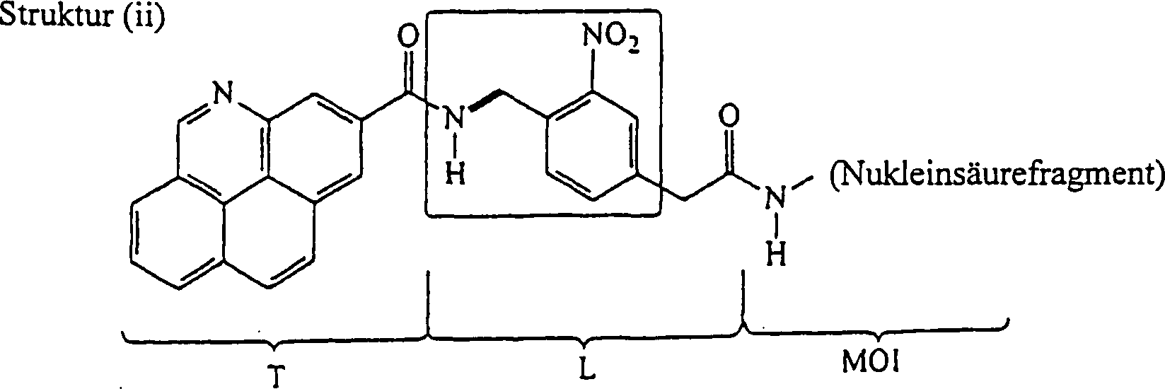
Es ist gut bekannt, daß Verbindungen mit einer ortho-Nitrobenzylamin-Einheit (siehe eingerahmte Atome in Struktur (ii)) photolytisch insofern instabil sind, als die Einwirkung aktinischer Strahlung einer spezifizierten Wellenlänge auf solche Verbindungen eine selektive Spaltung der Benzylamin-Bindung (siehe Bindung, die in Struktur (ii) mit einer dicken Linie bezeichnet ist) bewirkt. So hat Struktur (ii) dieselben T- und MOI-Gruppen wie Struktur (i) die Linker-Gruppe enthält jedoch mehrere Atome und Bindungen, innerhalb derer sich eine besonders labile Bindung befindet. Photoyse von Struktur (ii) setzt somit eine Markierungseinheit (T-enthaltende Einheit) vom Rest der Verbindung frei, wie unten dargestellt.It is well known that connections with an ortho-nitrobenzylamine unit (see boxed atoms in structure (ii)) are photolytically unstable insofar as the action actinic radiation of a specified wavelength such compounds selectively cleave the benzylamine bond (See bond that is marked with a thick line in structure (ii) is) effected. Structure (ii) has the same T and MOI groups as Structure (i), however, the linker group contains several atoms and Bonds within which there is a particularly unstable bond. Photoyse of structure (ii) thus sets a marking unit (T-containing unit) free from the rest of the connection as shown below.

![]()
![]()
Die Erfindung stellt somit Verbindungen zur Verfügung, die, bei Einwirkung geeigneter Spaltungsbedingungen, eine Spaltungsreaktion durchlaufen, so daß eine Markierungseinheit vom Rest der Verbindung freigesetzt wird. Verbindungen der Erfindung können als die Markierungseinheit, das MOI (oder eine Vorstufe dazu, Lh) und die labile(n) Bindung(en), die die zwei Gruppen miteinander verbindet (verbinden), beschrieben werden. Alternativ können die Verbindungen der Erfindung als die Komponenten beschrieben werden, aus denen sie gebildet sind. So können die Verbindungen wie folgt als das Reaktionsprodukt eines Markierungs-Reaktanten, eines Linker-Reaktanten und eines MOI-Reaktanten beschrieben werden.The invention thus provides compounds which, under the action of suitable cleavage conditions, undergo a cleavage reaction so that a labeling unit is released from the rest of the compound. Compounds of the invention can be described as the label moiety, the MOI (or a precursor to it, L h ), and the labile bond (s) connecting the two groups. Alternatively, the compounds of the invention can be described as the components from which they are formed. Thus, the compounds can be described as the reaction product of a labeling reactant, a linker reactant and an MOI reactant as follows.
Der Markierungs-Reaktant besteht
aus einem chemischen Griff (Th) und einer
variablen Komponente (Tvc), so daß der Markierungs-Reaktant
so betrachtet wird, daß er
die allgemeine Struktur hat:
Um diese Nomenklatur zu veranschaulichen,
kann Bezug genommen werden auf Struktur (iii), die einen Markieiungs-Reaktanten
zeigt, der verwendet werden kann, um die Verbindung von Struktur
(ii) herzustellen. Der Markierungs-Reaktant mit Struktur (iii) enthält eine
variable Markierungskomponente und einen Markierungsgriff, wie unten
dargestellt: 
![]()
![]()
In Struktur (iii) liefert der Markierungsgriff (-C(=O)-A) nur einen Weg für die Reaktion des Markierungs-Reaktanten mit dem Linker-Reaktanten, um eine T-L-Einheit zu bilden. Die Gruppe "A" in Struktur (iii) zeigt, daß die Carboxylgruppe sich in einem chemisch aktiven Zustand befindet, so daß sie für Kopplung mit anderen Griffen bereitsteht. "A" kann z. B. eine Hydroxylgruppe oder Pentafluorphenoxy sein, unter vielen anderen Möglichkeiten. Die Erfindung liefert eine große Anzahl möglicher Markierungsgriffe, die an eine variable Markierungskomponente gebunden werden können, wie im Detail unten diskutiert. Die variable Markierungskomponente ist somit ein Teil von "T" in der Formel T-L-X und wird auch Teil der Markierungseinheit sein, die sich aus der Reaktion bildet, die L spaltet.In structure (iii), the label handle (-C (= O) -A) provides only one way for the label reactant to react with the linker reactant to form a TL unit. Group "A" in structure (iii) shows that the carboxyl group is in a chemically active state so that it is ready for coupling with other handles. "A" can e.g. B. be a hydroxyl group or pentafluorophenoxy, among many other possibilities. The invention provides a large number of possible marking handles that are variable Marker components can be bound as discussed in detail below. The variable marker component is thus part of "T" in the TLX formula and will also be part of the marker unit that results from the reaction that cleaves L.
Wie ebenfalls unten genauer diskutiert,
wird die variable Markierungskomponente so genannt, weil es bei
der Herstellung von Sätzen
von Verbindungen gemäß der Erfindung
erwünscht
ist, daß Mitglieder
eines Satzes einzigartige variable Komponenten aufweisen, so daß die einzelnen
Mitglieder voneinander mit einer analytischen Technik unterschieden
werden können.
Als ein Beispiel kann die variable Markierungskomponente von Struktur
(iii) Mitglied des folgenden Satzes sein, in dem die Mitglieder
des Satzes durch ihre UV- oder Massenspektren unterschieden werden
können: 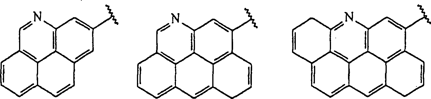
In ähnlicher Weise kann der Linker-Reaktant
anhand seiner chemischen Griffe (es gibt notwendigerweise wenigstens
zwei, von denen jeder als Lh bezeichnet
werden kann), beschrieben werden, die eine labile Linkerkomponente
flankieren, wobei die labile Linkerkomponente aus der erforderlichen
labilen Einheit (L2) und fakultativen labilen
Einheiten (L1 und L3)
besteht, wobei die fakultativen labilen Einheiten effektiv dazu
dienen, L2 von den Griffen Lh zu
trennen, und die erforderliche labile Einheit dazu dient, eine labile
Bindung innerhalb der labilen Linkerkomponente bereitzustellen.
So kann der Linker-Reaktant
so betrachtet werden, daß er
die allgemeine Formel hat:
Die Nomenklatur, die verwendet wird,
um den Linker-Reaktanten zu beschreiben, kann im Hinblick auf Struktur
(iv) veranschaulicht werden, die sich wieder von der Verbindung
von Struktur (ii) ableitet: 
Wie Struktur (iv) veranschaulicht, können Atome in mehr als eine funktionelle Rolle erfüllen. So dient, in Struktur (iv), der Benzylstickstoff als ein chemischer Griff, um zu ermöglichen, daß der Linker-Reaktant sich an den Markierungs-Reaktanten über eine Amidbildungsreaktion bindet, und dient anschließend auch insofern als ein notwendiger Teil der Struktur der labilen Einheit L2, als die Benzyl-Kohlenstoff-Stickstoff-Bindung besonders anfällig ist für photolytische Spaltung. Struktur (iv) veranschaulicht auch, daß ein Linker-Reaktant eine L3-Gruppe aufweisen kann (in diesem Fall eine Methylengruppe), obgleich er keine L1-Gruppe hat. In ähnlicher Weise können Linker-Reaktanten eine L1-Gruppe aufweisen, aber keine L3-Gruppe, oder können L1- und L3-Gruppen aufweisen oder können weder L1- noch L3-Gruppen aufweisen. In Struktur (iv) zeigt das Vorhandensein der Gruppe "P" neben der Carbonylgruppe, daß die Carbonylgruppe vor der Reaktion geschützt ist. Bei Vorgabe dieser Konfiguration kann die aktivierte Carboxylgruppe des Markierungs-Reaktanten (iii) sauber mit der Amingruppe des Linker-Reaktanten (iv) reagieren, um eine Amidbindung zu bilden und eine Verbindung der Formel T-L-Lh, zu ergeben.As structure (iv) illustrates, atoms can perform more than one functional role. Thus, in structure (iv), the benzyl nitrogen serves as a chemical handle to allow the linker reactant to bind to the label reactant via an amide formation reaction and subsequently also serves as a necessary part of the structure of the labile unit L 2 , as the benzyl-carbon-nitrogen bond is particularly susceptible to photolytic cleavage. Structure (iv) also illustrates that a linker reactant may have an L 3 group (in this case a methylene group), although it does not have an L 1 group. Similarly, linker reactants can have an L 1 group but no L 3 group, or can have L 1 and L 3 groups, or can have neither L 1 nor L 3 groups. In structure (iv), the presence of the group "P" next to the carbonyl group shows that the carbonyl group is protected from the reaction. Given this configuration, the activated carboxyl group of the labeling reactant (iii) can react properly with the amine group of the linker reactant (iv) to form an amide bond and give a compound of the formula TLL h .
Der MOI-Reaktant ist eine in geeigneter Weise reaktive Form eines interssierenden Moleküls. Wenn das interessierende Molekül ein Nukleinsäurefragment ist, ist ein geeigneter MOI-Reaktant ein Nukleinsäurefragment, das durch seine 5'-Hydroxylgruppe an eine Phosphodiestergruppe gebunden ist und anschließend an eine Alkylen-Kette, die in einer Aminogruppe endet. Diese Aminogruppe kann dann mit der Carbonylgruppe von Struktur (iv) reagieren (natürlich nach Entfernen der Schutzgruppe der Carbonylgruppe und vorzugsweise nach anschließender Aktivierung der Carbonylgruppe für die Reaktion mit der Amingruppe), um dadurch das MOI an den Linker zu binden.The MOI reactant is one suitable Wise reactive form of an interesting molecule. If that's interesting molecule a nucleic acid fragment is a suitable MOI reactant a nucleic acid fragment, that through its 5'-hydroxyl group is bound to a phosphodiester group and then to an alkylene chain that ends in an amino group. This amino group can then react with the carbonyl group of structure (iv) (naturally after Remove the protecting group from the carbonyl group and preferably after followed by Activation of the carbonyl group for reaction with the amine group) to thereby give the MOI to the linker to tie.
Wenn sie in einer chronologischen Reihenfolge betrachtet wird, kann die Erfindung so gesehen werden, daß sie einen Markierungs-Reaktanten (mit einem chemischen Markierungsgriff und einer variablen Markierungskomponente), einen Linker-Reaktanten (mit zwei chemischen Linkergriffen, einer erforderlichen labilen Einheit und 0–2 fakultativen labilen Einheiten) und einen MOI-Reaktanten (mit einer Komponente "Interessierendes Molekül" und einem chemischen Griff des interessierenden Moleküls) nimmt, um T-L-MOI zu bilden. So werden, um T-L-MOI zu bilden, entweder der Markierungs-Reaktant und der Linker-Reaktant zunächst miteinander umgesetzt, um T-L-Lh, zu liefern, und anschließend wird der MOI-Reaktant mit T-L-Lh umgesetzt, um T-L-MOI zu liefern, oder (weniger bevorzugt) werden der Linker-Reaktant und der MOI-Reaktant zunächst miteinander umgesetzt, um Lh-L-MOI zu liefern, und anschließend wird Lh-L-MOI mit dem Markierungs-Reaktanten umgesetzt, um T-L-MOI zu liefern. Aus Gründen der Bequemlichkeit werden Verbindungen mit der Formel T-L-MOI als der Markierungs-Reaktant, der Linker-Reaktant und der MOI-Reaktant beschrieben werden, die verwendet werden können, um solche Verbindungen zu bilden. Dieselben Verbindungen von Formel T-L-MOI könnten natürlich mit anderen (typischerweise aufwendigeren) Verfahren hergestellt werden und würden immer noch unter den Schutzumfang der erfinderischen T-L-MOI-Verbindungen fallen.When viewed in a chronological order, the invention can be seen to include a labeling reactant (with a chemical labeling handle and a variable labeling component), a linker reactant (with two chemical linkers, a required labile unit and 0 - 2 optional labile units) and an MOI reactant (with a "Molecule of Interest" component and a chemical handle of the molecule of interest) to form TL-MOI. Thus, to form TL-MOI, either the label reactant and the linker reactant are first reacted together to provide TLL h , and then the MOI reactant is reacted with TLL h to provide TL-MOI, or (less preferably) the linker reactant and the MOI reactant are first reacted together to provide Lh-L-MOI, and then Lh-L-MOI is reacted with the label reactant to provide TL-MOI. For convenience, compounds having the formula TL-MOI will be described as the labeling reactant, the linker reactant and the MOI reactant that can be used to form such compounds. The same compounds of formula TL-MOI could of course be made by other (typically more expensive) processes and would still fall within the scope of the inventive TL-MOI compounds.
In jedem Falle sorgt die Erfindung dafür, daß eine T-L-MOI-Verbindung Spaltungsbedingungen unterworfen wird, so daß eine Markierungseinheit vom Rest der Verbindung freigesetzt wird. Die Markierungseinheit wird wenigstens die variable Markierungskomponente umfassen und wird typischerweise zusätzlich einige oder alle Atome vom Markierungsgriff, einige oder alle Atome vom Linkergriff, die verwendet wurden, um das Markierungs-Reaktant an den Linker-Reaktanten zu binden, die fakultative labile Einheit L1, wenn diese Gruppe in T-L-MOI vorhanden war, umfassen und wird vielleicht einen Teil der erforderlichen labilert Einheit L2 enthalten, in Abhängigkeit von der genauen Struktur von L2 und der Natur der Spaltungschemie. Aus Gründen der Bequemlichkeit kann die Markierungseinheit als die T-enthaltende Einheit bezeichnet werden, weil T typischerweise den Hauptteil (im Hinblick auf die Masse) der Markierungseinheit ausmachen wird.In any event, the invention provides that a TL-MOI connection is subjected to cleavage conditions so that a marker is released from the rest of the connection. The labeling unit will include at least the variable labeling component and will typically additionally include some or all of the atoms from the marker handle, some or all of the atoms from the linker handle used to bind the label reactant to the linker reactants, the optional labile unit L 1 , if this group was present in TL-MOI, it may and may include part of the required labile unit L 2 , depending on the exact structure of L 2 and the nature of the cleavage chemistry. For convenience, the marker unit may be referred to as the T-containing unit because T will typically make up the bulk (in terms of mass) of the marker unit.
Nach dieser Einführung in einen Aspekt der vorliegenden Erfindung werden die verschiedenen Komponenten T, L und X im Detail beschrieben werden. Diese Beschreibung beginnt mit den folgenden Definitionen bestimmter Begriffe, die im weiteren bei der Beschreibung von T, L und X verwendet werden.After this introduction to one aspect of the present The various components T, L and X are invented in detail to be discribed. This description begins with the following Definitions of certain terms that are used in the description of T, L and X can be used.
Wie hierin verwendet, bedeutet der Begriff "Nukleinsäurefragment" ein Molekül, das komplementär zu einem ausgewählten Target-Nukleinsäuremolekül ist (d. h. komplementär zu dem gesamten Molekül oder einem Teil desselben) und aus der Natur gewonnen oder synthetisch oder rekombinant hergestellt werden kann, einschließlich nicht natürlich vorkommender Moleküle, und kann je nach Eignung in doppel- oder einzelsträngiger Form vorliegen; und schließt ein Oligonukleotid (z. B. DNA oder RNA), einen Primer, eine Sonde, ein Nukleinsäureanalog (z. B. PNA), ein Oligonukleotid, das in einer 5'-nach-3'-Richtung mit einer Polymerase verlängert ist, eine Nukleinsäure, die chemisch oder enzymatisch gespalten ist, eine Nukleinsäure, die mit einem Didesoxyterminator endet oder am 3'- oder 5'-Ende mit einer Verbindung abgedeckt ist, die Polymerisation am 5'- oder 3'-Ende verhindert, oder Kombinationen derselben ein. Die Komplimentarität eines Nukleinsäurefragments zu einem ausgewählten Target-Nukleinsäuremolekül bedeutet im allgemeinen, daß wenigstens etwa 70% spezifische Basenpaarung über die gesamte Länge des Fragments gezeigt wird. Vorzugsweise zeigt das Nukleinsäurefragment wenigstens etwa 80% spezifische Basenpaarung; und am bevorzugtesten wenigstens etwa 90%. Assays zum Bestimmen des prozentualen Mismatch (und somit der prozentualen spezifischen Basenpaarung) sind im Stand der Technik gut bekannt und beruhen auf dem prozentualen Mismatch als einer Funktion des Tm, wenn bezogen auf eine Kontrolle mit vollständiger Basenpaarung.As used herein, the Term "nucleic acid fragment" a molecule that is complementary to one chosen Is target nucleic acid molecule (i.e. H. complementary to the whole molecule or a part of it) and obtained from nature or synthetically or can be produced recombinantly, including not Naturally occurring molecules, and can be double or single-stranded depending on the suitability available; and closes an oligonucleotide (e.g. DNA or RNA), a primer, a probe, a nucleic acid analog (e.g. PNA), an oligonucleotide extended in a 5 'to 3' direction with a polymerase, a nucleic acid, which is chemically or enzymatically cleaved, a nucleic acid which ends with a dideoxy terminator or covered at the 3 'or 5' end with a compound is the polymerization at 5'- or 3 'end prevented or combinations thereof. The complimentarity of a nucleic acid fragment to a selected one Target nucleic acid molecule means in general that at least about 70% specific base pairing over the entire length of the Fragments is shown. The nucleic acid fragment preferably shows at least about 80% specific base pairing; and most preferred at least about 90%. Percentage Mismatch Assays (and thus the percentage specific base pairing) are in the state well known in the art and based on the percentage mismatch as a function of Tm when related to a complete base pairing control.
Wie hierin verwendet, bezieht sich der Begriff "Alkyl", allein oder in Kombination, auf einen gesättigten, geradekettigen oder verzweigtkettigen Kohlenwasserstoffrest, der von 1 bis 10, vorzugsweise von 1 bis 6 und bevorzugter von 1 bis 4 Kohlenstoffatome enthält. Beispiele für solche Reste schließen Methyl, Ethyl, n-Propyl, iso-Propyl, n-Butyl, iso-Butyl, sec.-Butyl, tert.-Butyl, Pentyl, iso-Amyl, Hexyl, Decyl und dergleichen ein, sind aber nicht hierauf beschränkt. Der Begriff "Alkylen" bezieht sich auf einen gesättigten, geradkettigen oder verzweigtkettigen Kohlenwasserstoffdirest, der von 1 bis 10, vorzugsweise von 1 bis 6 und bevorzugter von 1 bis 4 Kohlenstoffe enthält. Beispiele für solche einen Direst schließen Methylen, Ethylen (-CH2-CH2-), Propylen und dergleichen ein, sind aber nicht hierauf beschränkt.As used herein, the term "alkyl", alone or in combination, refers to a saturated, straight chain or branched chain hydrocarbon residue containing from 1 to 10, preferably from 1 to 6, and more preferably from 1 to 4 carbon atoms. Examples of such residues include, but are methyl, ethyl, n-propyl, iso-propyl, n-butyl, iso-butyl, sec-butyl, tert-butyl, pentyl, iso-amyl, hexyl, decyl and the like not limited to this. The term "alkylene" refers to a saturated, straight chain or branched chain hydrocarbon radical containing from 1 to 10, preferably from 1 to 6 and more preferably from 1 to 4 carbons. Examples of such a direst include, but are not limited to, methylene, ethylene (-CH 2 -CH 2 -), propylene, and the like.
Der Begriff "Alkenyl", allein oder in Kombination, bezieht sich auf einen geradkettigen oder verzweigtkettigen Kohlenwasserstoffrest mit wenigstens einer Kohlenstoff-Kohlenstoff-Doppelbindung in insgesamt von 2 bis 10, vorzugsweise von 2 bis 6 und bevorzugter von 2 bis 4 Kohlenstoffatomen. Beispiele für solche Reste schließen Ethenyl, E- und Z-Propenyl, Isopropenyl, E- und Z-Butenyl, E- und Z-Isobutenyl, E- und Z-Pentenyl, Decenyl und dergleichen ein, sind aber nicht hierauf beschränkt. Der Begriff "Alkenylen" bezieht sich auf einen geradkettigen oder verzweigtkettigen Kohlenwasserstoffdirest mit wenigstens einer Kohlenstoff-Kohlenstoff-Doppelbindung in insgesamt von 2 bis 10, vorzugsweise von 2 bis 6 und bevorzugter von 2 bis 4 Kohlenstoffatomen. Beispiele für solche Direste schließen Methyliden (=CH2), Ethyliden (-CH=CH-), Propyliden (-CH2-CH=CH-) und dergleichen ein, sind aber nicht hierauf beschränkt.The term "alkenyl", alone or in combination, refers to a straight-chain or branched-chain hydrocarbon radical with at least one carbon-carbon double bond in total from 2 to 10, preferably from 2 to 6 and more preferably from 2 to 4, carbon atoms. Examples of such groups include, but are not limited to, ethenyl, E- and Z-propenyl, isopropenyl, E- and Z-butenyl, E- and Z-isobutenyl, E- and Z-pentenyl, decenyl and the like. The term "alkenylene" refers to a straight or branched chain hydrocarbon radical having at least one carbon-carbon double bond in total from 2 to 10, preferably from 2 to 6 and more preferably from 2 to 4 carbon atoms. Examples of such radicals include, but are not limited to, methylidene (= CH 2 ), ethylidene (-CH = CH-), propylidene (-CH 2 -CH = CH-) and the like.
Der Begriff "Alkinyl", allein oder in Kombination, bezieht sich auf einen geradekettigen oder verzweigtkettigen Kohlenwasserstoffrest mit wenigstens einer Kohlenstoff-Kohlenstoff-Dreifachbindung in insgesamt von 2 bis 10, vorzugsweise von 2 bis 6 und bevorzugter von 2 bis 4 Kohlenstoffatomen. Beispiele für solche Reste schließen Ethinyl (Acetylenyl), Propinyl (Propargyl), Butinyl, Hexinyl, Decinyl und dergleichen ein, sind aber nicht hierauf beschränkt. Der Begriff "Alkinylen", allein oder in Kombination, bezieht sich auf einen geradkettigen oder verzweigtkettigen Kohlenwasserstoffdirest mit wenigstens einer Kohlenstoff-Kohlenstoff-Dreifachbindung in insgesamt von 2 bis 10, vorzugsweise von 2 bis 6 und bevorzugter von 2 bis 4 Kohlenstoffatomen. Beispiele für solche Reste schließen Ethinylen (-C≡C-), Propinylen (-CH2-C≡C-) und dergleichen ein, sind aber nicht hierauf beschränkt.The term "alkynyl", alone or in combination, refers to a straight-chain or branched-chain hydrocarbon radical with at least one carbon-carbon triple bond in total from 2 to 10, preferably from 2 to 6 and more preferably from 2 to 4 carbon atoms. Examples of such groups include, but are not limited to, ethynyl (acetylenyl), propynyl (propargyl), butynyl, hexynyl, decynyl, and the like. The term "alkynylene", alone or in combination, refers to a straight-chain or branched-chain hydrocarbon direst with at least one carbon-carbon triple bond in total from 2 to 10, preferably from 2 to 6 and more preferably from 2 to 4 carbon atoms. Examples of such radicals include, but are not limited to, ethynylene (-C≡C-), propinylene (-CH 2 -C≡C-) and the like.
Der Begriff "Cycloalkyl", allein oder in Kombination, bezieht sich auf eine gesättigte cyclische Anordnung von Kohlenstoffatomen, mit einer Anzahl von 3 bis 8 und vorzugsweise von 3 bis 6 Kohlenstoffatomen. Beispiele für solche Cycloalkylreste schließen Cyclopropyl, Cyclobutyl, Cyclopentyl, Cyclohexyl und dergleichen ein, sind aber nicht hierauf beschränkt. Der Begriff "Cycloalkylen" bezeichnet eine Direstform eines Cycloalkyls.The term "cycloalkyl", alone or in combination, refers on a saturated cyclic arrangement of carbon atoms, with a number of 3 to 8 and preferably 3 to 6 carbon atoms. Examples for such Close cycloalkyl residues Cyclopropyl, cyclobutyl, cyclopentyl, cyclohexyl and the like one, but are not limited to this. The term "cycloalkylene" denotes one Direct form of a cycloalkyl.
Der Begriff "Cycloalkenyl", allein oder in Kombination, bezieht sich auf einen cyclischen Kohlenstoffring, der von 4 bis 8, vorzugsweise 5 oder 6, Kohlenstoffatome und eine oder mehrere Doppelbindungen enthält. Beispiele für solche Cycloalkenylreste schließen Cyclopentenyl, Cyclohexenyl, Cyclopentadienyl und dergleichen ein, sind aber nicht hierauf beschränkt. Der Begriff "Cycloalkenylen" bezieht sich auf eine Direstform eines Cycloalkenyls.The term "cycloalkenyl", alone or in combination, refers refer to a cyclic carbon ring that is from 4 to 8, preferably 5 or 6, carbon atoms and one or more double bonds contains. examples for close such cycloalkenyl radicals Cyclopentenyl, cyclohexenyl, cyclopentadienyl and the like, but are not limited to this. The term "cycloalkenylene" refers to a direct form of a cycloalkenyl.
Der Begriff "Aryl" bezieht sich auf eine carbocyclische (vollständig aus Kohlenstoff und Wasserstoff bestehende) aromatische Gruppe, die ausgewählt ist aus der Gruppe, die aus Phenyl, Naphthyl, Indenyl, Indanyl, Azulenyl, Fluorenyl und Anthracenyl besteht; oder eine heterocyclische aromatische Gruppe, die ausgewählt ist aus der Gruppe, die aus Furyl, Thienyl, Pyridyl, Pyrrolyl, Oxazolyl, Thiazolyl, Imidazolyl, Pyrazolyl, 2-Pyrazolinyl, Pyrazolidinyl, Isoxazolyl, Isothiazolyl, 1,2,3-Oxadiazolyl, 1,2,3-Triazolyl, 1,3,4-Thiadiazolyl, Pyridazinyl, Pyrimidinyl, Pyrazinyl, 1,3,5-Triazinyl, 1,3,5-Trithianyl, Indolizinyl, Indolyl, Isoindolyl, 3H-Indolyl, Indolinyl, Benzo[b]furanyl, 2,3-Dihydrobenzofuranyl, Benzo[b]thiophenyl, 1H-Indazolyl, Benzimidazolyl, Benzthiazolyl, Purinyl, 4H-Chinolizinyl, Chinolinyl, Isochinolinyl, Cinnolinyl, Phthalazinyl, Chinazolinyl, Chinoxalinyl, 1,8-Naphthyridinyl, Pteridinyl, Carbazolyl, Acridinyl, Phenazinyl, Phenothiazinyl und Phenoxazinyl besteht.The term "aryl" refers relying on a carbocyclic (made entirely of carbon and hydrogen existing) aromatic group selected from the group consisting of from phenyl, naphthyl, indenyl, indanyl, azulenyl, fluorenyl and Anthracenyl exists; or a heterocyclic aromatic group, which is selected from the group consisting of furyl, thienyl, pyridyl, pyrrolyl, oxazolyl, Thiazolyl, imidazolyl, pyrazolyl, 2-pyrazolinyl, pyrazolidinyl, Isoxazolyl, isothiazolyl, 1,2,3-oxadiazolyl, 1,2,3-triazolyl, 1,3,4-thiadiazolyl, Pyridazinyl, pyrimidinyl, pyrazinyl, 1,3,5-triazinyl, 1,3,5-trithianyl, Indolizinyl, indolyl, isoindolyl, 3H-indolyl, indolinyl, benzo [b] furanyl, 2,3-dihydrobenzofuranyl, benzo [b] thiophenyl, 1H-indazolyl, benzimidazolyl, Benzothiazolyl, purinyl, 4H-quinolizinyl, quinolinyl, isoquinolinyl, Cinnolinyl, phthalazinyl, quinazolinyl, quinoxalinyl, 1,8-naphthyridinyl, pteridinyl, Carbazolyl, acridinyl, phenazinyl, phenothiazinyl and phenoxazinyl consists.
"Aryl"-Gruppen, wie in dieser Anmeldung definiert, können unabhängig einen bis vier Substituenten enthalten, die unabhängig ausgewählt sind aus der Gruppe, die aus Wasserstoff, Halogen, Hydroxyl, Amino, Nitro, Trifluormethyl, Trifluormethoxy, Alkyl, Alkenyl, Alkinyl, Cyano, Carboxy, Carboalkoxy, 1,2-Dioxyethylen, Alkoxy, Alkenoxy oder Alkinoxy, Alkylamino, Alkenylamino, Alkinylamino, aliphatischem oder aromatischem Acyl, Alkoxycarbonylamino, Alkylsulfonylamino, Morpholinocarbonylamino, Thiomorpholinocarbonylamino, N-Alkylguanidino, Aralkylaminosulfonyl; Aralkoxyalkyl; N-Aralkoxyharnstoff; N-Hydroxylharnstoff; N-Alkenylharnstoff; N,N-(Alkyl, Hydroxyl)harnstoff Heterocyclyl; Thioaryloxy-substituiertem Aryl; N,N-(Aryl, Alkyl)hydrazino; Ar'-substituiertem Sulfonylheterocyclyl; Aralkyl-substituiertem Heterocyclyl; Cycloalkyl- und Cycloalkenyl-substituiertem Heterocyclyl; Cycloalkyl-kondensiertem Aryl; Arlyoxy-substituiertem Alkyl; Heterocyclylamino; aliphatischem oder aromatischem Acylaminocarbonyl; aliphatischem oder aromatischem Acyl-substituiertem Alkenyl; Ar'-substituiertem Aminocarbonyloxy; Ar', Ar'-disubstituiertem Aryl; aliphatischem oder aromatischem Acyl-substituiertem Acyl; Cycloalkylcarbonylalkyl; Cycloalkyl-substituiertem Amino; Aryloxycarbonylalkyl; Phosphorodiamidylsäure oder -ester besteht."Aryl" groups as in defined in this application independently contain one to four substituents which are independently selected from the group consisting of hydrogen, halogen, hydroxyl, amino, nitro, Trifluoromethyl, trifluoromethoxy, alkyl, alkenyl, alkynyl, cyano, Carboxy, carboalkoxy, 1,2-dioxyethylene, alkoxy, alkenoxy or alkynoxy, Alkylamino, alkenylamino, alkynylamino, aliphatic or aromatic acyl, Alkoxycarbonylamino, alkylsulfonylamino, morpholinocarbonylamino, Thiomorpholinocarbonylamino, N-alkylguanidino, aralkylaminosulfonyl; aralkoxyalkyl; N-aralkoxyurea; N-hydroxylurea; N-alkenylurea; N, N- (alkyl, hydroxyl) urea heterocyclyl; Thioaryloxy substituted aryl; N, N- (aryl, alkyl) hydrazino; Ar 'substituted sulfonylheterocyclyl; Aralkyl-substituted heterocyclyl; cycloalkyl and cycloalkenyl substituted heterocyclyl; Cycloalkyl fused aryl; Arlyoxy-substituted alkyl; heterocyclylamino; aliphatic or aromatic acylaminocarbonyl; aliphatic or aromatic Acyl substituted alkenyl; Ar 'substituted aminocarbonyloxy; Ar ', Ar 'disubstituted aryl; aliphatic or aromatic acyl-substituted acyl; cycloalkylcarbonylalkyl; Cycloalkyl substituted amino; aryloxycarbonylalkyl; Phosphorodiamidylsäure or ester exists.
"Ar" ist eine carbocyclische oder heterocyclische Arylgruppe, wie oben definiert, mit einem bis drei Substituenten, die ausgewählt sind aus der Gruppe, die aus Wasserstoff, Halogen, Hydroxyl, Amino, Nitro, Trifluormethyl, Trifluormethoxy, Alkyl, Alkenyl, Alkinyl, 1,2-Dioxymethylen, 1,2-Dioxyethylen, Alkoxy, Alkenoxy, Alkinoxy, Alkylamino, Alkenylamino oder Alkinylamino, Alkylcarbonyloxy, aliphatischem oder aromatischem Acyl, Alkylcarbonylamino, Alkoxycarbonylamino, Alkylsulfonylamino, N-Alkyl- oder N,N-Dialkylharnstoff besteht."Ar" is a carbocyclic or heterocyclic aryl group as defined above with one to three substituents selected are from the group consisting of hydrogen, halogen, hydroxyl, amino, Nitro, trifluoromethyl, trifluoromethoxy, alkyl, alkenyl, alkynyl, 1,2-dioxymethylene, 1,2-dioxyethylene, alkoxy, alkenoxy, alkynoxy, alkylamino, alkenylamino or alkynylamino, alkylcarbonyloxy, aliphatic or aromatic acyl, Alkylcarbonylamino, alkoxycarbonylamino, alkylsulfonylamino, N-alkyl- or N, N-dialkylurea consists.
Der Begriff "Alkoxy", allein oder in Kombination, bezieht sich auf ein Alkyletherrest, wobei der Begriff "Alkyl" wie oben definiert ist. Beispiele für geeignete Alkyletherreste schließen Methoxy, Ethoxy, n-Propoxy, iso-Propoxy, n-Butoxy, iso-Butoxy, sec.-Butoxy, tert.-Butoxy und dergleichen ein, sind aber nicht hierauf beschränkt.The term "alkoxy", alone or in combination, refers refers to an alkyl ether radical, the term "alkyl" being as defined above. Examples of suitable ones Close alkyl ether residues Methoxy, ethoxy, n-propoxy, iso-propoxy, n-butoxy, iso-butoxy, sec.-butoxy, tert-butoxy and the like, but are not limited to this.
Der Begriff "Alkenoxy", allein oder in Kombination, bezieht sich auf einen Rest der Formel Alkenyl-O-, wobei der Begriff "Alkenyl" wie oben definiert ist, vorausgesetzt, daß der Rest kein Enolether ist. Beispiele für geeignete Alkenoxyreste schließen Allyloxy, E- und Z-3-Methyl-2-propenoxy und dergleichen ein, sind aber nicht hierauf beschränkt.The term "alkenoxy", alone or in combination, refers refer to a radical of the formula alkenyl-O-, the term "alkenyl" as defined above is provided that the Rest is not an enol ether. Examples of suitable alkenoxy radicals include allyloxy, E- and Z-3-methyl-2-propenoxy and the like, but are not limited to this.
Der Begriff "Alkinyloxy", allein oder in Kombination, bezieht sich auf einen Rest der Formel Alkinyl-O-, wobei der Begriff "Alkinyl" wie oben definiert ist, vorausgesetzt, daß der Rest kein Inolether ist. Beispiele für geeignete Alkinoxyreste schließen Propargyloxy, 2-Butinyloxy und dergleichen ein, sind aber nicht hierauf beschränkt.The term "alkynyloxy", alone or in combination, refers refer to a radical of the formula alkynyl-O-, the term "alkynyl" as defined above is provided that the Rest is not an inol ether. Examples of suitable alkyneoxy radicals include propargyloxy, 2-butynyloxy and the like, but are not limited to this.
Der Begriff "Thioalkoxy" bezieht sich auf einen Thioetherrest der Formel Alkyl-S-, wobei Alkyl wie oben definiert ist.The term "thioalkoxy" refers to a thioether residue of the formula alkyl-S-, where alkyl is as defined above.
Der Begriff "Alkylamino", allein oder in Kombination, bezieht sich auf einen Mono- oder Dialkyl-substituierten Aminorest (d. h. einen Rest der Formel Alkyl-NH- oder (Alkyl)2-N-), wobei der Begriff "Alkyl" wie oben definiert ist. Beispiele für geeignete Alkylaminoreste schließen Methylamino, Ethylamino, Propylamino, Isopropylamino, tert.-Butylamino, N,N-Diethylamino und dergleichen ein, sind aber nicht hierauf beschränkt.The term "alkylamino", alone or in combination, refers to a mono- or dialkyl-substi tuiert amino residue (ie a residue of the formula alkyl-NH- or (alkyl) 2 -N-), wherein the term "alkyl" is as defined above. Examples of suitable alkylamino radicals include, but are not limited to, methylamino, ethylamino, propylamino, isopropylamino, tert-butylamino, N, N-diethylamino and the like.
Der Begriff "Alkenylamino", allein oder in Kombination, bezieht sich auf einen Rest der Formel Alkenyl-NH- oder (Alkenyl)2N-, wobei der Begriff "Alkenyl" wie oben definiert ist, vorausgesetzt, daß der Rest kein Enamin ist. Ein Beispiel für solche Alkenylaminoreste ist der Allylaminorest.The term "alkenylamino", alone or in combination, refers to a residue of the formula alkenyl-NH- or (alkenyl) 2 N-, the term "alkenyl" being as defined above, provided that the residue is not an enamine. An example of such alkenylamino residues is the allylamino residue.
Der Begriff "Alkinylamino", allein oder in Kombination, bezieht sich auf einen Rest der Formel Alkinyl-NH- oder (Alkinyl)2N-, in der der Begriff "Alkinyl" wie oben definiert ist, vorausgesetzt, daß der Rest kein Inamin ist. Ein Beispiel für solche Alkinylaminoreste ist der Propargylaminorest.The term "alkynylamino", alone or in combination, refers to a residue of the formula alkynyl-NH- or (alkynyl) 2 N-, in which the term "alkynyl" is as defined above, provided that the residue is not an inamine , An example of such alkynylamino residues is the propargylamino residue.
Der Begriff "Amid" bezieht sich entweder auf -N(R1)-C(=O)- oder -C(=O)-N(R1)-, wobei R1 hierin so definiert ist, daß es Wasserstoff sowie andere Gruppen einschließt. Der Begriff "substituiertes Amid" bezieht sich auf die Situation, in der R1 nicht Wasserstoff ist, während der Begriff "unsubstituiertes Amid" sich auf die Situation bezieht, in der R1 Wasserstoff ist.The term "amide" refers to either -N (R 1 ) -C (= O) - or -C (= O) -N (R 1 ) -, where R 1 is defined herein to be hydrogen and others Includes groups. The term "substituted amide" refers to the situation in which R 1 is not hydrogen, while the term "unsubstituted amide" refers to the situation in which R 1 is hydrogen.
Der Begriff "Aryloxy", allein oder in Kombination, bezieht sich auf einen Rest der Formel Aryl-O-, wobei Aryl wie oben definiert ist. Beispiele für Aryloxyreste schließen Phenoxy, Naphthoxy, Pyridyloxy und dergleichen ein, sind aber nicht hierauf beschränkt.The term "aryloxy", alone or in combination, refers refers to a radical of the formula aryl-O-, where aryl is as defined above is. examples for Close aryloxy residues Phenoxy, naphthoxy, pyridyloxy and the like, but are not limited to this.
Der Begriff "Arylamino", allein oder in Kombination, bezieht sich auf einen Rest der Formel Aryl-NH-, in der Aryl wie oben definiert ist. Beispiele für Arylaminoreste schließen Phenylamino (Anilido), Naphthylamino, 2-, 3- und 4-Pyridylamino und dergleichen ein, sind aber nicht hierauf beschränkt.The term "arylamino", alone or in combination, refers refers to a radical of the formula aryl-NH-, in which aryl is as defined above is. examples for Close arylamino residues Phenylamino (anilido), naphthylamino, 2-, 3- and 4-pyridylamino and the like, but are not limited to this.
Der Begriff "Aryl-kondensiertes Cycloalkyl", allein oder in Kombination, bezieht sich auf einen Cycloalkylrest, der zwei benachbarte Atome mit einem Arylrest teilt, wobei die Begriffe "Cycloalkyl" und "Aryl" wie oben definiert sind. Ein Beispiel eines Aryl-kondensierten Cycloalkylrestes ist der Benzo-kondensierte Cyclobutylrest.The term "aryl-fused cycloalkyl", alone or in Combination, refers to a cycloalkyl group that has two neighboring ones Shares atoms with an aryl radical, the terms "cycloalkyl" and "aryl" as defined above are. An example of an aryl condensed Cycloalkyl is the benzo-fused cyclobutyl.
Der Begriff "Alkylcarbonylamino", allein oder in Kombination, bezieht sich auf einen Rest der Formel Alkyl-CONH, wobei der Begriff "Alkyl" wie oben definiert ist.The term "alkylcarbonylamino", alone or in combination, refers refers to a radical of the formula alkyl-CONH, the term "alkyl" as defined above is.
Der Begriff "Alkoxycarbonylamino", allein oder in Kombination, bezieht sich auf einen Rest der Formel Alkyl-OCONH-, wobei der Begriff "Alkyl" wie oben definiert ist.The term "alkoxycarbonylamino", alone or in combination, refers refer to a radical of the formula alkyl-OCONH-, the term "alkyl" as defined above is.
Der Begriff "Alkylsulfonylamino", allein oder in Kombination, bezieht sich auf einen Rest der Formel Alkyl-SO2NH-, wobei der Begriff "Alkyl" wie oben definiert ist.The term "alkylsulfonylamino", alone or in combination, refers to a radical of the formula alkyl-SO 2 NH-, the term "alkyl" being as defined above.
Der Begriff "Arylsulfonylamino", allein oder in Kombination, bezieht sich auf einen Rest der Formel Aryl-SO2NH-, wobei der Begriff "Aryl" wie oben definiert ist.The term "arylsulfonylamino", alone or in combination, refers to a radical of the formula aryl-SO 2 NH-, the term "aryl" being as defined above.
Der Begriff "N-Alkylharnstoff", allein oder in Kombination, bezieht sich auf einen Rest der Formel Alkyl-NH-CO-NH-, wobei der Begriff "Alkyl" wie oben definiert ist.The term "N-alkyl urea", alone or in combination, relates refer to a radical of the formula alkyl-NH-CO-NH-, the term "alkyl" as defined above is.
Der Begriff "N-Arylharnstoff", allein oder in Kombination, bezieht sich auf einen Rest der Formel Aryl-NH-CO-NH-, wobei der Begriff "Aryl" wie oben definiert ist.The term "N-arylurea", alone or in combination, relates refer to a radical of the formula aryl-NH-CO-NH-, the term "aryl" as defined above is.
Der Begriff "Halogen" bedeutet Fluor, Chlor, Brom und Iod.The term "halogen" means fluorine, chlorine, bromine and iodine.
Der Begriff "Kohlenwasserstoffrest" bezieht sich auf eine Anordnung von Kohlenstoff- und Wasserstoffatomen, die nur ein einziges Wasserstoffatom benötigt, um ein unabhängiges stabiles Molekül zu sein. Somit hat ein Kohlenwasserstoffrest eine offene Valenzstelle an einem Kohlenstoffatom, durch die der Kohlenwasserstoffrest an ein anderes Atom (andere Atome) gebunden werden kann. Alkyl, Alkenyl, Cycloalkyl, etc. sind Beispiele für Kohlenwasserstoffreste.The term "hydrocarbon residue" refers to an arrangement of carbon and hydrogen atoms that only one only one hydrogen atom needed, to an independent stable molecule to be. A hydrocarbon residue thus has an open valence point on a carbon atom through which the hydrocarbon residue is attached another atom (other atoms) can be bound. Alkyl, alkenyl, Cycloalkyl, etc. are examples of hydrocarbon residues.
Der Begriff "Kohlenwasserstoffdirest" bezieht sich auf eine Anordnung von Kohlenstoff- und Wasserstoffatomen, die zwei Wasserstoffatome benötigt, um ein unabhängiges stabiles Molekül zu sein. Somit hat ein Kohlenwasserstoffrest zwei offene Valenzstellen an einem oder zwei Kohlenstoffatomen, durch die der Kohlenwasserstoffrest an ein anderes Atom (andere Atome) gebunden werden kann. Alkylen, Alkenylen, Alkinylen, Cycloalkylen, etc. sind Beispiele für Kohlenwasserstoffdireste.The term "hydrocarbon direct" refers to an arrangement of carbon and hydrogen atoms, the two Hydrogen atoms needed to be an independent stable molecule to be. A hydrocarbon residue thus has two open valence points on one or two carbon atoms through which the hydrocarbon residue can be bound to another atom (other atoms). alkylene, Alkenylene, alkynylene, cycloalkylene, etc. are examples of direct hydrocarbon radicals.
Der Begriff "Hydrocarbyl" bezieht sich auf jede stabile Anordnung, die vollständig aus Kohlenstoff und Wasserstoff besteht, mit einer einzelnen Valenzstelle, an der sie an eine andere Einheit gebunden ist, und schließt somit Reste ein, die als Alkyl, Alkenyl, Alkinyl, Cycloalkyl, Cycloalkenyl, Aryl (ohne Einbeziehung von Heteroatomen in den Arylring), Arylalkyl, Alkylaryl und dergleichen ein. Kohlenwasserstoffrest ist ein anderer Name für Hydrocarbyl.The term "hydrocarbyl" refers to any stable arrangement the complete consists of carbon and hydrogen, with a single valence point, to which it is bound to another unit, and thus closes Radicals which are alkyl, alkenyl, alkynyl, cycloalkyl, cycloalkenyl, Aryl (without inclusion of heteroatoms in the aryl ring), arylalkyl, Alkylaryl and the like. Hydrocarbon residue is another name for hydrocarbyl.
Der Begriff "Hydrocarbylen" bezieht sich auf jede stabile Anordnung, die vollständig aus Kohlenstoff und Wasserstoff besteht, mit zwei Valenzstellen, an der sie an andere Einheiten gebunden ist, und schließt somit Alkylen, Alkenylen, Alkinylen, Cycloalkylen, Cycloalkenylen, Arylen (ohne Einbeziehung von Heteroatomen in den Arylring), Arylalkylen, Alkylarylen und dergleichen ein. Kohlenwasserstoffdirest ist ein anderer Name für Hydrocarbylen.The term "hydrocarbylene" refers to any stable arrangement the complete consists of carbon and hydrogen, with two valence points, to which it is bound to other units, and thus closes Alkylene, alkenylene, alkynylene, cycloalkylene, cycloalkenylene, arylene (without inclusion of heteroatoms in the aryl ring), arylalkylene, Alkylarylene and the like. Hydrocarbon direct is a another name for hydrocarbylene.
Der Begriff "Hydrocarbyl-O-hydrocarbylen" bezieht sich auf eine Hydrocarbyl-Gruppe, die an ein Sauerstoffatom gebunden ist, wobei das Sauerstoffatom in ähnlicher Weise an eine Hydrocarbylen-Gruppe an einer der zwei Valenzstellen gebunden ist, an der die Hydrocarbylen-Gruppe an andere Einheiten gebunden ist. Die Begriffe "Hydrocarbyl-S-hydrocarbylen", "Hydrocarbyl-NH-hydrocarbylen" und "Hydrocarbyl-Amid-hydrocarbylen" haben äquivalente Bedeutungen, wobei Sauerstoff durch Schwefel, -NH- bzw. eine Amidgruppe ersetzt worden ist.The term "hydrocarbyl-O-hydrocarbylene" refers to a hydrocarbyl group attached to an oxygen atom, where the oxygen atom is similarly attached to a hydrocarbylene group at one of the two valence sites to which the hydrocarbylene group attaches other units is bound. The terms "hydrocarbyl-S-hydrocarbylene", "hydrocarbyl-NH-hydrocarbylene" and "hydrocarbyl-amide-hydro carbylene "have equivalent meanings, where oxygen has been replaced by sulfur, -NH- or an amide group.
Der Begriff N-(Hydrocarbyl)hydrocarbylen bezieht sich auf eine Hydrocarbylen-Gruppe, wobei eine der zwei Valenzstellen an ein Stickstoffatom gebunden ist und dieses Stickstoffatom gleichzeitig an einen Wasserstoff und eine Hydrocarbyl-Gruppe gebunden ist.The term N- (hydrocarbyl) hydrocarbylene refers to a hydrocarbylene group, one of the two Valence points is bound to a nitrogen atom and this nitrogen atom bound simultaneously to a hydrogen and a hydrocarbyl group is.
Der Begriff N,N-Di(hydrocarbyl)hydrocarbylen bezieht sich auf eine Hydrocarbylengruppe, wobei eine der zwei Valenzstellen an ein Stickstoffatom gebunden ist und dieses Stickstoffatom gleichzeitig an zwei Hydrocarbyl-Gruppen gebunden ist.The term N, N-Di (hydrocarbyl) hydrocarbylene refers to a hydrocarbylene group with one of the two valence sites is bound to a nitrogen atom and this nitrogen atom at the same time is attached to two hydrocarbyl groups.
Der Begriff "Hydrocarbylacyl-hydrocarbylen" bezieht sich auf eine Hydrocarbyl-Gruppe, die durch eine Acyl(-C(≡O)-)-Gruppe an eine der zwei Valenzstellen einer Hydrocarbylen-Gruppe gebunden ist.The term "hydrocarbyl acyl hydrocarbylene" refers to a hydrocarbyl group which is attached to one of the two by an acyl (-C (≡O) -) group Valence points of a hydrocarbylene group is bound.
Die Begriffe "Heterocyclylhydrocarbyl" und "Heterocyclyl" beziehen sich auf eine stabile cyclische Anordnung von Atomen, die Kohlenstoffatome und bis zu vier Atome (als Heteroatome bezeichnet) einschließt, die ausgewählt sind aus Sauerstoff, Stickstoff, Phosphor und Schwefel. Die cyclische Anordnung kann in der Form eines monocyclischen Ringes mit 3–7 Atomen oder eines bicyclischen Ringes mit 8–11 Atomen vorliegen. Die Ringe können gesättigt oder ungesättigt sein (einschließlich aromatischer Ringe) und können fakultativ Benzo-kondensiert sein. Stickstoff- und Schwefelatomen im Ring können in jeder oxidierten Form vorliegen, einschließlich der quaternisierten Form von Stickstoff. Ein Heterocyclylhydrocarbyl kann an irgendeinem endocyclischen Kohlenstoff oder Heteroatom gebunden sein, das zur Schaffung einer stabilen Struktur führt. Bevorzugte Heterocyclylhydrocarbyle schließen 5- bis 7-gliedrige monocyclische Heterocyclen ein, die ein oder zwei Stickstoff-Heteroatome enthalten.The terms "heterocyclylhydrocarbyl" and "heterocyclyl" refer to a stable cyclic arrangement of atoms, the carbon atoms and includes up to four atoms (referred to as heteroatoms) that selected are made of oxygen, nitrogen, phosphorus and sulfur. The cyclic The arrangement can be in the form of a monocyclic ring with 3-7 atoms or a bicyclic ring with 8-11 atoms. The Rings can saturated or unsaturated be (including aromatic rings) and can optionally be benzo-condensed. Nitrogen and sulfur atoms can in the ring be in any oxidized form, including the quaternized form of nitrogen. A heterocyclyl hydrocarbyl can be attached to any endocyclic Carbon or heteroatom bound to create one stable structure. preferred Close heterocyclyl hydrocarbyls 5- to 7-membered monocyclic heterocycles containing one or contain two nitrogen heteroatoms.
Ein substituiertes Heterocyclylhydrocarbyl bezieht sich auf ein Heterocyclyhydrocarbyl, wie oben definiert, wobei wenigstens ein Ringatom davon an einen angegebenen Substituenten gebunden ist, der sich vom Ring weg erstreckt.A substituted heterocyclyl hydrocarbyl refers to a heterocyclyhydrocarbyl as defined above, wherein at least one ring atom thereof is attached to a specified substituent bound, which extends away from the ring.
Bei Bezugnahme auf Hydrocarbyl- und Hydrocarbylen-Gruppen bezieht sich der Begriff "Derivate von irgendeinem der vorstehenden, wobei ein oder mehr Wasserstoffe durch eine gleiche Anzahl Fluoride ersetzt ist" auf Moleküle, die Kohlenstoff-, Wasserstoff- und Fluoridatome enthalten, aber keine anderen Atome.When referring to hydrocarbyl and Hydrocarbylene groups refers to the term "derivatives of any of the foregoing, taking one or more hydrogens through an equal number of fluorides is replaced "on molecules which contain carbon, hydrogen and fluoride atoms, however no other atoms.
Der Begriff "aktivierter Ester" ist ein Ester, der eine "Abgangsgruppe" enthält, die leicht durch ein Nukleophil verdrängbar ist, wie etwa ein Amin und Alkohol oder ein Thiol-Nukleophil. Solche Abgangsgruppen sind gut bekannt und schließen, ohne Beschränkung, N-Hydroxysuccinimid, N-Hydroxybenzotriazol, Halogen (Halogenide), Alkoxy einschließlich Tetrafluorphenolaten, Thioalkoxy und dergleichen ein. Der Begriff "geschützter Ester" bezieht sich auf eine Estergruppe, die maskiert oder in anderer Weise unreaktiv ist. Siehe z. B. Greene, "Protecting Groups In Organic Synthesis".The term "activated ester" is an ester that contains a "leaving group" which is easily displaceable by a nucleophile, such as an amine and alcohol or a thiol nucleophile. Such leaving groups are well known and conclude without restriction N-hydroxysuccinimide, N-hydroxybenzotriazole, halogen (halides), alkoxy including tetrafluorophenolates, Thioalkoxy and the like. The term "protected ester" refers to an ester group which is masked or otherwise unreactive. See e.g. B. Greene, "Protecting Groups In Organic Synthesis ".
Angesichts der obigen Definitionen können weitere chemische Begriffe, die in dieser Anmeldung verwendet werden, von den Fachleuten leicht verstanden werden. Begriffe können allein oder in irgendeiner Kombination derselben verwendet werden. Die bevorzugten und weiter bevorzugten Kettenlängen der Reste gelten für alle solche Kombinationen.Given the definitions above can other chemical terms used in this application easy to understand by professionals. Terms alone or used in any combination thereof. The preferred and further preferred chain lengths of the radicals apply to all such Combinations.
A. ERZEUGUNG MARKIERTER NUKLEINSÄUREFRAGMENTEA. PRODUCTION OF MARKED Nucleic Acid Fragments
Wie oben angegeben, stellt ein Aspekt der vorliegenden Erfindung ein allgemeines Schema zur DNA-Sequenzierung bereit, das die Verwendung von mehr als 16 Markierungen in jeder Spur ermöglicht; bei kontinuierlichem Nachweis können die Markierungen nachgewiesen und die Sequenz gelesen werden, wenn die Trennung nach Größe abläuft, ebenso wie bei herkömmlicher Sequenzierung auf Fluoreszenz-Basis. Dieses Schema ist anwendbar auf alle DNA-Sequenzierungstechniken, die auf Trennung der markierten Moleküle nach Größe beruhen. Geeignete Markierungen und Linker zur Verwendung in der vorliegenden Erfindung sowie Verfahren zur Sequenzierung von Nukleinsäuren sind unten detaillierter diskutiert.As stated above, there is one aspect a general scheme for DNA sequencing of the present invention willing to use more than 16 marks in each Track allows; with continuous detection can the markings are detected and the sequence read when the Separation by size expires, as well like conventional ones Fluorescence-based sequencing. This scheme is applicable on all DNA sequencing techniques that are based on separation of the labeled Molecules after Size based. Suitable markers and linkers for use in the present Invention and methods for sequencing nucleic acids are discussed in more detail below.
1. Markierungen1. Markings
"Markierung", wie hierin verwendet, bezieht sich allgemein auf eine chemische Einheit, die verwendet wird, um ein "interessierendes Molekül" in eindeutiger Weise zu identifizieren, und bezieht sich, genauer gesagt, auf die variable Markierungskomponente sowie auf alles, was in dem Markierungs-Reaktanten, der Markierungs-Komponente und/oder der Markierungs-Einheit am dichtesten daran gebunden ist."Tag" as used herein generally refers to a chemical entity that is used about an "interesting Molecule "in a clear way to identify, and more specifically relates to the variable Labeling component as well as everything in the labeling reactant, the marking component and / or the marking unit closest is bound to it.
Eine Markierung, die in der vorliegenden Erfindung nützlich ist, besitzt mehrere Eigenschaften:A marker in the present Invention useful has several properties:
- 1) Sie kann von allen anderen Markierungen unterschieden werden. Diese Unterscheidung von anderen chemischen Einheiten basiert auf ihren spektroskopischen Eigenschaften d. h. in der Massenspektroskopie.1) It can be distinguished from all other markings become. This distinction is based on other chemical entities on their spectroscopic properties d. H. in mass spectroscopy.
- 2) Die Markierung kann nachgewiesen werden, wenn sie mit 10–22 bis 10–6 Mol vorliegt.2) The label can be detected if it is present with 10 –22 to 10 –6 mol.
- 3) Die Markierung besitzt einen chemischen Griff, durch den sie an das MOI gebunden werden kann, welches die Markierung in eindeutiger Weise identifizieren soll. Die Bindung kann direkt an das MOI erfolgen oder indirekt durch eine "Linker"-Gruppe.3) The marking has a chemical handle through which it can be linked to the MOI, which uniquely identifies the marker Way to identify. The link can be made directly to the MOI or indirectly through a "linker" group.
- 4) Die Markierung ist chemisch stabil gegenüber allen Manipulationen, denen sie unterworfen wird, einschließlich Bindung und Spaltung vom MOI, und allen Manipulationen des MOI, während die Markierung daran gebunden ist.4) The label is chemically stable to all manipulations that it is subjected to, including Binding and cleavage of the MOI, and all manipulations of the MOI, while the mark is bound to it.
- 5) Die Markierung beeinflußt die Manipulationen, die am MOI durchgeführt werden, während die Markierung daran gebunden ist, nicht in signifikanter Weise. Wenn die Markierung z. B. an ein Oligonukleotid gebunden ist, darf die Markierung irgendwelche Hybridisierungs- oder enzymatischen Reaktionen (z. B. PCR-Sequenzierungsreaktionen), die am Nukleotid durchgeführt werden, nicht signifikant beeinflussen. In ähnlicher Weise darf, wenn die Markierung an einen Antikörper gebunden ist, sie die Antigen-Erkennung durch den Antikörper nicht signifikant beeinflussen.5) The marking affects the manipulations that are carried out at the MOI while the Label attached to it is not significant. If the mark z. B. is bound to an oligonucleotide, the Label any hybridization or enzymatic reactions (e.g. PCR sequencing reactions) performed on the nucleotide not significantly affect. Similarly, if the Labeling on an antibody is bound, it does not recognize the antigen by the antibody significantly affect.
Eine Markierungs-Einheit, die mit einem bestimmten spektroskopischen oder potentiometrischen Verfahren nachgewiesen werden soll, sollte Eigenschaften besitzen, die die Empfindlichkeit und Spezifität des Nachweises mit diesem Verfahren erhöht. Typischerweise wird die Markierungs-Einheit diese Eigenschaften haben, weil sie in die variable Markierungs-Komponente hineinkonstruiert worden sind, die typischerweise den Hauptteil der Markierungs-Einheit darstellen wird. In der folgenden Diskussion bezieht sich die Verwendung des Wortes "Markierung" typischerweise auf die Markierungs-Einheit (d. h. das Spaltprodukt, das die variable Markierungs-Komponente enthält), kann jedoch auch so betrachtet werden, daß es die variable Markierungs-Komponente selbst bezeichnet, weil diese der Teil der Markierungs-Einheit ist, der typischerweise dafür verantwortlich ist, die eindeutig nachweisbaren Eigenschaften bereitzustellen. In Verbindungen der Formel T-L-X wird der "T"-Teil die variable Markierungs-Komponente enthalten, der mit Massenspektrometrie charakterisiert wird, der "T"-Teil von T-L-X wird daher als Tms bezeichnet. In ähnlicher Weise kann das Spaltprodukt von T-L-X, das T enthält, als die Tms-enthaltende Einheit bezeichnet werden. Die folgenden spektroskopischen und potentiometrischen Verfahren können verwendet werden, um Tms-enthaltende Einheiten zu charakterisieren.A labeling unit that is to be detected using a specific spectroscopic or potentiometric method should have properties that increase the sensitivity and specificity of the detection using this method. Typically, the marker will have these properties because they have been engineered into the variable marker component that will typically be the major part of the marker. In the following discussion, the use of the word "tag" typically refers to the tag unit (ie the fission product containing the variable tag component), but can also be considered to refer to the variable tag component itself, because this is the part of the marking unit that is typically responsible for providing the clearly demonstrable properties. In compounds of the formula TLX, the "T" part will contain the variable labeling component that is characterized by mass spectrometry, the "T" part of TLX is therefore referred to as T ms . Similarly, the TLX cleavage product containing T may be referred to as the T ms -containing unit. The following spectroscopic and potentiometric methods can be used to characterize T ms containing units.
a. Eigenschaften von MS-Markierungena. Properties of MS markings
Der Marker ist durch Massenspektrometrie analysierbar (d. h. ist eine MS-lesbare Markierung, hierin auch als eine MS-Markierung oder "Tms-enthaltende Einheit" bezeichnet). Das wesentliche Merkmal der Markierung ist, daß sie ionisiert werden kann. Es ist somit ein bevorzugtes Element bei der Konstruktion von MS-lesbaren Markierungen, darin eine chemische Funktionalität einzubauen, die unter Ionisationsbedingungen im MS eine positive oder negative Ladung tragen kann. Dieses Merkmal verleiht verbesserte Effizienz der Ionenbildung und höhere Gesamtempfindlichkeit des Nachweises, insbesondere bei Elektrosprayionisation. Die chemische Funktionalität, die eine ionisierte Ladung unterstützt, kann sich von Tms oder L oder beiden ableiten. Faktoren, die die relative Empfindlichkeit eines durch Massenspektrometrie nachzuweisenden Analyten erhöhen kann, sind in z. B. Sunner, J., et al., Anal. Chem. 60: 1300–1307 (1988), diskutiert.The marker can be analyzed by mass spectrometry (ie is an MS-readable label, also referred to herein as an MS label or "T ms -containing unit"). The essential feature of the label is that it can be ionized. It is therefore a preferred element in the construction of MS-readable markings to incorporate a chemical functionality which can carry a positive or negative charge under ionization conditions in the MS. This feature gives improved efficiency of ion formation and higher overall sensitivity of the detection, especially with electrospray ionization. The chemical functionality that supports an ionized charge can be derived from T ms or L or both. Factors which can increase the relative sensitivity of an analyte to be detected by mass spectrometry are described in e.g. B. Sunner, J., et al., Anal. Chem. 60: 1300-1307 (1988).
Eine bevorzugte Funktionalität, um das Tragen einer negativen Ladung zu erleichtern, ist eine organische Säure, wie etwa phenolisches Hydroxyl, Carbonsäure, Phosphonat, Phosphat, Tetrazol, Sulfonylharnstoff Perfluoralkohol und Sulfonsäure.A preferred functionality to do that Ease of carrying a negative charge is an organic one Acid, such as phenolic hydroxyl, carboxylic acid, phosphonate, phosphate, Tetrazole, sulfonylurea, perfluoroalcohol and sulfonic acid.
Bevorzugte Funktionalität, um das Tragen einer positiven Ladung unter Ionisationsbedingungen zu erleichtern, sind aliphatische oder aromatische Amine. Beispiele für funktionelle Amingruppen, die erhöhte Nachweisbarkeit von MS-Markierungen ergeben, schließen quartäre Amine (d. h. Amine, die vier Bindungen, jede zu Kohlenstoffatomen, aufweisen, siehe Aebersold, U.S.-Patent Nr. 5,240,859) und tertiäre Amine ein (d. h. Amine, die drei Bindungen, jede zu Kohlenstoffatomen, aufweisen, was C=N-C-Gruppen einschließt, wie sie in Pyridin vorhanden sind, siehe Hess et al., Anal. Biochem. 224: 373, 1995; Bures et al., Anal. Biochem. 224–364, 1995). Gehinderte tertiäre Amine sind besonders bevorzugt. Tertiäre und quartäre Amine können Alkyl oder Aryl sein. Eine Tms-enthaltende Einheit muß wenigstens eine ionisierbare Spezies tragen, kann aber mehr als eine ionisierbare Spezies besitzen. Der bevorzugte Ladungszustand ist eine einzelne ionisierte Spezies pro Markierung.Preferred functionality to facilitate carrying a positive charge under ionization conditions are aliphatic or aromatic amines. Examples of functional amine groups that provide increased detectability of MS labels include quaternary amines (ie, amines that have four bonds, each to carbon atoms, see Aebersold, U.S. Patent No. 5,240,859) and tertiary amines (ie, amines that three bonds, each to carbon atoms, including C = NC groups as present in pyridine, see Hess et al., Anal. Biochem. 224: 373, 1995; Bures et al., Anal. Biochem. 224 -364, 1995). Hindered tertiary amines are particularly preferred. Tertiary and quaternary amines can be alkyl or aryl. A T ms -containing unit must carry at least one ionizable species, but can have more than one ionizable species. The preferred state of charge is a single ionized species per label.
Demgemäß ist es bevorzugt, daß jede Tms-enthaltende Einheit (und jede variable Markierungs-Komponente) nur ein einziges gehindertes Amin oder eine einzige organische Säuregruppe enthält.Accordingly, it is preferred that each T ms -containing unit (and variable label component) contain only a single hindered amine or organic acid group.
Geeignete Amin-enthaltende Reste,
die Teil der Tms-enthaltenden Einheit bilden
können,
schließen
die folgenden ein: 

Die Identifizierung einer Markierung durch Massenspektrometrie beruht vorzugsweise auf ihrem Verhältnis Molekülmasse zu Ladung (m/z). Der bevorzugte Molekülmassenbereich für MS-Markierungen ist von etwa 100 bis 2.000 Daltons, und vorzugsweise hat die Tms-enthaltende Einheit eine Masse von wenigstens etwa 250 Daltons, bevorzugter wenigstens etwa 300 Daltons und noch bevorzugter wenigstens etwa 350 Daltons. Es im allgemeinen schwierig für Massenspektrometer, zwischen Einheiten zu unterscheiden, die Molekülionen unter etwa 200–250 Daltons haben (in Abhängigkeit von dem genauen Instrument), und so haben bevorzugte Tms-enthaltende Einheiten der Erfindung Massen oberhalb dieses Bereiches.Identification of a label by mass spectrometry is preferably based on its molecular weight to charge ratio (m / z). The preferred molecular weight range for MS tags is from about 100 to 2,000 daltons, and preferably the T ms -containing unit has a mass of at least about 250 daltons, more preferably at least about 300 daltons, and even more preferably at least about 350 daltons. It is generally difficult for mass spectrometers to distinguish between units that have molecular ions below about 200-250 daltons (depending on the exact instrument), and so preferred T ms -containing units of the invention have masses above this range.
Wie oben erklärt, kann die Tms-enthaltende Einheit andere Atome als diejenigen enthalten, die in der variablen Markierungs-Komponente vorhanden sind und tatsächlich ändere als in Tms selbst vorhanden sind. Demgemäß kann die Masse von Tms selbst niedriger sein als etwa 250 Daltons, so lange die Tms-enthaltende Einheit eine Masse von wenigstens etwa 250 Daltons hat. So kann die Masse von Tms von 15 (d. h. einem Methylrest) bis etwa 10.000 Daltons reichen und reicht vorzugsweise von etwa 100 bis etwa 5.000 Daltons und reicht bevorzugter von etwa 200 bis etwa 1.000 Daltons.As explained above, the T ms -containing unit may contain atoms other than those that are present in the variable marker component and actually change than are present in T ms itself. Accordingly, the mass of T ms may itself be less than about 250 daltons as long as the T ms -containing unit has a mass of at least about 250 daltons. Thus, the mass of T ms can range from 15 (ie, a methyl residue) to about 10,000 daltons, and preferably ranges from about 100 to about 5,000 daltons, and more preferably ranges from about 200 to about 1,000 daltons.
Es ist relativ schwierig, Markierungen durch Massenspektrometrie zu unterscheiden, wenn diese Markierungen Atome enthalten, die mehr als ein Isotop in signifikanter Menge aufweisen. Demgemäß enthalten T-Gruppen, die für massenspektroskopische Identifizierung (Tms-Gruppen) gedacht sind, Kohlenstoff, wenigstens eines von Wasserstoff und Fluorid und fakultative Atome, die ausgewählt sind aus Sauerstoff, Stickstoff, Schwefel, Phosphor und Iod. Obgleich weitere Atome im Tms vorhanden sein können, kann ihr Vorhandensein die Analyse der Massenspektrometerdaten etwas schwieriger machen. Vorzugsweise haben die Tms-Gruppen nur Kohlenstoff-, Stickstoff- und Sauerstoffatome, zusätzlich zu Wasserstoff und/oder Fluorid.It is relatively difficult to distinguish markings using mass spectrometry if these Mar Cations contain atoms that have more than one isotope in a significant amount. Accordingly, T groups intended for mass spectroscopic identification (T ms groups) contain carbon, at least one of hydrogen and fluoride, and optional atoms selected from oxygen, nitrogen, sulfur, phosphorus and iodine. Although there may be more atoms in the T ms , their presence can make analysis of the mass spectrometer data somewhat more difficult. Preferably the T ms groups have only carbon, nitrogen and oxygen atoms in addition to hydrogen and / or fluoride.
Fluorid ist ein fakultatives, aber bevorzugtes Atom für die Einbeziehung in eine Tms-Gruppe. Im Vergleich zu Wasserstoff ist Fluorid natürlich viel schwerer. So führt das Vorhandensein von Fluoridatomen statt Wasserstoffatomen zu Tms-Gruppen mit höherer Masse, wodurch ermöglicht wird, daß die Tms-Gruppe eine Masse von mehr als 250 Daltons erreicht und übersteigt, was wünschenswert ist, wie oben erläutert. Zusätzlich verleiht der Ersatz von Wasserstoff durch Fluorid der Tms-enthaltenden Einheit größere Flüchtigkeit, und größere Flüchtigkeit des Analyten erhöht die Empfindlichkeit, wenn Massenspektrometrie als das Nachweisverfahren verwendet wird.Fluoride is an optional but preferred atom for inclusion in a T ms group. Fluoride is of course much heavier than hydrogen. Thus, the presence of fluoride atoms instead of hydrogen atoms results in higher mass T ms groups, allowing the T ms group to reach and exceed a mass greater than 250 daltons, which is desirable, as discussed above. In addition, the replacement of hydrogen with fluoride imparts greater volatility to the T ms -containing unit, and greater volatility of the analyte increases sensitivity when mass spectrometry is used as the detection method.
Die Molekülformel von Tms fällt in den Bereich C1-5 00N0 -100O0-100O0- 10P0 - 10HαFβIδ, wobei die Summe von α, β und δ ausreichend ist, um die sonst ungesättigten Valenzen der C-, N-, O-, S- und P-Atome abzusättigen. Die Bezeichnung C1-500N0-100O0-100O0- 10P 0- 10HαFβIδ bedeutet, daß Tms wenigstens ein Kohlenstoffatom enthält und jede Zahl von 1 bis 500 Kohlenstoffatome enthalten kann, zusätzlich dazu, daß es so viel wie 100 Stickstoffatome ("N0-" bedeutet, daß Tms keinerlei Stickstoffatome enthalten muß), und so viel wie 100 Sauerstoffatome und so viel wie 10 Schwefelatome und so viel wie 10 Phosphoratome enthält. Die Symbole α, β und δ stellen die Anzahl von Wasserstoff-, Fluorid- und Iodidatomen in Tms dar, wobei jeweils zwei dieser Zahlen Null sein können und wobei die Summe dieser Zahlen der Gesamtheit der sonst ungesättigten Valenzen der C-, N-, O-, S- und P-Atome entspricht. Vorzugsweise hat Tms eine Molekülformel, die in den Bereich C1-50N0-10 O 0-10HαFβ fällt, wobei die Summe von α und β der Anzahl von Wasserstoff- bzw. Fluoridatomen entspricht, die in der Einheit vorhanden sind.The molecular formula of Tms falls in the Area C1-5 00N0 -100O0-100O0- 10P0 - 10HαFβIδ. where the sum of α, β and δ is sufficient is to the otherwise unsaturated Saturate valences of the C, N, O, S and P atoms. The designation C1-500N0-100O0-100O0- 10P 0- 10HαFβIδ means the Stms contains at least one carbon atom and each Can contain from 1 to 500 carbon atoms, in addition to that it as much as 100 nitrogen atoms ("N0-"means that Tms does not have to contain any nitrogen atoms), and as much as 100 oxygen atoms and as much as 10 sulfur atoms and contains as much as 10 phosphorus atoms. The symbols α, β and δ represent the Number of hydrogen, fluoride and iodide atoms in Tms represents, where two of these numbers can be zero and the sum of these Numbers of the total of the otherwise unsaturated valences of the C, N, Corresponds to O, S and P atoms. Preferably Tms a Molecular formula in the area C1-50N0-10 O 0-10HαFβ falls, with the sum of α and β the number of hydrogen or fluoride atoms corresponds to that in the unit available.
Zusätzlich zu den obigen Eigenschaften ist es bevorzugt, daß die Markierung eine modulare chemische Struktur besitzt. Dies hilft bei der Konstruktion einer großen Anzahl von strukturell verwandten Markierungen unter Verwendung der Techniken der kombinatorischen Chemie. Die Tms-Gruppe hat z. B. wünschenswerterweise mehrere Eigenschaften. Sie enthält wünschenswerterweise eine funktionelle Gruppe, die einen einzelnen ionisierten Ladungszustand unterstützt, wenn die Tms-enthaltende Einheit Massenspektrometrie unterzogen wird (einfacher als eine "Massenspektrometrieempfindlichkeitsverstärker"-Gruppe oder MSSE bezeichnet). Auch kann sie wünschenswerterweise als ein Mitglied in einer Familie von Tms-enthaltenden Einheiten dienen, in der die Mitglieder der Familie jedes ein unterschiedliches Masse/Ladungs-Verhältnis haben, jedoch ungefähr dieselbe Empfindlichkeit im Massenspektrometer aufweisen. Mitglieder der Familie haben somit wünschenswerterweise denselben MSSE. Um die Schaffung von Familien von Verbindungen zu ermöglichen, hat es sich als geeignet erwiesen, Markierungs-Reaktanten über ein modulares Syntheseschema zu erzeugen, so daß die Markierungs-Komponenten selbst als Module-umfassend angesehen werden können.In addition to the above properties, it is preferred that the label has a modular chemical structure. This helps in the construction of a large number of structurally related labels using combinatorial chemistry techniques. The T ms group has e.g. B. Desirably, several properties. It desirably contains a functional group that supports a single ionized state of charge when the T ms -containing unit is subjected to mass spectrometry (more simply referred to as a "mass spectrometry sensitivity amplifier" group or MSSE). Also, it may desirably serve as a member of a family of T ms containing units, in which the members of the family each have a different mass / charge ratio but have approximately the same sensitivity in the mass spectrometer. Family members thus desirably have the same MSSE. To enable families of compounds to be created, it has been found appropriate to generate labeling reactants via a modular synthesis scheme so that the labeling components themselves can be considered to be module-wide.
In einem bevorzugten modularen Ansatz
für die
Struktur der Tms-Gruppe hat Tms die
Formel
Die modulare Struktur T2-(J-T3)n- liefert einen geeigneten Zugang zu Familien von T-L-X-Verbindungen, in denen jedes Mitglied der Familie eine verschiedene T-Gruppe besitzt. Wenn T z. B. Tms ist und jedes Familienmitglied wünschenswerterweise denselben MSSE besitzt, kann eine der T3-Gruppen diese MSSE-Struktur liefern. Um für Variabilität zwischen Mitgliedern einer Familie im Hinblick auf die Masse von Tms zu sorgen, kann die T2-Gruppe unter den Familienmitgliedern variiert werden. Ein Familienmitglied kann z. B. T2 = Methyl haben, während ein anderes T2 = Ethyl hat und ein anderes T2 = Propyl hat, etc.The modular structure T 2 - (JT 3 ) n - provides suitable access to families of TLX connections in which each member of the family has a different T group. If T z. B. T ms and each family member desirably has the same MSSE, one of the T 3 groups can provide this MSSE structure. To provide variability between members of a family in terms of the mass of T ms , the T 2 group can be varied among the family members. A family member can e.g. B. T 2 = methyl, while another T 2 = ethyl and another T 2 = propyl, etc.
Um "grobe" oder große Sprünge in der Masse bereitzustellen,
kann eine T3-Gruppe konstruiert werden,
die in signifikantem Maße
(z. B. eine oder mehrere hundert) Masseeinheiten zu T-L-X hinzufügt. Solch eine
T3-Gruppe kann als eine Molekulargewichtsbereicheinstellungsgruppe
("WRA") bezeichnet werden.
Eine WRA ist ziemlich nützlich,
wenn man mit einem einzigen Satz von T2-Gruppen
arbeitet, die Massen haben werden, die sich über einen beschränkten Bereich
erstrecken. Ein einzelner Satz von T2-Gruppen
kann verwendet werden, um Tms-Gruppen mit
einem weiten Massebereich zu schaffen, einfach indem eine oder mehrere WRA-T3-Gruppen in die Tms einbezogen
werden. Als ein einfaches Beispiel liefert somit, wenn ein Satz
von T2-Gruppen einen Massebereich von 250–340 Daltons
für die
Tms bereitstellt, das Hinzufügen einer
einzigen WRA mit einer beispielhaften Zahl von 100 Daltons, als
eine T3-Gruppe Zugang zum Massebereich 350–440 Daltons,
während
derselbe Satz von T2-Gruppen verwendet wird.
In ähnlicher
Weise stellt das Hinzufügen
von zwei MWA-Gruppen mit 100 Daltons (jede als eine T3-Gruppe)
Zugang zum Massebereich von 450–540
Daltons bereit, wobei dieses inkrementale Hinzufügen von WRA-Gruppen fortgesetzt werden kann, um
Zugang zu einem sehr großen
Massebereich für
die Tms-Gruppe zu liefern. Bevorzugte Verbindungen
der Formel T2-(J-T3-)n-L-X haben die Formel RVWC-(RWRA)w-RMSSE-L-X,
in der VWC eine "T2"-Gruppe
ist und jede der WRA- und MSSE-Gruppen "T3"-Gruppen sind. Diese
Struktur ist in
In der Formel T2-(J-T3-)n- sind T2 und T3 vorzugsweise ausgewählt aus Hydrocarbyl, Hydrocarbyl-O-hydrocarbylen, Hydrocarbyl-S-hydrocarbylen, Hydrocarbyl-NH-hydrocarbylen, Hydrocarbyl-amid-hydrocarbylen, N-(Hydrocarbyl hydrocarbylen, N,N-Di(hydrocarbyl)hydrocarbylen, Hydrocarbylacylhydrocarbylen, Heterocyclylhydrocarbyl, wobei das (die) Heteroatom(e) ausgewählt ist (sind) aus Sauerstoff, Stickstoff, Schwefel und Phosphor, substituiertem Heterocyclylhydrocarbyl, wobei das (die) Heteroatom(e) ausgewählt ist (sind) aus Sauerstoff, Stickstoff, Schwefel und Phosphor und die Substituenten ausgewählt sind aus Hydrocarbyl, Hydrocarbyl-O-hydrocarbylen, Hydrocarbyl-NH-hydrocarbylen, Hydrocarbyl-S-hydrocarbylen, N-(Hydrocarbyl)hydrocarbylen, N,N-Di(hydrocarbyl)hydro'carbylen und Hydrocarbylacyl-hydrocarbylen. Zusätzlich kann T2 und/oder T3 ein Derivat irgendeiner der zuvor aufgelisteten potentiellen T2/T3-Gruppen sein, wobei ein oder mehrere Wasserstoffe durch Fluoride ersetzt sind.In the formula T 2 - (JT 3 -) n - T 2 and T 3 are preferably selected from hydrocarbyl, hydrocarbyl-O-hydrocarbylene, hydrocarbyl-S-hydrocarbylene, hydrocarbyl-NH-hydrocarbylene, hydrocarbyl-amide-hydrocarbylene, N- (Hydrocarbyl hydrocarbylene, N, N-di (hydrocarbyl) hydrocarbylene, hydrocarbylacyl hydrocarbylene, heterocyclyl hydrocarbyl, where the hetero atom (s) is (are) selected from oxygen, nitrogen, sulfur and phosphorus, substituted heterocyclyl hydrocarbyl, where the hetero atom (s) (e) is (are) selected from oxygen, nitrogen, sulfur and phosphorus and the substituents are selected from hydrocarbyl, hydrocarbyl-O-hydrocarbylene, hydrocarbyl-NH-hydrocarbylene, hydrocarbyl-S-hydrocarbylene, N- (hydrocarbyl) hydrocarbylene, N , N-Di (hydrocarbyl) hydro'carbylene and hydrocarbyl acyl hydrocarbylene In addition, T 2 and / or T 3 may be a derivative of any of the potential T 2 / T 3 groups listed above, with one or more hydrogens being replaced by fluorides nd.
Ebenfalls im Hinblick auf die Formel T2-(J-T3-)n- hat ein bevorzugtes T3 die Formel -G(R2)-, in der G eine C1-6-Alkylenkette mit einem einzelnen R2-Substituenten ist. So kann, wenn G Ethylen (-CH2-CH2-) ist, einer der zwei Ethylen-Kohlenstoffe einen R2-Substituenten tragen und R2 ist ausgewählt aus Alkyl, Alkenyl, Alkinyl, Cycloalkyl, Aryl-kondensiertem Cycloalkyl, Cycloalkenyl, Aryl, Aralkyl, Aryl-substituiertem Alkenyl oder Alkinyl, Cycloalkyl-substituiertem Alkyl, Cycloalkenyl-substituiertem Cycloalkyl, Biaryl, Alkoxy, Alkenoxy, Alkinoxy, Aralkoxy, Aryl-substituiertem Alkenoxy oder Alkinoxy, Alkylamino, Alkenylamino oder Alkinylamino, Aryl-substituiertem Alkylamino, Aryl-substituiertem Alkenylamino oder Alkinylamino, Aryloxy, Arylamino, N-Alkylharnstoff-substituiertem Alkyl, N-Arylharnstoff-substituiertem Alkyl, Alkylcarbylamino-substituiertem Alkyl, Aminocarbonyl-substituiertem Alkyl, Heterocyclyl, Heterocyclyl-substituiertem Alkyl, Heterocyclyl-substituiertem Amino, Carboxyalkyl-substituiertem Aralkyl, Oxocarbocyclyl-kondensiertem Aryl und Heterocyclylalkyl; Cycloalkenyl, Aryl-substituiertem Alkyl und Aralkyl, Hydroxy-substituiertem Alkyl, Alkoxy-substituiertem Alkyl, Aralkoxy-substituiertem Alkyl, Alkoxy-substituiertem Alkyl, Aralkoxy-substituiertem Alkyl, Amino-substituiertem Alkyl, (Aryl-substituiertem Alkyloxycarbonylamino)-substituiertem Alkyl, Thiol-substituiertem, Alkylsulfonyl-substituiertem Alkyl, (Hydroxy-substituiertem Alkylthio)-substituiertem Alkyl, Thioalkoxy-substituiertem Alkyl, Hydrocarbylacylamino-substituiertem Alkyl, Heterocyclylocylamino-substituiertem Alkyl, Hydrocarbyl-substituiertem-heterocyclylacylamino-substituiertem Alkyl, Alkylsulfonylamino-substituiertem Alkyl, Arylsulfonylamino-substituiertem Alkyl, Morpholinoalkyl, Thiomorpholinoalkyl, Morpholinocarbonyl-substituiertem Alkyl, Thiomorpholinocarbonyl-substituiertem Alkyl, [N-(Alkyl, Alkenyl oder Alkinyl)- oder N,N-[Dialkyl, Dialkenyl, Dialkinyl oder (Alkyl, Alkenyl)-amino]carbonyl-substituiertem Alkyl, Heterocyclylaminocarbonyl, Heterocyclylalkylenaminocarbonyl, Heterocyclylaminocarbonyl-substituiertem Alkyl, Heterocyclylalkylenaminocarbonyl-substituiertem Alkyl, N,N-[Dialkyl]alkylenaminocarbonyl, N,N-[Dialkyl]alkylenaminocarbonyl-substituiertem Alkyl, Alkyl-substituiertem Heterocyclylcarbonyl, Alkyl-substituiertem Heterocyclylcarbonylalkyl, Carboxyl-substituiertem Alkyl, Dialkylamino-substituiertem Acylaminoalkyl und Aminosäureseitenketten, ausgewählt aus Arginin, Asparagin, Glutamin, S-Methylcystein, Methionin und entsprechenden Sulfoxid- und Sulfonderivaten davon, Glycin, Leucin, Isoleucin, allo-Isoleucin, tert.-Leucin, Norleucin, Phenylalanin, Tyrosin, Tryptophan, Prolin, Alanin, Ornithin, Histidin, Glutamin, Valin, Threonin, Serin, Asparaginsäure, beta-Cyanoalanin und allo-Threonin; Alinyl und Heterocyclylcarbonyl, Aminocarbonyl, Amido, Mono- oder Dialkylaminocarbonyl, Mono- oder Diarylaminocarbonyl, Alkylarylaminocarbonyl, Diarylaminocarbonyl, Mono- oder Diacylaminocarbonyl, aromatischem oder aliphatischem Acyl, Alkyl, fakultativ substituiert mit Substituenten, die ausgewählt sind aus Amino, Carboxy, Hydroxy, Mercapto, Mono- oder Dialkylamino, Mono- oder Diarylamino, Alkylarylamino, Diarylamino, Mono- oder Diacylamino, Alkoxy, Alkenoxy, Aryloxy, Thioalkoxy, Thioalkenoxy, Thioalkinoxy, Thioaryloxy und Heterocyclyl.Also with respect to the formula T 2 - (JT 3 -) n - a preferred T 3 has the formula -G (R 2 ) -, in which G is a C 1-6 alkylene chain with a single R 2 substituent. Thus, when G is ethylene (-CH 2 -CH 2 -), one of the two ethylene carbons has an R 2 substituent and R 2 is selected from alkyl, alkenyl, alkynyl, cycloalkyl, aryl-fused cycloalkyl, cycloalkenyl, Aryl, aralkyl, aryl-substituted alkenyl or alkynyl, cycloalkyl-substituted alkyl, cycloalkenyl-substituted cycloalkyl, biaryl, alkoxy, alkenoxy, alkynoxy, aralkoxy, aryl-substituted alkenoxy or alkynoxy, alkylamino, alkenylamino or alkynylamino, aryl-substituted alkylamino, aryl -substituted alkenylamino or alkynylamino, aryloxy, arylamino, N-alkylurea-substituted alkyl, N-arylurea-substituted alkyl, alkylcarbylamino-substituted alkyl, aminocarbonyl-substituted alkyl, heterocyclyl, heterocyclyl-substituted alkyl, heterocyclyloxy-substituted aryl, substituted amino , Oxocarbocyclyl-fused aryl and heterocyclylalkyl; Cycloalkenyl, aryl-substituted alkyl and aralkyl, hydroxy-substituted alkyl, alkoxy-substituted alkyl, aralkoxy-substituted alkyl, alkoxy-substituted alkyl, aralkoxy-substituted alkyl, amino-substituted alkyl, (aryl-substituted alkyloxycarbonylamino) -substituted alkyl, thiol -substituted, alkylsulfonyl-substituted alkyl, (hydroxy-substituted alkylthio) -substituted alkyl, thioalkoxy-substituted alkyl, hydrocarbylacylamino-substituted alkyl, heterocyclylocylamino-substituted alkyl, hydrocarbyl-substituted-heterocyclylacylamino-substituted alkyl, alkylsulfonylino, alkylsulfonylino Alkyl, morpholinoalkyl, thiomorpholinoalkyl, morpholinocarbonyl-substituted alkyl, thiomorpholinocarbonyl-substituted alkyl, [N- (alkyl, alkenyl or alkynyl) - or N, N- [dialkyl, dialkenyl, dialkinyl or (alkyl, alkenyl) -amino] carbonyl-substituted Alkyl, heterocyclylaminocarbonyl, heterocyclylalkyleneaminocarbonyl , Heterocyclylaminocarbonyl-substituted alkyl, heterocyclylalkyleneaminocarbonyl-substituted alkyl, N, N- [dialkyl] alkyleneaminocarbonyl, N, N- [dialkyl] alkyleneaminocarbonyl-substituted alkyl, alkyl-substituted heterocyclylcarbonyl, alkyl-substituted heterocyclyl-carbonylalkyl, carb Acylaminoalkyl and amino acid side chains, selected from arginine, asparagine, glutamine, S-methylcysteine, methionine and corresponding sulfoxide and sulfone derivatives thereof, glycine, leucine, isoleucine, allo-isoleucine, tert.-leucine, norleucine, phenylalanine, tyrosine, tryptophan, prptophan, proline Alanine, ornithine, histidine, glutamine, valine, threonine, serine, aspartic acid, beta-cyanoalanine and allo-threonine; Alinyl and heterocyclylcarbonyl, aminocarbonyl, amido, mono- or dialkylaminocarbonyl, mono- or diarylaminocarbonyl, alkylarylaminocarbonyl, diarylaminocarbonyl, mono- or diacylaminocarbonyl, aromatic or aliphatic acyl, alkyl, optionally substituted with substituents, carboxy, selected from amino, carboxy, from hydroxino, selected from amino, carboxy , Mono- or dialkylamino, mono- or diarylamino, alkylarylamino, diarylamino, mono- or diacylamino, alkoxy, alkenoxy, aryloxy, thioalkoxy, thioalkenoxy, thioalkinoxy, thioaryloxy and heterocyclyl.
Eine bevorzugte Verbindung der Formel
T2-(J-T3-)n-L-X hat die Struktur: 

In einer weiter bevorzugten Ausführungsform
hat eine Verbindung der Formel T2-(J-T3-)n-L-X die Struktur: 
Eine weitere bevorzugte Verbindung
mit der Formel T2-(J-T3-)n-L-X hat die besondere Struktur: 
In den obigen Strukturen, die eine
T5-Gruppe besitzen, ist -Amid-T5 vorzugsweise
eine der folgenden Gruppen, die geeigneterweise hergestellt werden,
indem organische Säuren
mit freien Aminogruppen umgesetzt werden, die an "G" gebunden sind: 


In drei bevorzugten Ausführungsformen
der Erfindung hat T-L-MOI die Struktur: 

In Strukturen, wie oben angegeben, die eine T2-C(=O)-N(R1)-Gruppe enthalten, kann diese Gruppe dadurch gebildet werden, daß ein Amin der Formel HN(R1)- mit einer organischen Säure umgesetzt wird, die aus den folgenden ausgewählt ist, die nur beispielhaft sind und keine erschöpfende Liste möglicher organischer Säuren darstellen: Ameisensäure, Essigsäure, Propiolsäure, Propionsäure, Fluoressigsäure, 2-Butinsäure, Cyclopropancarbonsäure, Buttersäure, Methoxyessigsäure, Difluoressigsäure, 4-Pentinsäure, Cyclobutancarbonsäure, 3,3-Dimethylacrylsäure, Valeriansäure, N,N-Dimethylglycin, N-Formyl-Gly-OH, Ethoxyessigsäure, (Methylthio)essigsäure, Pyrrol-2-carbonsäure, 3-Furancarbonsäure, Isoxazol-5-carbonsäure, trans-3-Hexensäure, Trifluoressigsäure, Hexansäure, Ac-Gly-OH, 2-Hydroxy-2-methylbuttersäure, Benzoesäure, Nicotinsäure, 2-Pyrazincarbonsäure, 1-Methyl-2-pyrrolcarbonsäure, 2-Cyclopenten-1-essigsäure, Cyclopentylessigsäure, (S)-(–)-2-Pyrrolidon-5-carbonsäure, N-Methyl-L-prolin, Heptansäure, Ac-b-Ala-OH, 2-Ethyl-2-hydroxybuttersäure, 2-(2-Methoxyethoxy)essigsäure, p-Tolylsäure, 6-Methylnicotinsäure, 5- Methyl-2-pyrazincarbonsäure, 2,5-Dimethylpyrrol-3-carbonsäure, 4-Fluorbenzoesäure, 3,5-Dimethylisoxazol-4-carbonsäure, 3-Cyclopentylpropionsäure, Octansäure, N,N-Dimethylsuccinamidsäure, Phenylpropiolsäure, Zimtsäure, 4-Ethylbenzoesäure, p-Anissäure; 1,2,5-Trimethylpyrrol-3-carbonsäure, 3-Fluor-4-methylbenzoesäure, Ac-DL-Propargylglycin, 3-(Trifluormethyl)buttersäure, 1-Piperidinpropionsäure, N-Acetylprolin, 3,5-Difluorbenzoesäure, Ac-L-Val-OH, Indol-2-carbonsäure, 2-Benzofurancarbonsäure, Benzotriazol-5-carbonsäure, 4-n-Propylbenzoesäure, 3-Dimethylaminobenzoesäure, 4-Ethoxybenzoesäure, 4-(Methylthio)benzoesäure, N-(2-Furoyl)glycin, 2-(Methylthio)nicotinsäure, 3-Fluor-4-methoxybenzoesäure, Tfa-Gly-OH, 2-Naphthoesäure, Chinaldinsäure, Ac-L-Ile-OH, 3-Methylinden-2-carbonsäure, 2-Chinoxalincarbonsäure, 1-Methylindol-2-carbonsäure, 2,3,6-Trifluorbenzoesäure, N-Formyl-L-Met-OH, 2-[2-(2-Methoxyethoxy)ethoxy]essigsäure, 4-n-Butylbenzoesäure, N-Benzoylglycin, 5-Fluorindol-2-carbonsäure, 4-n-Propoxybenzoesäure, 4-Acetyl-3,5-dimethyl-2-pyrrolcarbonsäure, 3,5-Dimethoxybenzoesäure, 2,6-Dimethoxynicotinsäure, Cyclohexanpentansäure, 2-Naphthylessigsäure, 4-(1H-Pyrrol-1-yl)benzoesäure, Indol-3-propionsäure, m-Trifluormethylbenzoesäure, 5-Methoxyindol-2-carbonsäure, 4-Pentylbenzoesäure, Bz-b-Ala-OH, 4-Diethylaminobenzoesäure, 4-n-Butoxybenzoesäure, 3-Methyl-5-CF3-isoxazol-4-carbonsäure, (3,4-Dimethoxyphenyl)essigsäure, 4-Biphenylcarbonsäure, Pivaloyl-Pro-OH, Octanoyl-Gly-OH, (2-Naphthoxy)essigsäure, Indol-3-buttersäure, 4-(Trifluormethyl)phenylessigsäure, 5-Methoxyindol-3-essigsäure, 4-(Trifluormethoxy)benzoesäure, Ac-L-Phe-OH, 4-Pentyloxybenzoesäure, Z-Gly-OH14-Carboxy-N-(fur-2-ylmethyl)pyrrolidin-2-on, 3,4-Diethoxybenzoesäure, 2,4-Dimethyl-5-CO2Et-pyrrol-3-carbonsäure, N-(2-Fluorphenyl)succinamidsäure, 3,4,5-Trimethoxybenzoesäure, N-Phenylanthranilsäure, 3-Phenoxybenzoesäure, Nonanoyl-Gly-OH, 2-Phenoxypyridin-3-carbonsäure, 2,5-Dimethyl-l-phenylpyrrol-3-carbonsäure, trans-4-(Trifluormethyl)zimtsäure, (5-Methyl-2-phenyloxazol-4-yl)essigsäure, 4-(2-Cyclohexenyloxy)benzoesäure, 5-Methoxy-2-methylindol-3-essigsäure, trans-4-Cotinincarbonsäure, Bz-5-Aminovaleriansäure, 4-Hexyloxybenzoesäure, N-(3-Methoxyphenyl)succinamidsäure, Z-Sar-OH, 4-(3,4-Dimethoxyphenyl)buttersäure, Ac-o-Fluor-DL-Phe-OH, N-(4-Fluorphenyl)glutaminsäure, 4'-Ethyl-4-biphenylcarbonsäure, 1,2,3,4-Tetrahydroacridincarbonsäure, 3-Phenoxyphenylessigsäure, N-(2,4-Difluorphenyl)succinamidsäure, N-Decanoyl-Gly-OH, (+)-6-Methoxy-a-methyl-2-naphthalinessigsäure, 3-(Trifluormethoxy)zimtsäure, N-Formyl- DL-Trp-OH, (R)-(+)-a-Methoxy-a-(trifluormethyl)phenylessigsäure, Bz-DL-Leu-OH, 4-(Trifluormethoxy)phenoxyessigsäure, 4-Heptyloxybenzoesäure, 2,3,4-Trimethoxyzimtsäure, 2,6-Dimethoxybenzoyl-Gly-OH, 3-(3,4,5-Trimethoxyphenyl)propionsäure, 2,3,4,5,6-Pentafluorphenoxyessigsäure, N-(2,4-Difluorphenyl)glutaminsäure, N-Undecanoyl-Gly-OH, 2-(4-Fluorbenzoyl)benzoesäure, 5-Trifluormethoxyindol-2-carbonsäure, N-(2,4-Difluorphenyl)diglycolamidsäure, Ac-L-Trp-OH, Tfa-L-Phenylglycin-OH, 3-Iodbenzoesäure, 3-(4-n-Pentylbenzoyl)propionsäure, 2-Phenyl-4-chinolincarbonsäure, 4-Octyloxybenzoesäure, Bz-L-Met-OH, 3,4,5-Triethoxybenzoesäure, N-Lawoyl-Gly-OH, 3,5-Bis(trifluormethyl)benzoesäure, Ac-5-Methyl-DL-Trp-OH, 2-Iodphenylessigsäure, 3-Iod-4-methylbenzoesäure, 3-(4-n-Hexylbenzoyl)propionsäure, N-Hexanoyl-L-Phe-OH, 4-Nonyloxybenzoesäure, 4'-(Trifluormethyl)-2-biphenylcarbonsäure, Bz-L-Phe-OH, N-Tridecanoyl-Gly-OH, 3,5-Bis(trifluormethyl)phenylessigsäure, 3-(4-n-Heptylbenzoyl)propionsäure, N-Heptanoyl-L-Phe-OH, 4-Decyloxybenzoesäure, N-(α,α,α-Trifluor-m-tolyl)anthranilsäure, Nifluminsäure, 4-(2-Hydroxyhexafluorisopropyl)benzoesäure, N-Myristoyl-Gly-OH, 3-(4-n-Octylbenzoyl)propionsäure, N-Octanoyl-L-Phe-OH, 4-Undecyloxybenzoesäure, 3-(3,4,5-Trimethoxyphenyl)propionyl-Gly-OH, 8-Iodnaphthoesäure, N-Pentadecanoyl-Gly-OH, 4-Dodecyloxybenzoesäure, N-Palmitoyl-Gly-OH und N-Stearoyl-Gly-OH. Diese organischen Säuren sind von einer der folgenden Firmen erhältlich: Advanced ChemTech, Louisville, KY; Bachem Bioscience Inc., Tonance, CA; Calbiochem-Novabiochem Corp., San Diego, CA; Farchan Laboratories Inc., Gainesville FL; Lancaster Synthesis, Windham, NH; und MayBridge Chemical Company (c/o Ryan Scientific), Columbia, SC. Die Kataloge dieser Firmen verwenden die Abkürzungen, die oben verwendet werden, um die Säuren zu identifizieren.In structures, as indicated above, which contain a T 2 -C (= O) -N (R 1 ) group, this group can be formed by reacting an amine of the formula HN (R 1 ) with an organic acid which is selected from the following, which are only exemplary and are not an exhaustive list of possible organic acids: formic acid, acetic acid, propiolic acid, propionic acid, fluoroacetic acid, 2-butynic acid, cyclopropanecarboxylic acid, butyric acid, methoxyacetic acid, difluoroacetic acid, 4-pentynoic acid, cyclo butane carboxylic acid, 3,3-dimethylacrylic acid, valeric acid, N, N-dimethylglycine, N-formyl-gly-OH, ethoxyacetic acid, (methylthio) acetic acid, pyrrole-2-carboxylic acid, 3-furancarboxylic acid, isoxazole-5-carboxylic acid, trans-3 -Hexene acid, trifluoroacetic acid, hexanoic acid, Ac-Gly-OH, 2-hydroxy-2-methylbutyric acid, benzoic acid, nicotinic acid, 2-pyrazine carboxylic acid, 1-methyl-2-pyrrolecarboxylic acid, 2-cyclopentene-1-acetic acid, cyclopentylacetic acid, (S) - (-) - 2-pyrrolidone-5-carboxylic acid, N-methyl-L-proline, heptanoic acid, Ac-b-Ala-OH, 2-ethyl-2-hydroxybutyric acid, 2- (2-methoxyethoxy) acetic acid, p- Tolylic acid, 6-methylnicotinic acid, 5-methyl-2-pyrazinecarboxylic acid, 2,5-dimethylpyrrole-3-carboxylic acid, 4-fluorobenzoic acid, 3,5-dimethylisoxazole-4-carboxylic acid, 3-cyclopentylpropionic acid, octanoic acid, N, N-dimethylsuccinamic acid, Phenylpropiolic acid, cinnamic acid, 4-ethylbenzoic acid, p-anisic acid; 1,2,5-trimethylpyrrole-3-carboxylic acid, 3-fluoro-4-methylbenzoic acid, Ac-DL-propargylglycine, 3- (trifluoromethyl) butyric acid, 1-piperidinepropionic acid, N-acetylproline, 3,5-difluorobenzoic acid, Ac-L -Val-OH, indole-2-carboxylic acid, 2-benzofuran carboxylic acid, benzotriazole-5-carboxylic acid, 4-n-propylbenzoic acid, 3-dimethylaminobenzoic acid, 4-ethoxybenzoic acid, 4- (methylthio) benzoic acid, N- (2-furoyl) glycine , 2- (methylthio) nicotinic acid, 3-fluoro-4-methoxybenzoic acid, Tfa-Gly-OH, 2-naphthoic acid, quinaldic acid, Ac-L-Ile-OH, 3-methylinden-2-carboxylic acid, 2-quinoxaline carboxylic acid, 1- Methyl indole-2-carboxylic acid, 2,3,6-trifluorobenzoic acid, N-formyl-L-Met-OH, 2- [2- (2-methoxyethoxy) ethoxy] acetic acid, 4-n-butylbenzoic acid, N-benzoylglycine, 5- Fluoroindole-2-carboxylic acid, 4-n-propoxybenzoic acid, 4-acetyl-3,5-dimethyl-2-pyrrolecarboxylic acid, 3,5-dimethoxybenzoic acid, 2,6-dimethoxynicotinic acid, cyclohexane pentanoic acid, 2-naphthylacetic acid, 4- (1H-pyrrole -1-yl) benzoic acid, indole-3-propionic acid, m-trifl uormethylbenzoic acid, 5-methoxyindole-2-carboxylic acid, 4-pentylbenzoic acid, Bz-b-Ala-OH, 4-diethylaminobenzoic acid, 4-n-butoxybenzoic acid, 3-methyl-5-CF3-isoxazole-4-carboxylic acid, (3.4 -Dimethoxyphenyl) acetic acid, 4-biphenylcarboxylic acid, pivaloyl-Pro-OH, octanoyl-Gly-OH, (2-naphthoxy) acetic acid, indole-3-butyric acid, 4- (trifluoromethyl) phenylacetic acid, 5-methoxyindole-3-acetic acid, 4 - (Trifluoromethoxy) benzoic acid, Ac-L-Phe-OH, 4-pentyloxybenzoic acid, Z-Gly-OH 1 4-carboxy-N- (fur-2-ylmethyl) pyrrolidin-2-one, 3,4-diethoxybenzoic acid, 2 , 4-Dimethyl-5-CO 2 Et-pyrrole-3-carboxylic acid, N- (2-fluorophenyl) succinamic acid, 3,4,5-trimethoxybenzoic acid, N-phenylanthranilic acid, 3-phenoxybenzoic acid, nonanoyl-Gly-OH, 2- Phenoxypyridine-3-carboxylic acid, 2,5-dimethyl-l-phenylpyrrole-3-carboxylic acid, trans-4- (trifluoromethyl) cinnamic acid, (5-methyl-2-phenyloxazol-4-yl) acetic acid, 4- (2-cyclohexenyloxy ) benzoic acid, 5-methoxy-2-methylindole-3-acetic acid, trans-4-cotinine carboxylic acid, Bz-5-aminovaleric acid , 4-hexyloxybenzoic acid, N- (3-methoxyphenyl) succinamic acid, Z-Sar-OH, 4- (3,4-dimethoxyphenyl) butyric acid, Ac-o-fluoro-DL-Phe-OH, N- (4-fluorophenyl) glutamic acid, 4'-ethyl-4-biphenylcarboxylic acid, 1,2,3,4-tetrahydroacridine carboxylic acid, 3-phenoxyphenylacetic acid, N- (2,4-difluorophenyl) succinamic acid, N-decanoyl-Gly-OH, (+) - 6- Methoxy-a-methyl-2-naphthaleneacetic acid, 3- (trifluoromethoxy) cinnamic acid, N-formyl-DL-Trp-OH, (R) - (+) - a-methoxy-a- (trifluoromethyl) phenylacetic acid, Bz-DL- Leu-OH, 4- (trifluoromethoxy) phenoxyacetic acid, 4-heptyloxybenzoic acid, 2,3,4-trimethoxycinnamic acid, 2,6-dimethoxybenzoyl-Gly-OH, 3- (3,4,5-trimethoxyphenyl) propionic acid, 2,3, 4,5,6-pentafluorophenoxyacetic acid, N- (2,4-difluorophenyl) glutamic acid, N-undecanoyl-Gly-OH, 2- (4-fluorobenzoyl) benzoic acid, 5-trifluoromethoxyindole-2-carboxylic acid, N- (2,4 -Difluorophenyl) diglycolamic acid, Ac-L-Trp-OH, Tfa-L-phenylglycine-OH, 3-iodobenzoic acid, 3- (4-n-pentylbenzoyl) propionic acid, 2-phenyl-4-quinolinecarboxylic acid, 4-octyloxybe nzoic acid, Bz-L-Met-OH, 3,4,5-triethoxybenzoic acid, N-lawoyl-Gly-OH, 3,5-bis (trifluoromethyl) benzoic acid, Ac-5-methyl-DL-Trp-OH, 2- Iodophenylacetic acid, 3-iodo-4-methylbenzoic acid, 3- (4-n-hexylbenzoyl) propionic acid, N-hexanoyl-L-Phe-OH, 4-nonyloxybenzoic acid, 4 '- (trifluoromethyl) -2-biphenylcarboxylic acid, Bz-L- Phe-OH, N-tridecanoyl-Gly-OH, 3,5-bis (trifluoromethyl) phenylacetic acid, 3- (4-n-heptylbenzoyl) propionic acid, N-heptanoyl-L-Phe-OH, 4-decyloxybenzoic acid, N- ( α, α, α-trifluoro-m-tolyl) anthranilic acid, niflumic acid, 4- (2-hydroxyhexafluoroisopropyl) benzoic acid, N-myristoyl-Gly-OH, 3- (4-n-octylbenzoyl) propionic acid, N-octanoyl-L- Phe-OH, 4-undecyloxybenzoic acid, 3- (3,4,5-trimethoxyphenyl) propionyl-Gly-OH, 8-iodonaphthoic acid, N-pentadecanoyl-Gly-OH, 4-dodecyloxybenzoic acid, N-palmitoyl-Gly-OH and N stearoyl-Gly-OH. These organic acids are available from one of the following companies: Advanced ChemTech, Louisville, KY; Bachem Bioscience Inc., Tonance, CA; Calbiochem-Novabiochem Corp., San Diego, CA; Farchan Laboratories Inc., Gainesville FL; Lancaster Synthesis, Windham, NH; and MayBridge Chemical Company (c / o Ryan Scientific), Columbia, SC. These companies' catalogs use the abbreviations used above to identify the acids.
f. Kombinatorische Chemie als ein Mittel zur Herstellung von Markierungenf. Combinatorial chemistry as a means of making marks
Kombinatorische Chemie ist eine Art von Synthesestrategie, die zur Herstellung großer chemischer Bibliotheken führt (siehe z. B. PCT-Anmeldung Veröffentlichungs-Nr. WO 94/08051). Diese kombinatorischen Bibliotheken können als Markierungen zur Identifizierung von interessierenden Molekülen (MOIs) verwendet werden. Kombinatorische Chemie kann definiert werden als die systematische und wiederholte, kovalente Verknüpfung eines Satzes unterschiedlicher "Bausteine" variierender Struktur miteinander, um eine große Anzahl verschiedener Moleküleinheiten zu liefern. Bausteine können viele Formen annehmen, sowohl natürlich vorkommende als auch synthetische, wie etwa Nukleophile, Elektrophile, Diene, Alkylierungs- oder Acylierungsmittel, Diamine, Nukleotide, Aminosäuren, Zucker, Lipide, organische Monomere, Synthons und Kombinationen der obigen. Chemische Reaktionen, die verwendet werden, um die Bausteine zu verknüpfen, können Alkylierung, Acylierung, Oxidation, Reduktion, Hydrolyse, Substitution, Elimination, Addition, Zyklisierung, Kondensation und dergleichen einschließen. Dieser Prozeß kann Bibliotheken von Verbindungen erzeugen, die oligomer, nicht-oligomer oder Kombinationen davon sind. Wenn oligomer, können die Verbindungen verzweigt, unverzweigt oder zyklisch sein. Beispiele für oligomere Strukturen, die mit kombinatorischen Verfahren hergestellt werden können, schließen Oligopeptide, Oligonukleotide, Oligosaccharide, Polylipide, Polyester, Polyamide, Polyurethane, Polyharnstoffe, Polyether, Poly(phosphorderivate), z. B. Phosphate, Phosphonate, Phosphoramide, Phosphonamide, Phosphite, Phosphinamide, etc., und Poly(schwefelderivate), z. B. Sulfone, Sulfonate, Sulfate, Sulfonamide, Sulfenamide, etc., ein.Combinatorial chemistry is a type of synthesis strategy that leads to the creation of large chemical libraries (see, e.g., PCT application publication number WO 94/08051). These combinatorial libraries can be used as labels to identify molecules of interest (MOIs). Combinatorial chemistry can be defined as the systematic and repeated, covalent linkage of a set of different "building blocks" of varying structure to one another to provide a large number of different molecular units. Building blocks can take many forms, both naturally occurring and synthetic, such as nucleophiles, electrophiles, dienes, alkylation or acylation medium, diamines, nucleotides, amino acids, sugars, lipids, organic monomers, synthons and combinations of the above. Chemical reactions used to link the building blocks can include alkylation, acylation, oxidation, reduction, hydrolysis, substitution, elimination, addition, cyclization, condensation and the like. This process can create libraries of compounds that are oligomeric, non-oligomeric, or combinations thereof. If oligomeric, the compounds can be branched, unbranched or cyclic. Examples of oligomeric structures that can be produced by combinatorial methods include oligopeptides, oligonucleotides, oligosaccharides, polylipids, polyesters, polyamides, polyurethanes, polyureas, polyethers, poly (phosphorus derivatives), e.g. As phosphates, phosphonates, phosphoramides, phosphonamides, phosphites, phosphinamides, etc., and poly (sulfur derivatives), for. B. sulfones, sulfonates, sulfates, sulfonamides, sulfenamides, etc., a.
Ein üblicher Typ von oligomerer kombinatorischer Bibliothek ist die kombinatorische Peptid-Bibliothek. Kürzliche Innovationen in der Peptidchemie und Molekularbiologie haben Bibliotheken ermöglicht, die aus einigen zehn bis einigen hundert von Millionen verschiedenen Peptidsequenzen bestehen, die hergestellt und verwendet werden. Solche Bibliotheken können in drei große Kategorien unterteilt werden. Eine Kategorie von Bibliotheken umfaßt die chemische Synthese von löslichen, nicht an einen Träger gebundenen Peptidbibliotheken (z. B. Houghten et al., Nature 354: 84, 1991). Eine zweite Kategorie umfaßt die chemische Synthese von trägergebundenen Peptidbibliotheken, präsentiert auf festen Trägern, wie etwa Kunststoffnadeln, Harzperlen oder Baumwolle (Geysen et al., Mol. Immunol. 23: 709, 1986; Lam et al., Nature 354: 82, 1991; Eichler und Houghten, Biochemistry 32: 11035, 1993). In diesen ersten zwei Kategorien sind die Bausteine typischerweise L-Aminosäuren, D-Aminosäuren, nicht-natürliche Aminosäuren oder irgendeine Mischung oder Kombination davon. Eine dritte Kategorie verwendet molekularbiologische Ansätze, um Peptide oder Proteine auf der Oberfläche filamentöser Phagenteilchen oder Plasmide herzustellen (Scott und Craig, Curr. Opinion Biotech. 5: 40, 1994). Lösliche, nicht an Träger gebundene Peptidbibliotheken scheinen für eine Reihe von Anwendungen geeignet zu sein, einschließlich Verwendung als Markierungen. Das verfügbare Repertoire von chemischen Diversitäten in Peptidbibliotheken kann durch solche Schritte wie Permethylierung ausgedehnt werden (Ostresh et al., Proc. Natl. Acad. Sci., USA 91: 11138, 1994).A common type of oligomeric combinatorial library is the combinatorial peptide library. recent Libraries have innovations in peptide chemistry and molecular biology allows from tens to hundreds of millions of different ones Peptide sequences exist that are made and used. Such libraries can in three big ones Categories are divided. One category of libraries includes chemical Synthesis of soluble, not to a carrier bound peptide libraries (e.g. Houghten et al., Nature 354: 84, 1991). A second category includes the chemical synthesis of carrier-bound Peptide libraries, presented on solid supports, such as plastic needles, resin beads or cotton (Geysen et al., Mol. Immunol. 23: 709, 1986; Lam et al., Nature 354: 82, 1991; Eichler and Houghten, Biochemistry 32: 11035, 1993). In those first Two categories are typically the building blocks L-amino acids, D-amino acids, or non-natural amino acids any mixture or combination thereof. A third category uses molecular biological approaches to peptides or proteins on the surface filamentous Prepare phage particles or plasmids (Scott and Craig, Curr. Opinion biotech. 5:40, 1994). soluble, not on carriers bound peptide libraries appear for a number of applications to be suitable, including Use as markers. The available repertoire of chemical diversities in peptide libraries can be done through such steps as permethylation (Ostresh et al., Proc. Natl. Acad. Sci., USA 91: 11138, 1994).
Zahlreiche Varianten von kombinatorischen Peptidbibliotheken sind möglich, in denen die Peptid-Rückgratkette modifiziert wird und/oder die Amidbindungen durch mimetische Gruppen ersetzt worden sind. Amid-mimetische Gruppen, die verwendet werden können, schließen Harnstoffe, Urethane und Carbonylmethylengruppen ein. Die Umstrukturierung der Rückgratkette, so daß Seitenketten von den Amid-Stickstoffen jeder Aminosäure statt von den Alpha-Kohlenstoffatomen ausgehen, ergeben Bibliotheken von Verbindungen, die als Peptoide bekannt sind (Simon et al., Proc. Natl. Acad. Sci., USA 89: 9367, 1992).Numerous variants of combinatorial Peptide libraries are possible in which the peptide backbone chain is modified and / or the amide bonds by mimetic groups have been replaced. Amide-mimetic groups that are used can, conclude Ureas, urethanes and carbonylmethylene groups. The restructuring the backbone chain, so that side chains from the amide nitrogen of each amino acid instead of the alpha carbon atoms emanate libraries of compounds called peptoids (Simon et al., Proc. Natl. Acad. Sci., USA 89: 9367, 1992).
Ein weiterer üblicher Typ von oligomerer kombinatorischer Bibliothek ist die kombinatorische Oligonukleotidbibliothek, in der die Bausteine irgendeine Form von natürlich vorkommenden oder nicht-natürlichen Nukleotid- oder Polysaccharid-Derivaten sind, einschließlich die Fälle, in denen verschiedene organische und anorganische Gruppen die Phosphatverknüpfung ersetzen können und Stickstoff oder Schwefel Sauerstoff in einer Etherverknüpfung ersetzen können (Schneider et al., Biochem. 34: 9599, 1995; Freier et al., J. Med. Chem. 38: 344, 1995; Frank, J. Biotechnology 41: 259, 1995; Schneider et al., veröffentlichte PCT WO 942052; Ecker et al., Nucleic Acids Res. 21: 1853, 1993).Another common type of oligomer combinatorial library is the combinatorial oligonucleotide library, in which the building blocks are some form of naturally occurring or non-natural Are nucleotide or polysaccharide derivatives, including those Cases, in which different organic and inorganic groups replace the phosphate linkage can and replace nitrogen or sulfur oxygen in an ether linkage can (Schneider et al., Biochem. 34: 9599, 1995; Freier et al., J. Med. Chem. 38: 344, 1995; Frank, J. Biotechnology 41: 259, 1995; cutter et al., published PCT WO 942052; Ecker et al., Nucleic Acids Res. 21: 1853, 1993).
Vor kurzem ist die kombinatorische Herstellung von Sammlungen nicht-oligomerer, kleiner Molekülverbindungen beschrieben worden (DeWitt et al., Proc. Natl. Acad. Sci. USA 90: 690, 1993; Bunin et al., Proc. Natl. Acad. Sci., USA 91: 4708, 1994). Strukturen, die für die Einstellung von Kleinmolekül-Bibliotheken geeignet sind, umfassen eine breite Vielfalt organischer Moleküle, z. B. Heterozyklen, Aromaten, Alizyklen, Aliphaten, Steroide, Antibiotika, Enzyminhibitoren, Liganden, Hormone, pharmazeutische Wirkstoffe, Alkaloide, Opioide, Terpene, Porphyrine, Toxine, Katalysatoren sowie Kombinationen davon.Recently, the combinatorial Production of collections of non-oligomeric, small molecular compounds (DeWitt et al., Proc. Natl. Acad. Sci. USA 90: 690, 1993; Bunin et al., Proc. Natl. Acad. Sci., USA 91: 4708, 1994). Structures for the setting of small molecule libraries are suitable include a wide variety of organic molecules, e.g. B. Heterocycles, aromatics, alicycles, aliphatics, steroids, antibiotics, Enzyme inhibitors, ligands, hormones, active pharmaceutical ingredients, Alkaloids, opioids, terpenes, porphyrins, toxins, catalysts as well Combinations of these.
g. Spezifische Verfahren zur kombinatorischen Synthese von MarkierungenG. Specific procedures for combinatorial synthesis of labels
Zwei Verfahren zur Herstellung und
Verwendung eines unterschiedlichen Satzes Amin-enthaltender MS-Markierungen sind unten
umrissen. In beiden Verfahren wird Festphasensynthese eingesetzt,
um simultane parallele Synthese einer großen Anzahl markierter Linker
zu ermöglichen,
unter Verwendung der Techniken der kombinatorischen Chemie. Beim
ersten Verfahren führt
die letztendliche Abspaltung der Markierung vom Oligonukleotid zur
Freisetzung eines Carbonsäureamids.
Beim zweiten Verfahren erzeugt die Abspaltung der Markierung eine
Carbonsäure.
Die chemischen Komponenten und Verknüpfungselemente, die in diesen
Verfahren verwendet werden, werden wie folgt abgekürzt:
Die Abfolge der Schritte in Verfahren
1 ist wie folgt: 

Die Abfolge der Schritte in Verfahren
2 ist wie folgt: 

h. Phosphoramidit und verwandte Verfahren zur Marker-SyntheseH. Phosphoramidite and related methods for marker synthesis
Festphasesynthese von natürlichen Polymeren wurde ursprünglich gleichzeitig durch Merrifield (Merrifield, 1963) und für Peptidchemie entwickelt und anschließend durch Letsinger (Letsinger und Mahadevan 1965) an die Oligonukleotidsynthese angepaßt. Das Konzept weist vier grundsätzliche Aspekte auf: I. Das Oligonukleotid wird synthetisiert, während es kovalent an einen festen Träger gebunden ist; II. Überschüssige lösliche geschützte Nukleotide und Kopplungsreagenz können eine Reaktion fast bis zur Vervollständigung antreiben; III. Die Reaktion wird in einem einzelnen Reagenz durchgeführt, um mechanische Verluste aufgrund der festen Träger-Manipulation zu verringern, was eine Synthese mit geringen Mengen an Ausgangsmaterialien ermöglicht; und IV. Die heterogenen Reaktionen werden standardisiert und diese Verfahren werden leicht automatisiert.Solid phase synthesis of natural polymers was originally developed simultaneously by Merrifield (Merrifield, 1963) and for peptide chemistry and then adapted to oligonucleotide synthesis by Letsinger (Letsinger and Mahadevan 1965). The concept has four basic aspects: I. The Oli gonucleotide is synthesized while covalently bound to a solid support; II. Excess soluble protected nucleotides and coupling reagent can drive a reaction almost to completion; III. The reaction is carried out in a single reagent to reduce mechanical losses due to the solid carrier manipulation, which enables synthesis with small amounts of starting materials; and IV. The heterogeneous reactions are standardized and these procedures are easily automated.
Das am meisten verbreitete Verfahren zur Synthese von Oligonukleotiden ist der Phosphit-Triester-Ansatz. Ein weiteres jedoch weniger verbreitetes Verfahren der Synthese ist der H-Phosphonat-Ansatz.The most common method the phosphite-triester approach is used to synthesize oligonucleotides. Another less common method of synthesis is the H-phosphonate approach.
Der Phosphit-Triester-Ansatz zur Oligonukleotid-SyntheseThe phosphite-triester approach for oligonucleotide synthesis
Die Entwicklung dieses Verfahrens wurde 1975 begonnen, als Letsinger (Letsinger et al. 1975) das symmetrische Posphit-Reagenz Methoxyphospodichloridit einführten. Obwohl die Kopplungszeiten dramatisch verringert wurden, weist diese Verbindung den Nachteil auf, daß sie zu reaktiv ist, was die Handhabung bei Raumtemperatur sehr schwierig macht und die Lagerung von Phophitmonomeren sogar bei –10°C unmöglich macht. Die Reaktion mit geschützten Nukleotiden führt zu der Herstellung von großen Mengen von symmetrischem 3'-3'-Dimer (Letsinger et al., 1982).The development of this process was started in 1975 when Letsinger (Letsinger et al. 1975) developed the symmetric Phosphite reagent introduced methoxyphospodichloridite. Although the coupling times have been reduced dramatically, this connection has the disadvantage on that she is too reactive, which makes handling at room temperature very difficult and makes the storage of phosphite monomers impossible even at -10 ° C. The reaction with protected Leads nucleotides for the production of large Amounts of symmetrical 3'-3 'dimer (Letsinger et al., 1982).
1981 löste die Einführung des neuen phosphitylierenden Mittels N,N-Dimethylaminomethoxyphosphin (Beaucage und Caruthers 1981) nicht nur das Problem der Bildung von 3'-3'-Dimeren während der Phophitylierung, sondern führte auch zur Erzeugung von Desoxyribonukleosid-Phosphitderivaten, die bis zu einem bestimmten Ausmaß stabil gegenüber Sauerstoff und atmosphärischer Luftfeuchtigkeit bei Raumtemperatur sind. Als die am meisten brauchbare Verbindung erwies sich N,N-Diisopropylamin (Adams et al. 1983, McBride und Caruthers 1983) das leicht auf Silicagelsäulen gereinigt werden kann und das als trockene Pulver bei Raumtemperatur stabil ist.1981 triggered the introduction of the new phosphitylating agent N, N-dimethylaminomethoxyphosphine (Beaucage and Caruthers 1981) not only the problem of the formation of 3'-3 'dimers during the Phophitylation, but led also for the generation of deoxyribonucleoside phosphite derivatives which stable to a certain extent across from Oxygen and atmospheric Are at room temperature. As the most usable Compound proved to be N, N-diisopropylamine (Adams et al. 1983, McBride and Caruthers 1983) which can be easily cleaned on silica gel columns and that is stable as a dry powder at room temperature.
Die durch dieses Verfahren verwendete 5'-Schutzgruppe ist das Dimethoxytrityl (DMT). Diese Gruppe wird vollständig durch Behandlung mit Trichloressigsäure (1–3% w/v) in Dichlormethan in weniger als einer Minute abgespalten. Sobald die Schutzgruppe entfernt ist, ist das freie 5'-Hydroxyl zur Kopplung an den nächsten Nukleosidbaustein erhältlich.The one used by this method 5'-protecting group is the dimethoxytrityl (DMT). This group is completely through Treatment with trichloroacetic acid (1-3% w / v) split off in dichloromethane in less than a minute. As soon as the protective group is removed, is the free 5'-hydroxyl for coupling to the next nucleoside building block available.
Anders als die Phosphodichloridite in Letsingers Ansatz und das Posphomonochloridit/Tetrazol in der Ursprungsarbeit von Caruthers (Caruthers et al. 1980) können die Phosphoramidite nicht direkt mit einer freien 5'-Hydroxylfunktion auf einer wachsenden Kette reagieren. Sie müssen zunächst durch Behandlung mit einer schwachen Säure, wie zum Beispiel Tetrazol, aktiviert werden. Tetrazol wurde als eine zweifache Rolle spielend gezeigt (Berner et al., 1990; Dahl et al., 1990): sie protoniert die Dialklyaminogruppe der Phosphoramiditfunktion und wirkt als nächstes als ein Nukleophil wodurch ein sehr reaktives Tetrazolphosphan-Intermediat erzeugt wird. Die Kopplungsreaktionen mit diesen Desoxynukleosid-Phosphoramiditreagenzien sind sehr schnell (weniger als 2 Minuten) und fast quantitativ.Unlike the phosphodichloridites in Letsinger's approach and the Posphomonochloridit / Tetrazol in the original work from Caruthers (Caruthers et al. 1980) the phosphoramidites cannot directly with a free 5'-hydroxyl function react on a growing chain. You must first go through treatment with a weak one Acid, such as tetrazole. Tetrazole was called played a dual role (Berner et al., 1990; Dahl et al., 1990): it protonates the dialklyamino group of the phosphoramidite function and works next as a nucleophile which creates a very reactive tetrazolphosphane intermediate becomes. The coupling reactions with these deoxynucleoside phosphoramidite reagents are very fast (less than 2 minutes) and almost quantitative.
Weil die Kopplungsreaktion in einer begrenzten Zeitdauer nicht quantitativ sein kann, wird bei jedem Kopplungsschritt ein kleiner Prozentsatz von verkürzten Sequenzen hergestellt. Diese Reaktionsfehlschläge enthalten 5'-Hydroxyle. Falls diesen falschen Sequenzen ermöglicht würde, weiter zu reagieren, wäre es schwierig, das Produkt aus dem Sequenzgemisch zu isolieren. Dieses Problem wird größtenteils durch maskieren der verbleibenden freien 5'-Hydroxyle durch Acetylierung überwunden. Dieser Maskierungsschritt wird mit dem starken Acetylierungsreagenz N-Acetyldimethylaminopyridiniumion erreicht, das sich bei Reaktion von äquimolaren Mengen von Essigsäureanhydrid und 4-Dimethylaminopyridin (DMAP) bildet. Die Reaktion ist in 0,5 Minuten fast quantitativ. Unter der Verwendung von N-Methylimidazol anstelle von DMAP wird das Oligonukleotid verbesserte biologische Eigenschaften aufweisen (Fenance et al., 1989).Because the coupling reaction in one limited amount of time cannot be quantitative at each coupling step a small percentage of shortened Sequences produced. These reaction failures contain 5'-hydroxyls. If allows these wrong sequences would, would be to react further it is difficult to isolate the product from the sequence mixture. This Problem becomes mostly overcome by masking the remaining free 5'-hydroxyls by acetylation. This masking step is done with the strong acetylating reagent N-Acetyldimethylaminopyridiniumion reached, which is the reaction of equimolar Amounts of acetic anhydride and forms 4-dimethylaminopyridine (DMAP). The reaction is in 0.5 Minutes almost quantitative. Using N-methylimidazole instead of DMAP, the oligonucleotide is improved biological Have properties (Fenance et al., 1989).
Die neu gebildete Phosphit-Internukleotidverbindung ist instabil und sowohl gegenüber saurer als auch basischer Spaltung empfindlich. Daher wird der trivalente Phosphitester nach der Maskierung zu einem stabilen pentavalenten Phosphattriester oxidiert. Jod wird als ein mildes Oxidationsmittel in basischer Tetrahydrofwanlösung mit Wasser als dem Sauerstoff-Donor verwendet. Die Reaktionen sind extrem schnell, wobei sie in 30 Sekunden quantitativ ist. Die Oxidation vervollständigt den Nukleotidadditionszyklus. Die Kettenverlängerung kann nach Entfernen der Dimethoxytritylgruppe und dem 5'-Ende der wachsenden Kette und dem wiederholen eines weiteren Zyklus von der Nukleotidaddition fortschreiten. Die Vervollständigung der Abspaltung von dem Träger und die gleichzeitige Phasen- und Phosphatentschützung werden durch Behandlung mit konzentriertem Ammoniumhydroxyd erreicht.The newly formed phosphite internucleotide compound is unstable and both towards acidic as well as basic cleavage sensitive. Hence the trivalent Phosphite ester after masking to a stable pentavalent Phosphate triesters oxidized. Iodine is considered a mild oxidizer in basic tetrahydrofwan solution used with water as the oxygen donor. The reactions are extremely fast, being quantitative in 30 seconds. The oxidation completed the nucleotide addition cycle. The chain extension can be removed the dimethoxytrityl group and the 5 'end of the growing chain and repeat another cycle of nucleotide addition. The completion separation from the carrier and the simultaneous phase and phosphate deprotection are through treatment achieved with concentrated ammonium hydroxide.
H-Phosphonat-Verfahren der Oligonukleotid-SyntheseH-phosphonate methods of oligonucleotide synthesis
Die Verwendung eines Nukleosid-H-Phosphonats wurde zuerst durch Todd und Mitarbeiter berichtet (Hall et al. 1957). Danach blieb die H-Phosphonat-Chemie bis in die 80er Jahre unerforscht. 1985 und 1986 wurde das Wiederaufleben dieses Ansatzes durch Garegg et al. (1985, 1986a, 1986b) und Froehler et al. (1986a und 1986b) eingeführt.The use of a nucleoside H-phosphonate was first reported by Todd and co-workers (Hall et al. 1957). After that, H-phosphonate chemistry remained unexplored until the 1980s. In 1985 and 1986 the resurgence of this approach by Garegg et al. (1985, 1986a, 1986b) and Froehler et al. (1986a and 1986b).
In diesem Verfahren ist das aktivierbare Monomer ein 5'-DNT-Base-geschütztes-Nukleosid-3'-Hydrogenphosphonat. In diesen Monomeren macht die Anwesenheit der H-Phosphonatgruppe den Phosphatschutz überflüssig.In this procedure this is activatable Monomer is a 5'-DNT base protected nucleoside 3'-hydrogen phosphonate. In these monomers makes the presence of the H-phosphonate group no need for phosphate protection.
Die selben Basenschutzgruppen werden verwendet, wie im Phosphat-Triester-Ansatz. Daher ist die Schutzstrategie dieselbe für die Hydroxyl- und die exocyclischen Amine auf den Heterocyclen der Entschützung.The same base protection groups will be used as in the phosphate-triester approach. Hence the protection strategy the same for the hydroxyl and exocyclic amines on the heterocycles of Deprotection.
H-Phosphonat-Synthese-Zyklus. Es gibt keinen Oxidationsschritt während der Kettenverlängerung in der Oliognukleotid-Synthese gemäß den H-Phosphonat-Verfahren. Die Oxidation wird am Ende der Synthese durchgeführt.H-phosphonate synthesis cycle. It gives no oxidation step during the chain extension in oligonucleotide synthesis according to the H-phosphonate method. The oxidation is carried out at the end of the synthesis.
Der Kopplungsprozess der H-Phosphonat-Synthese wird durch ein gehindertes Acylchlorid aktiviert und das gebildete Anhydrid wird dazu verwendet, um mit einem freien Oligonukleotid 5'-Hydroxylende zu reagieren, wobei ein H-Phosphonat-Analog der Internukleotid-Brücke gebildet wird. Die Ausbeuten sind ungefähr 96–99%. Von Pivaloylchlorid und 1-Adamantancarbonylchlorid (Andrus et al., 1988) wurde berichtet, daß dies die besten Aktivatoren sind. Jedoch werden einige Nebenreaktionen zwischen den kondensierende Reagenzien und den Ausführungsmaterial während der Kondensation beobachtet und dies führt zu abnehmenden Ausbeuten der gewünschten Verbindung. Insbesondere führte die Präaktivierung eines nukleosidischen 3'-H-Phosphonats, gefolgt von der Addition an einer OH-Komponente, die gewöhnlicherweise bei der Synthese auf Polymerträgern stattfindet, zu geringeren Ausbeuten der H-Phosphonat-Diester. Die andere Seite der Reaktion ist eine Modifikation von heterozyklischen Basen von Nukleotiden (Acylierung oder Phosphitylierung von Guanin und Thymin), während der Kondensation). Das Maskierungsreagenz der Phosphoramidit-Chemie (Essigsäureanhydrid/N- Methylimidazol) ist für den H-Phosphonat-Ansatz nicht geeignet. Cyanoethyl-H-Phosphonat (Gaffney und Jones, 1988) oder Iso-Propyl-H-Phosphonat (Andres et al., 1988), aktiviert durch Acetylchlorid oder PFPC, kann verwendet werden.The coupling process of H-phosphonate synthesis is activated by a hindered acyl chloride and the formed Anhydride is used to deal with a free oligonucleotide 5'-hydroxyl to react, forming an H-phosphonate analog of the internucleotide bridge becomes. The yields are approximately 96-99%. Of Pivaloyl chloride and 1-adamantane carbonyl chloride (Andrus et al., 1988) it has been reported that this is the best activators are. However, there are some side reactions between the condensing reagents and the execution material during the Condensation is observed and this leads to decreasing yields the desired one Connection. In particular led the pre-activation a nucleoside 3'-H-phosphonate, followed by the addition to an OH component, which is usually in the synthesis on polymer supports takes place, to lower yields of H-phosphonate diesters. The other side of the reaction is a modification of heterocyclic Bases of nucleotides (acylation or phosphitylation of guanine and thymine) while condensation). The masking reagent of phosphoramidite chemistry (acetic anhydride / N-methylimidazole) is for the H-phosphonate approach not suitable. Cyanoethyl H-phosphonate (Gaffney and Jones, 1988) or iso-propyl-H-phosphonate (Andres et al., 1988), activated by acetyl chloride or PFPC, can be used.
Nach Vervollständigung der Sequenz werden alle H-Phosphonat-Bindungen gleichzeitig zu Phosphodiesterverbindungen oxidiert. Anstelle der Oxidation mit Jod sind H-Phosphonat-Desoxyribonukleotide in der Lage, in DNA-Analoga zu Phosphorthioaten, Phosphoramidaten oder Phosphotriestern konvertiert zu werden (Froehler, 1986c, Froehler et al., 1988). Die Vorteile dieses Verfahrens sind eine erhöhte Monomerstabilität, die Präparation von 35Smarkierten Oligonukleotiden (Stein et al., 1990) und die Möglichkeit der Wiederverwendung des Überschusses von aktiviertem Nukleosid, das nicht reagiert hat (Seliger und Rösch, 1990). H-Phosphonate sind käuflich erhältlich von Glen Research (Herndon, VA).After completion of the sequence, all H-phosphonate bonds are oxidized simultaneously to phosphodiester compounds. Instead of oxidation with iodine, H-phosphonate deoxyribonucleotides are able to be converted in DNA analogs to phosphorothioates, phosphoramidates or phosphotriesters (Froehler, 1986c, Froehler et al., 1988). The advantages of this method are increased monomer stability, the preparation of 35 labeled oligonucleotides (Stein et al., 1990) and the possibility of reusing the excess of activated nucleoside that did not react (Seliger and Rösch, 1990). H-phosphonates are commercially available from Glen Research (Herndon, VA).
Die vorliegender Erfindung stellt Marker zur Verfügung die leicht in die oben beschriebenen Standardoligonukleotidsynthesen inkorporiert werden können. Die vorliegende Erfindung stellt einen CMST-Marker zur Anbringung an eine Nukleinsäure während der Festphase-Synthese zur Verfügung. Der CMST-Marker ist ein Phosphoramidit eines CMST.The present invention provides Markers available which are easily incorporated into the standard oligonucleotide syntheses described above can be incorporated. The present invention provides a CMST marker for attachment to a nucleic acid while solid phase synthesis to disposal. The CMST marker is a phosphoramidite of a CMST.
Ein Verfahren gemäß der vorliegenden Erfindung schließt die Anbringung eines CMST-Markers an eine Nukleinsäure während Festphase-Synthese ein. Genauer gesagt wird ein Phosphoramidit eines CMST an ein Träger-gebundenes Oligodesoxynukleotid kondensiert. Die Anpassung von Festphase-Phosphoramidit-Chemie an CMST-Technologie vereinfacht die Reinigung und erleichtert die Automatisierung des Verfahrens, wodurch der Durchsatz erhöht wird. Dieses ermöglicht die einfache Herstellung der markierten Moleküle.A method according to the present invention includes the attachment of a CMST marker to a nucleic acid while Solid phase synthesis. More specifically, a phosphoramidite becomes one CMST bound to a carrier Oligodeoxynucleotide condensed. The adaptation of solid phase phosphoramidite chemistry CMST technology makes cleaning easier and easier Automate the process, increasing throughput. This enables the simple production of the labeled molecules.
Wie oben beschrieben wurde das Phosphoramiditverfahren der Oligonukleotid-Synthese automatisiert und wird weit verwendet. Ein Phosphoramidit eines CMST-Markers stellt ein bequemes Verfahren der Markierung von Oligonukleotiden mit CMST-Markern zur Verfügung. Gemäß der vorliegenden Erfindung wird ein Phosphoramidit eines CMST- Markers an eine Oligonukleotidkette kondensiert, die an einen festen Träger gebunden wurde. Der Träger kann von jeder für die Festphase von Nukleinsäuren geeigneten Art sein. Das Polynukleosid kann ein Ribonukleosid- oder ein Desoxyribonuklosid-Phosphodiester sein. Auch können Polynuklosid-Analoga, einschließlich, jedoch nicht begrenzt auf, Phosphorthioate, Methylphosphonate, usw., verwendet werden.The phosphoramidite process was described as above automated oligonucleotide synthesis and is widely used. A phosphoramidite from a CMST marker is a convenient method labeling of oligonucleotides with CMST markers. According to the present Invention is a phosphoramidite of a CMST marker on an oligonucleotide chain condensed, which was bound to a solid support. The carrier can from everyone for the solid phase of nucleic acids appropriate type. The polynucleoside can be a ribonucleoside or be a deoxyribonucloside phosphodiester. Polynucloside analogues, including, but not limited to, phosphorothioates, methylphosphonates, etc., be used.
Ein CMST-Phosphoamidid gemäß der vorliegenden Erfindung kann von der generellen Struktur TMS-L-X sein, wobei TMS durch Massenspektrometrie nachgewiesen wird, L ein photochemischer Linker ist und x eine Gruppe ist, die ein Koppeln an Polynukleosid an einem festen Träger ermöglicht.A CMST phosphoamidide according to the present invention can be of the general structure T MS -LX, where T MS is detected by mass spectrometry, L is a photochemical linker and x is a group that enables coupling to polynucleoside on a solid support.
Die automatisierte Synthese von Oligonukleotiden
verwendete Phosphoramidit-Chemie1 und eine Vielzahl
von Phosphoamidit-Reagenzien (z. B. Biotin2)
wurden entwickelt, die vorteilhafterweise auf dieser Chemie beruhen,
um Oligonukleotide zu markieren (siehe
Ein vorgeschlagenes Schema, das von
der bereits hergestellten Markersäure ausgeht, ist in
2. Linker2. Left
Eine "Linker"-Komponente (oder L), wie hierin verwendet, bedeutet entweder eine direkte kovalente Bindung oder eine organische chemische Gruppe, die verwendet wird, um eine "Markierung" (oder T) an ein "interessierendes Molekül" (oder MOI) durch kovalente chemische Bindungen zu binden. Zusätzlich ist die direkte Bindung selbst oder eine oder mehrere Bindungen innerhalb der Linker-Komponente unter Bedingungen spaltbar, die ermöglichen, daß T vom Rest der T-L-X-Verbindung (einschließlich der MOI-Komponente) freigesetzt (mit anderen Worten abgespalten) wird. Die variable Markierungskomponente, die innerhalb von T vorhanden ist, sollte gegenüber den Spaltungsbedingungen stabil sein. Vorzugsweise kann die Spaltung schnell durchgeführt werden; innerhalb weniger Minuten und vorzugsweise innerhalb etwa 15 Sekunden oder weniger.A "linker" component (or L) as used herein means either a direct covalent bond or an organic one chemical group that is used to attach a "label" (or T) to a "molecule of interest" (or MOI) bind covalent chemical bonds. In addition there is the direct bond itself or one or more bonds within the linker component cleavable under conditions that allow T from the rest of the T-L-X compound (including the MOI component) released (in other words, split off) becomes. The variable marker component that exists within T. is, should be opposite be stable under the cleavage conditions. Preferably, the cleavage done quickly become; within a few minutes and preferably within about 15 seconds or less.
Im allgemeinen wird ein Linker verwendet, um jede aus einem großen Satz von Markierungen mit jedem aus einem ähnlich großen Satz von MOIs zu verknüpfen. Typischerweise wird eine einzige Markierungs-Linker-Kombination mit jedem MOI verknüpft (um verschiedene T-L-MOI zu ergeben), aber in einigen Fällen kann mehr als eine Markierungs-Linker-Kombination mit jedem individuellen MOI verknüpft werden (um verschiedene (T-L)n-MOI zu ergeben). In einer weiteren Ausführungsform der vorliegenden Erfindung werden zwei oder mehr Markierungen an einen einzigen Linker durch mehrere, unabhängige Stellen auf dem Linker gebunden, und diese Mehrfachmarkierungs-Linker-Kombination wird dann an ein einzelnes MOI gebunden (um verschiedene (T)n-L-MOI zu ergeben).Generally a linker is used to each one from a big one Link set of tags to everyone from a similar sized set of MOIs. typically, a single marker-linker combination is associated with each MOI (um different T-L-MOI to yield), but in some cases can do more than one marker-linker combination with each individual MOI linked (to give different (T-L) n-MOI). In another embodiment In the present invention, two or more marks are applied a single linker bound by multiple independent bodies on the linker, and this multi-marker linker combination is then on single MOI bound (to give different (T) n-L-MOI).
Nach verschiedenen Manipulationen des Satzes markierter MOIs werden spezielle chemische und/oder physikalische Bedingungen verwendet, um eine oder mehrere kovalente Bindungen im Linker zu spalten, was zur Freisetzung der Markierungen von den MOIs führt. Die spaltbare(n) Bindungen) kann (können) einige derselben Bindungen sein, die gebildet wurden, als die Markierung, der Linker und das MOI miteinander verknüpft wurden, oder nicht. Die Konstruktion des Linkers wird zum großen Teil die Bedingungen bestimmen, unter denen die Spaltung durchgeführt werden kann. Demgemäß können Linker durch die Spaltungsbedingungen identifiziert werden, gegenüber denen sie besonders empfindlich sind. Wenn ein Linker photolabil ist (d. h. zur Spaltung durch Einwirkung von aktinischer Strahlung neigt), kann dem Linker die Bezeichnung Lhυ gegeben werden. In ähnlicher Weise können die Bezeichnungen LSäure, LBase, L[O], L[R], Lenz, Lelc, LΔ und Lss verwendet werden, um Linker zu bezeichnen, die besonders empfindlich sind gegenüber der Spaltung durch Säure, Base, chemische Oxidation, chemische Reduktion, die katalytische Aktivität eines Enzyms (einfacher "Enzym"), elektrochemische Oxidation oder Reduktion, erhöhte Temperatur ("thermisch") bzw. Thiolaustausch.After various manipulations of the set of labeled MOIs, special chemical and / or physical conditions are used to break one or more covalent bonds in the linker, which leads to the release of the labels from the MOIs. The cleavable bond (s) may be some of the same bonds that were formed when the label, linker and MOI were linked, or not. The construction of the linker will largely determine the conditions under which the cleavage can be carried out. Accordingly, linkers can be identified by the cleavage conditions to which they are particularly sensitive. If a linker is photolabile (ie tends to cleave due to the action of actinic radiation), the linker can be given the name L hυ . Similarly, the terms L acid , L base , L [O] , L [R] , L enz , L elc , L Δ and L ss can be used to designate linkers that are particularly sensitive to acid cleavage , Base, chemical oxidation, chemical reduction, the catalytic activity of an enzyme (simple "enzyme"), electrochemical oxidation or reduction, elevated temperature ("thermal") or thiol exchange.
Bestimmte Linker-Typen sind gegenüber einem einzigen Typ von Spaltungsbedingung labil, wohingegen andere gegenüber mehreren Typen von Spaltungsbedingungen labil sind. Zusätzlich kann jede der Markierungs-Bindungsstellen in Linkern, die mehrere Markierungen binden können (um (T)n-L-MOI-Strukturen zu ergeben), gegenüber unterschiedlichen Spaltungsbedingungen labil sein. Bei einem Linker mit zwei daran gebundenen Markierungen kann z. B. eine der Markierungen nur gegenüber Base labil sein und die andere nur gegenüber Photolyse.Certain types of linkers are against one single type of cleavage condition unstable, whereas others are unstable to several Types of cleavage conditions are unstable. In addition, each of the label binding sites in linkers that can bind multiple labels (around (T) n-L-MOI structures to surrender) different cleavage conditions. With a linker with two markings attached to it, e.g. B. one of the marks only opposite Base to be unstable and the other only to photolysis.
Ein Linker, der in der vorliegenden Erfindung nützlich ist, besitzt mehrere Eigenschaften:A linker in the present Invention useful has several properties:
- 1) Der Linker besitzt einen chemischen Griff (Lh), durch den er mit einem MOI verknüpft werden kann.1) The linker has a chemical handle (L h ) through which it can be linked to an MOI.
- 2) Der Linker besitzt einen zweiten, separaten chemischen Griff (Lh), durch den die Markierung an den Linker gebunden wird. Wenn mehrere Markierungen an einen einzigen Linker gebunden werden ((T)n-L-MOI-Strukturen), dann existiert für jede Markierung ein separater Griff.2) The linker has a second, separate chemical handle (L h ) through which the label is bound to the linker. If several labels are bound to a single linker ((T) nL-MOI structures), then there is a separate handle for each label.
- 3) Der Linker ist gegenüber allen Manipulationen stabil, denen er unterworfen wird, mit Ausnahme der Bedingungen, die eine Spaltung ermöglichen, so daß eine T-enthaltende Einheit vom Rest der Verbindung, einschließlich MOI, freigesetzt wird. Somit ist der Linker während der Bindung der Markierung an den Linker, der Bindung des Linkers an das MOI und allen Manipulationen des MOIs, während die Markierung und der Linker (T-L) daran gebunden sind, stabil.3) The linker is opposite stable to all manipulations to which it is subjected, with the exception of the conditions that allow cleavage so that a T-containing one Unit is released from the rest of the link, including MOI. So the linker is during the binding of the label to the linker, the binding of the linker to the MOI and all manipulations of the MOI, during the marking and the Linker (T-L) bound to it, stable.
- 4) Der Linker beeinflußt die Manipulationen, die am MOI durchgeführt werden, während das T-L daran gebunden ist, nicht signifikant. Wenn das T-L z. B. an ein Oligonukleotid gebunden ist, darf das T-L irgendwelche Hybridisierungs- oder enzymatischen Reaktionen (z. B. PCR), die am Oligonukleotid durchgeführt werden, nicht signifikant beeinflussen. In ähnlicher Weise darf, wenn das T-L an einen Antikörper gebunden ist, es nicht die Antigen-Erkennung durch den Antikörper beeinflussen.4) The linker influences the manipulations that are carried out at the MOI while the T-L bound to it is not significant. If the T-L z. B. on an oligonucleotide is bound, the T-L may undergo any hybridization or enzymatic reactions (e.g. PCR) on the oligonucleotide carried out will not significantly affect. Similarly, if that T-L to an antibody is bound, it does not affect the antigen recognition by the antibody.
- 5) Die Abspaltung der Markierung vom Rest der Verbindung tritt in einer in hohem Maße kontrollierten Art und Weise ein, unter Verwendung physikalischer oder chemischer Verfahren, die die Nachweisbarkeit der Markierung nicht nachteilig beeinflussen.5) The tag separates from the rest of the connection to a great extent controlled way, using physical or chemical methods that increase the detectability of the label not adversely affect.
Für jeden gegebenen Linker ist es bevorzugt, daß der Linker mit einer breiten Vielfalt von MOIs verknüpfbar ist und daß eine breite Vielfalt von Markierungen mit dem Linker verknüpfbar ist. Eine solche Flexibilität ist vorteilhaft, weil sie erlaubt, daß eine Bibliothek von T-L-Konjugaten, wenn sie erst einmal hergestellt ist, mit verschiedenen unterschiedlichen Sätzen von MOIs verwendet werden kann.For for any given linker, it is preferred that the linker have a broad Diversity of MOIs can be linked is and that one wide variety of markings can be linked to the linker. Such flexibility is advantageous because it allows a library of T-L conjugates, once it's made, with different different ones records can be used by MOIs.
Wie oben erläutert, hat ein bevorzugter
Linker die Formel
in der jedes Lh ein reaktiver Griff ist, der verwendet werden kann, um den Linker mit einem Markierungs-Reaktanten und einem Reaktanten aus einem interessierenden Molekül zu verknüpfen. L2 ist ein wesentlicher Teil des Linkers, weil L2 dem Linker Labilität verleiht. L1 und L3 sind fakultative Gruppen, die wirkungsvoll dazu dienen, L2 von den Griffen Lh zu trennen.in which each L h is a reactive handle that can be used to link the linker to a label reactant and a reactant of a molecule of interest. L 2 is an essential part of the linker because L 2 gives the linker instability. L 1 and L 3 are optional groups that effectively serve to separate L 2 from the handles L h .
L1 (das definitionsgemäß näher an T ist als L3) dient dazu, T von der erforderlichen labilen Einheit L2 zu trennen. Diese Trennung kann nützlich sein, wenn die Spaltungsreaktion besonders reaktive Spezies (z. B. freie Radikale) erzeugt, die zufällige Veränderungen in der Struktur der T-enthaltenden Einheit bewirken könnten. Da die Spaltungsstelle weiter von der T-enthaltenden Einheit getrennt wird, besteht eine verringerte Wahrscheinlichkeit, daß reaktive Spezies, die an der Spaltungsstelle gebildet werden, die Struktur der T-enthaltenden Einheit aufbrechen werden. Auch können, da die Atome in L1 typischerweise in der T-enthaltenden Einheit vorhanden sein werden, diese L1-Atome der T-enthaltenden Einheit eine wünschenswerte Qualität verleihen. Wenn die T-enthaltende Einheit z. B. eine Tms-enthaltende ist und ein gehindertes Amin wünschenswerterweise als Teil der Struktur der Tms-enthaltenden Einheit vorhanden ist (um z.B. als ein MSSE zu dienen), kann das gehinderte Amin in der labilen L1-Einheit vorhanden sein.L 1 (which by definition is closer to T than L 3 ) serves to separate T from the required unstable unit L 2 . This separation can be useful if the cleavage reaction produces particularly reactive species (e.g. free radicals) that could cause random changes in the structure of the T-containing moiety. As the cleavage site is further separated from the T-containing moiety, there is a reduced likelihood that reactive species formed at the cleavage site will disrupt the structure of the T-containing moiety. Also, since the atoms in L 1 will typically be present in the T-containing unit, these L 1 atoms can give the T-containing unit a desirable quality. If the T-containing unit e.g. B. is a T ms -containing and a hindered amine is desirably present as part of the structure of the T ms -containing unit (e.g. to serve as a MSSE), the hindered amine may be present in the labile L 1 unit.
In anderen Fällen können L1 und/oder L3 in einer Linkerkomponente vorhanden sein, weil der kommerzielle Lieferant eines Linkers sich entschieden hat, den Linker in einer Form mit solch einer L1- und/oder L3-Gruppe zu vertreiben. In solch einem Fall besteht kein Nachteil darin, Linker mit L1- und/oder L3-Gruppen zu verwenden (so lange diese Gruppen die Spaltungsreaktion nicht hemmen), selbst dann, wenn sie den Verbindungen, die sie umfassen, keine besonderen Leistungsvorteile verleihen. Somit erlaubt die vorliegende Erfindung, daß L1- und/oder L3-Gruppen in der Linkerkomponente vorhanden sind.In other cases, L 1 and / or L 3 may be present in a linker component because the commercial supplier of a linker has chosen to sell the linker in a form with such an L 1 and / or L 3 group. In such a case, there is no disadvantage in using linkers with L 1 and / or L 3 groups (as long as these groups do not inhibit the cleavage reaction), even if they do not confer any particular performance advantages on the compounds comprising them , Thus, the present invention allows L 1 and / or L 3 groups to be present in the linker component.
L1- und/oder L3-Gruppen können eine direkte Bindung sein (wobei in diesem Falle die Gruppe effektiv nicht vorhanden ist), eine Hydrocarbylen-Gruppe (z. B. Alkylen, Arylen, Cycloalkylen, etc.), -O-Hydrocarbylen (z. B. -O-CH2-, O-CH2CH(CH3)-, etc.) oder Hydrocarbylen-(O-hydrocarbylen)w , wobei w eine ganze Zahl aus dem Bereich von 1 bis etwa 10 ist (z. B. -CH2-O-Ar-, -CH2-(O-CH2CH2)4-, etc.).L 1 and / or L 3 groups can be a direct bond (in which case the group is effectively not present), a hydrocarbylene group (e.g. alkylene, arylene, cycloalkylene, etc.), -O- Hydrocarbylene (e.g. -O-CH 2 -, O-CH 2 CH (CH 3 ) -, etc.) or hydrocarbylene- (O-hydrocarbylene) w , where w is an integer ranging from 1 to about 10 is (e.g. -CH 2 -O-Ar-, -CH 2 - (O-CH 2 CH 2 ) 4 -, etc.).
Mit dem Auftauchen der Festphasensynthese ist eine große Menge an Literatur zu Linkem entstanden, die gegenüber spezifischen Reaktionsbedingungen labil sind. Bei der typischen Festphasensynthese wird ein fester Träger durch einen labilen Linker an eine reaktive Stelle gebunden, und ein zu synthetisierendes Molekül wird an der reaktiven Stelle erzeugt. Wenn das Molekül vollständig synthetisiert ist, wird das Konstrukt aus festem Träger, Linker und Molekül Spaltungsbedingungen unterworfen, die das Molekül vom festen Träger freisetzen. Die labilen Linker, die zur Verwendung in diesem Zusammenhang entwickelt worden sind (oder die in diesem Zusammenhang verwendet werden können), können auch ohne weiteres als der Linker-Reaktant in der vorliegenden Erfindung verwendet werden.With the appearance of solid phase synthesis is a big one Amount of literature on links emerged that is specific to Reaction conditions are unstable. In typical solid phase synthesis becomes a solid support bound to a reactive site by an unstable linker, and becomes a molecule to be synthesized generated at the reactive point. When the molecule is fully synthesized, it will the solid support construct, Linker and molecule Subject to cleavage conditions that release the molecule from the solid support. The unstable linker that is designed for use in this context (or that can be used in this context) may also readily as the linker reactant in the present invention be used.
Lloyd-Williams, P., et al., "Convergent Solid-Phase Peptide Synthesis", Tetrahedron Report Nr. 347, 49(48): 11065–11133 (1993) liefert eine ausführliche Diskussion von Linkern, die gegenüber aktinischer Strahlung (d. h. Photolyse) sowie sauren, basischen und anderen Spaltungsbedingungen labil sind. Zusätzliche Informationsquellen über labile Linker sind leicht erhältlich.Lloyd-Williams, P., et al., "Convergent Solid Phase Peptide Synthesis ", Tetrahedron Report No. 347, 49 (48): 11065-11133 (1993) provides one detailed Discussion of linkers that are sensitive to actinic radiation (i.e. H. Photolysis) as well as acidic, basic and other cleavage conditions are unstable. additional Sources of information about unstable linkers are readily available.
Wie oben beschrieben, werden unterschiedliche Linkerkonstruktionen unter unterschiedlichen spezifischen physikalischen oder chemischen Bedingungen Spaltbarkeit ("Labilität") verleihen. Beispiele für Bedingungen, die dazu dienen, verschiedene Linkerkonstruktionen zu spalten, schließen saure, basische, Oxidations-, Reduktions-, Fluorid-, Thiolaustausch-, Photolyse- und enzymatische Bedingungen ein.As described above, will be different Linker constructions under different specific physical or chemical conditions to give fissibility ("lability"). Examples of conditions which serve to split different linker constructions, close acidic, basic, oxidation, reduction, fluoride, thiol exchange, photolysis and enzymatic conditions.
Beispiele für spaltbare Linker, die die allgemeinen Kriterien für Linker erfüllen, die oben aufgelistet sind, werden den Fachleuten gut bekannt sein und schließen diejenigen ein, die im Katalog zu finden sind, der von Pierce (Rockford, IL) erhältlich ist. Beispiele schließen ein:Examples of cleavable linkers that the general criteria for Meet left, those listed above will be well known to those skilled in the art and close ones that can be found in the catalog by Pierce (Rockford, IL) available is. Close examples on:
- – Ethylenglycobis(succinimidylsuccinat) (EGS), ein aminreaktives Vernetzungsreagens, das mit Hydroxylamin spaltbar ist (1 M bei 37°C für 3–6 Stunden); - ethylene glycobis (succinimidyl succinate) (EGS), an amine-reactive cross-linking reagent with hydroxylamine is fissile (1 M at 37 ° C for 3-6 hours);
- – Disuccinimidyltartrat (DST) und Sulfo-DST, die aminreaktive Vernetzungsreagentien sind, spaltbar mit 0,015 M Natriumperiodat;- disuccinimidyl tartrate (DST) and sulfo-DST, which are amine-reactive cross-linking reagents, cleavable with 0.015 M sodium periodate;
- – Bis[2-(succinimidyloxycarbonyloxy)ethyl]sulfon (BSOCOES) und Sulfo-BSOCOES, die aminreaktive Vernetzungsmittel sind, spaltbar durch Base (pH 11,6);Bis [2- (succinimidyloxycarbonyloxy) ethyl] sulfone (BSOCOES) and Sulfo-BSOCOES, which are amine-reactive crosslinking agents, cleavable by base (pH 11.6);
- – 1,4-Di[3'-(2'-pyridyldithio(propionamido))butan (DPDPB), ein Pyridyldithiol-Vernetzer, der durch Thiolaustausch oder Reduktion spaltbar ist;- 1,4-Di [3 '- (2'-pyridyldithio (propionamido)) butane (DPDPB), a pyridyldithiol crosslinker, which is cleavable by thiol exchange or reduction;
- – N-[4-(p-Azidosalicylamido)-butyl]-3'-(2'-pyridyldithio)propionamid (APDP), ein Pyridyldithiol-Vernetzer, der durch Thiolaustausch oder Reduktion spaltbar ist;- N- [4- (p-Azidosalicylamido) butyl] -3 '- (2'-pyridyldithio) propionamide (APDP), a pyridyldithiol crosslinker that can be replaced by thiol exchange or Reduction is fissile;
- – Bis-[beta-4-(azidosalicylamido)ethyl]-disulfid, ein photoreaktiver Vernetzer, der durch Thiolaustausch oder Reduktion spaltbar ist;Bis- [beta-4- (azidosalicylamido) ethyl] disulfide, a photoreactive crosslinker, which is obtained by thiol exchange or reduction is fissile;
- N-Succinimidyl-(4-azidophenyl)-1,3'-dithiopropionat (SADP), ein photoreaktiver Vernetzer, der durch Thiolaustausch oder Reduktion spaltbar ist;N-succinimidyl- (4-azidophenyl) -1,3'-dithiopropionate (SADP), a photoreactive crosslinker, which was developed by Thio oil exchange or reduction is fissile;
- – Sulfosuccinimidyl-2-(7-azido-4-methylcumarin-3-acetamid)ethyl-1,3'-dithiopropionat (SAED), ein photoreaktiver Vernetzer, der durch Thiolaustausch oder Reduktion spaltbar ist; - Sulfosuccinimidyl-2- (7-azido-4-methylcoumarin-3-acetamide) ethyl 1,3'-dithiopropionate (SAED), a photoreactive Crosslinker that can be cleaved by thiol exchange or reduction;
- – Sulfosuccinimidyl-2-(m-azido-o-nitrobenzamido)-ethyl-1,3'-dithiopropionat (SAND), ein photoreaktiver Vernetzer, der durch Thiolaustausch oder Reduktion spaltbar ist.- Sulfosuccinimidyl-2- (m-azido-o-nitrobenzamido) ethyl 1,3'-dithiopropionate (SAND), a photoreactive crosslinker which can be obtained by thiol exchange or Reduction is fissile.
Andere Beispiele für spaltbare Linker und die Spaltungsbedingungen, die verwendet werden können, um Markierungen freizusetzen, sind wie folgt. Eine Silyl-Verknüpfungsgruppe kann durch Fluorid oder unter sauren Bedingungen gespalten werden. Eine 3-, 4-, 5- oder 6-substituierte 2-Nitrobenzyloxy- oder 2-, 3-, 5- oder 6-substituierte 4-Nitrobenzyloxy-Verknüpfungsgruppe kann durch eine Photonenquelle (Photolyse) gespalten werden. Eine 3-, 4-, 5- oder 6-substituierte 2-Alkoxyphenoxy- oder 2-, 3-, 5- oder 6-substituierte 4-Alkoxyphenoxy-Verknüpfungsgruppe kann durch Ce(NH4)2(NO3)6 (Oxidation) gespalten werden. Ein NCO2(Urethan)-Linker kann durch Hydroxid (Base), Säure oder LiAlH4 (Reduktion) gespalten werden. Eine 3-Pentenyl-, 2-Butenyl- oder 1-Butenyl-Verknüpfungsgruppe kann durch O3, OsO4/IO4 - oder KMnO4 (Oxidation) gespalten werden. Eine 2-[3-, 4- oder 5-substituierte Furyl]oxy-Verknüpfungsgruppe kann durch O2, Br2, MeOH oder Säure gespalten werden.Other examples of cleavable linkers and the cleavage conditions that can be used to release labels are as follows. A silyl linking group can be cleaved by fluoride or under acidic conditions. A 3-, 4-, 5- or 6-substituted 2-nitrobenzyloxy or 2-, 3-, 5- or 6-substituted 4-nitrobenzyloxy linking group can be cleaved by a photon source (photolysis). A 3-, 4-, 5- or 6-substituted 2-alkoxyphenoxy or 2-, 3-, 5- or 6-substituted 4-alkoxyphenoxy linking group can be substituted by Ce (NH 4 ) 2 (NO 3 ) 6 (oxidation ) be split. An NCO 2 (urethane) linker can be cleaved by hydroxide (base), acid or LiAlH 4 (reduction). A 3-pentenyl, 2-butenyl or 1-butenyl linking group can be cleaved by O 3 , OsO 4 / IO 4 - or KMnO 4 (oxidation). A 2- [3-, 4- or 5-substituted furyl] oxy linking group can be cleaved by O 2 , Br 2 , MeOH or acid.
Bedingungen für die Spaltung weiterer labiler Verknüpfungsgruppen schließen ein: tert.-Alkyloxy-Verknüpfungsgruppen können durch Säure gespalten werden; Methyl(dialkyl)methoxy- oder 4-substituierte 2-Alkyl-l,3-dioxan-2-yl-Verknüpfungsgruppen können durch H3O+ gespalten werden; 2-Silylethoxy-Verknüpfungsgruppen können durch Fluorid oder durch Säure gespalten werden; 2-(X)-Ethoxy (wobei X = Keto, Esteramid, Cyano, NO2, Sulfid, Sulfoxid, Sulfon ist)-Verknüpfungsgruppen können unter alkalischen Bedingungen gespalten werden; 2-, 3-, 4-, 5- oder 6-substituierte Benzyloxy-Verknüpfungsgruppen können durch Säure oder unter reduktiven Bedingungen gespalten werden; 2-Butenyloxy-Verknüpfungsgruppen können durch (Ph3P)3RhCl(H) gespalten werden; 3-, 4-, 5- oder 6-substituierte 2-Bromphenoxy-Verknüpfungsgruppen können durch Li, Mg oder BuLi gespalten werden; Methylthiomethoxy-Verknüpfungsgruppen können durch Hg2+ gespalten werden; 2-(X)-Ethyloxy(wobei X = ein Halogen)-Verknüpfungsgruppen können durch Zn oder Mg gespalten werden; 2-Hydroxyethyloxy-Verknüpfungsgruppen können durch Oxidation (z. B. mit Pb(OAc)4) gespalten werden.Conditions for the cleavage of further labile linking groups include: tert-alkyloxy linking groups can be cleaved by acid; Methyl (dialkyl) methoxy or 4-substituted 2-alkyl-l, 3-dioxan-2-yl linking groups can be cleaved by H 3 O + ; 2-silylethoxy linking groups can be cleaved by fluoride or by acid; 2- (X) -ethoxy (where X = keto, ester amide, cyano, NO 2 , sulfide, sulfoxide, sulfone) linking groups can be cleaved under alkaline conditions; 2-, 3-, 4-, 5- or 6-substituted benzyloxy linking groups can be cleaved by acid or under reductive conditions; 2-butenyloxy linking groups can be cleaved by (Ph 3 P) 3 RhCl (H); 3-, 4-, 5- or 6-substituted 2-bromophenoxy linking groups can be cleaved by Li, Mg or BuLi; Methylthiomethoxy linking groups can be cleaved by Hg 2+ ; 2- (X) -ethyloxy (where X = a halogen) linking groups can be cleaved by Zn or Mg; 2-Hydroxyethyloxy linking groups can be cleaved by oxidation (e.g. with Pb (OAc) 4 ).
Bevorzugte Linker sind diejenigen, die durch Säure oder Photolyse gespalten werden. Mehrere der säurelabilen Linker, die für Festphasen-Peptidsynthese entwickelt worden sind, sind zur Verknüpfung von Markierungen mit MOIs nützlich. Einige dieser Linker sind bei einem kürzlich erschienenen Überblick von Lloyd-Williams et al. beschrieben (Tetrahedron 49: 11065–11133, 1993). Ein nützlicher Linker-Typ beruht auf p-Alkoxybenzylalkoholen, von denen zwei, 4-Hydroxymethylphenoxyessigsäure und 4-(4-Hydroxymethyl-3-methoxyphenoxy)buttersäure, von Advanced ChemTech (Louisville, KY) kommerziell erhältlich sind. Beide Linker können über eine Esterbindung mit dem Benzylalkohol an eine Markierung gebunden werden und über eine Amidbindung mit der Carbonsäure an ein aminhaltiges MOI. Markierungen, die durch diese Moleküle gebunden sind, werden vom MOI mit variierenden Konzentrationen in Trifluoressigsäure freigesetzt. Die Spaltung dieser Linker führt zur Freisetzung einer Carbonsäure auf der Markierung. Säurespaltung von Markierungen, die durch verwandte Linker gebunden sind, wie etwa 2-Dimethoxy-4'-(carboxymethyloxy)-benzhydrylamin (erhältlich von Advanced ChemTech in FMOCgeschützter Form), führt zur Freisetzung eines Carbonsäureamids auf der freigesetzten Markierung.Preferred linkers are those by acid or be split by photolysis. Several of the acid labile linkers used for solid phase peptide synthesis have been developed to link markings with MOIs useful. Some of these linkers are in a recent review by Lloyd-Williams et al. (Tetrahedron 49: 11065-11133, 1993). A useful one Linker type is based on p-alkoxybenzyl alcohols, two of which, 4-hydroxymethylphenoxyacetic acid and 4- (4-hydroxymethyl-3-methoxyphenoxy) butyric acid, from Advanced ChemTech (Louisville, KY) are commercially available. Both Linkers can use a Ester bond with the benzyl alcohol are bound to a label and over an amide bond with the carboxylic acid to an amine containing MOI. Labels bound by these molecules are released by the MOI with varying concentrations in trifluoroacetic acid. The split of these linkers leads to release a carboxylic acid on the mark. acid cleavage of tags bound by related linkers, such as about 2-dimethoxy-4 '- (carboxymethyloxy) benzhydrylamine (available from Advanced ChemTech in FMOC-protected Form) to release a carboxylic acid amide on the released mark.
Die photolabilen Linker, die für diese Anwendung nützlich sind, sind ebenfalls größtenteils für die Festphasen-Peptidsynthese entwickelt worden (siehe Übersicht von Lloyd-Williams). Diese Linker beruhen üblicherweise auf 2-Nitrobenzylestern oder 2-Nitrobenzylamiden. Zwei Beispiele für photolabile Linker, über die vor kurzem in der Literatur berichtet worden ist, sind 4-(4-(1-FMOC-Amino)ethyl)-2-methoxy-5-nitrophenoxy)butansäure (Holmes und Jones, J. Org. Chem. 60: 2318–2319, 1995) und 3-(FMOC-Amino)-3-(2-nitrophenyl)propionsäure (Brown et al., Molecular Diversity 1: 4–12, 1995). Beide Linker können über die Carbonsäure an ein Amin auf dem MOI gebunden werden. Die Bindung der Markierung an den Linker erfolgt durch Bildung eines Amids zwischen einer Carbonsäure auf der Markierung und dem Amin auf dem Linker. Die Spaltung photolabiler Linker erfolgt üblicherweise mit UV-Licht mit 350 nm Wellenlänge bei Intensitäten und Zeiträumen, die den Fachleuten bekannt sind. Die Spaltung der Linker führt zu einer Freisetzung eines primären Amids auf der Markierung. Beispiele für photospaltbare Linker schließen Nitrophenylglycinester, exo- und endo-2-Benzonorborneylchloride und Methansulfonate und 3-Amino-3(2-nitrophenyl)propionsäure ein. Beispiele für enzymatische Spaltung schließen Esterasen, die Esterbindungen spalten werden, Nuklearen, die Phosphodiester spalten werden, Proteasen, die Peptidbindungen spalten werden, etc. ein.The photolabile linker for this Application useful are also mostly for the Solid phase peptide synthesis has been developed (see overview by Lloyd-Williams). These linkers are usually based on 2-nitrobenzyl esters or 2-nitrobenzylamides. Two examples of photolabile linkers above Recently reported in the literature are 4- (4- (1-FMOC-amino) ethyl) -2-methoxy-5-nitrophenoxy) butanoic acid (Holmes and Jones, J. Org. Chem. 60: 2318-2319, 1995) and 3- (FMOC-amino) -3- (2-nitrophenyl) propionic acid (Brown et al., Molecular Diversity 1: 4-12, 1995). Both linkers can use the Carboxylic acid an amine to be bound on the MOI. The binding of the marker on the linker occurs by forming an amide between a carboxylic acid the marker and the amine on the linker. The cleavage more photolabile Linker is usually done with UV light with 350 nm wavelength at intensities and periods, which are known to the experts. The split of the linkers leads to a Release of a primary Amids on the marker. Examples of photo-cleavable linkers include nitrophenylglycine esters, exo and endo-2-benzonorborneyl chlorides and methanesulfonates and 3-amino-3 (2-nitrophenyl) propionic acid. examples for close enzymatic cleavage Esterases that will break ester bonds, nuclear ones, the phosphodiesters will cleave, proteases that will cleave peptide bonds, etc. on.
Eine bevorzugte Linkerkomponente
besitzt eine ortho-Nitrobenzyl-Struktur, wie unten dargestellt: 
Eine weitere bevorzugte Linkerkomponente
mit einem chemischen Griff Lh hat die folgende
Struktur:

3. Interessierendes Molekül (MOI)3. Interesting Molecule (MOI)
Beispiele für MOIs schließen Nukleinsäuren oder Nukleinsäureanaloge (z. B. PNA), Fragmente von Nukleinsäuren (d. h. Nukleinsäurefragmente), synthetische Nukleinsäuren oder Fragmente, Oligonukleotide (z. B. DNA oder RNA), Proteine, Peptide, Antikörper oder Antikörperfragmente, Rezeptoren, Rezeptorliganden, Teile eines Ligandenpaares, Cytokine, Hormone, Oligosaccharide, synthetische organische Moleküle, pharmazeutische Wirkstoffe und Kombinationen derselben ein.Examples of MOIs include nucleic acids or nucleic acid analogs (e.g. PNA), fragments of nucleic acids (i.e. nucleic acid fragments), synthetic nucleic acids or fragments, oligonucleotides (e.g. DNA or RNA), proteins, Peptides, antibodies or antibody fragments, Receptors, receptor ligands, parts of a ligand pair, cytokines, Hormones, oligosaccharides, synthetic organic molecules, pharmaceutical Active ingredients and combinations thereof.
Bevorzugte MOIs schließen Nukleinsäurefragmente ein. Bevorzugte Nukleinsäurefragmente sind Primersequenzen, die zu den Vektoren vorhandenen Sequenzen komplementär sind, wobei die Vektoren zur Basensequenzierung verwendet werden. Vorzugsweise ist ein Nukleinsäurefragment direkt oder indirekt an eine Markierung an einem anderen als dem 3'-Ende des Fragments gebunden; am bevorzugtesten am 5'-Ende des Fragmentes. Nukleinsäurefragmente können gekauft oder hergestellt werden auf der Basis genetischer Datenbanken (z. B. Dib et al., Nature 380: 152–154, 1996 und CEPH Genotype Database, http://www.cephb.fr) und von kommerziellen Anbietern (z. B. Promega, Madison, WI).Preferred MOIs include nucleic acid fragments on. Preferred nucleic acid fragments are primer sequences that exist for the vectors complementary where the vectors are used for base sequencing. A nucleic acid fragment is preferred directly or indirectly to a marker on another than that 3 'end of the fragment bound; most preferably at the 5 'end of the fragment. nucleic acid fragments can are bought or manufactured on the basis of genetic databases (e.g. Dib et al., Nature 380: 152-154, 1996 and CEPH Genotype Database, http://www.cephb.fr) and from commercial providers (e.g. B. Promega, Madison, WI).
Wie hierin verwendet, schließt MOI Derivate eines MOIs ein, die eine Funktioalität enthalten, die nützlich ist bei der Verknüpfung des MOIs mit einer T-L-Lh-Verbindung. Ein Nukleinsäurefragment, das einen Phosphodiester am 5'-Ende aufweist, wobei der Phosphodiester auch an ein Alkylenamin gebunden ist, ist z. B. ein MOI. Solch ein MOI ist in z. B. U.S.-Patent 4,762,779 beschrieben, das hierin durch Bezugnahme mit einbezogen wird. Ein Nukleinsäurefragment mit einer internen Modifikation ist ebenfalls ein MOI. Eine beispielhafte interne Modifikation eines Nukleinsäurefragmentes ist, daß die Base (z. B. Adenin, Guanin, Cytosin, Thymidin, Uracil) modifiziert worden ist, um eine reaktive funktionelle Gruppe hinzuzufügen. Solche intern modifizierten Nukleinsäurefragmente sind kommerziell erhältlich von z. B. Glen Research, Herndon, VA. Eine weitere beispielhafte interne Modifikation eines Nukleinsäurefragments ist, daß ein abasisches Phosphoramidat verwendet wird, um einen modifizierten Phosphodiester zu synthetisieren, der zwischen einem Zucker und einer Phosphatgruppe eines Nukleinsäurefragments angeordnet ist. Das abasische Phosphoramidat enthält eine reaktive Gruppe, die ermöglicht, daß ein Nukleinsäurefragment, das diese Phosphoramidat-abgeleitete Einheit enthält, mit einer weiteren Einheit verknüpft wird z. B. mit einer T-L-Lh-Verbindung. Solche abasischen Phosphoramidate sind kommerziell erhältlich von z. B. Clonetech Laboratories, Inc., Palo Alto, CA.As used herein, MOI includes derivatives of an MOI that contain functionality that is useful in linking the MOI to a TLL h compound. A nucleic acid fragment which has a phosphodiester at the 5 'end, the phosphodiester also being bound to an alkylene amine, is, for. B. an MOI. Such a MOI is in e.g. BUS Patent 4,762,779, which is incorporated herein by reference. A nucleic acid fragment with an internal modification is also a MOI. An exemplary internal modification of a nucleic acid fragment is that the base (e.g. adenine, guanine, cytosine, thymidine, uracil) has been modified to add a reactive functional group. Such internally modified nucleic acid fragments are commercially available from e.g. B. Glen Research, Herndon, VA. Another exemplary internal modification of a nucleic acid fragment is that an abasic phosphoramidate is used to synthesize a modified phosphodiester located between a sugar and a phosphate group of a nucleic acid fragment. The abasic phosphoramidate contains a reactive group which enables a nucleic acid fragment containing this phosphoramidate-derived unit to be linked to another unit, e.g. B. with a TLL h connection. Such Abasic phosphorami dates are commercially available from e.g. B. Clonetech Laboratories, Inc., Palo Alto, CA.
4. Chemische Griffe (Lh)4. Chemical handles (L h )
Ein chemischer Griff ist eine stabile, aber reaktive Atomanordnung, die als ein Teil eines ersten Moleküls vorhanden ist, wo der Griff eine chemische Reaktion mit einem komplementären chemischen Griff durchlaufen kann, der als Teil eines zweiten Moleküls vorhanden ist, um eine kovalente Bindung zwischen den zwei Molekülen zu bilden. Der chemische Griff kann z. B. eine Hydroxylgruppe sein und der komplementäre chemische Griff kann eine Carbonsäuregruppe (oder ein aktiviertes Derivat, z. B. ein Hydrofluoroarylester) sein, woraufhin eine Reaktion zwischen diesen zwei Griffen eine kovalente Bindung (spezifisch eine Estergruppe) bildet, die die zwei Moleküle miteinander verknüpft.A chemical handle is a stable, but reactive atomic arrangement that exists as part of a first molecule is where the handle has a chemical reaction with a complementary chemical Handle that is present as part of a second molecule is to form a covalent bond between the two molecules. The chemical handle can e.g. B. be a hydroxyl group and complementary chemical handle can be a carboxylic acid group (or an activated one Derivative, e.g. B. a hydrofluoroaryl ester), whereupon a reaction a covalent bond between these two handles (specific an ester group) that connects the two molecules.
Chemische Griffe können in einer großen Anzahl von kovalente Bindungen bildenden Reaktionen verwendet werden, die für die Bindung von Markierungen an Linker und Linker in MOIs geeignet sind. Solche Reaktionen schließen Alkylierung (z. B. um Ether, Thioether zu bilden), Acylierung (z. B. um Ester, Amide, Carbamate, Harnstoffe, Thioharnstoffe zu bilden), Phosphorylierung (z. B. um Phosphate, Phosphonate, Phosphoramide, Phosphonamide zu bilden), Sulfonylierung (z. B. um Sulfonate, Sulfonamide zu bilden), Kondensation (z. B. um Imine, Oxime, Hydrazone zu bilden), Silylierung, Disulfidbildung und Erzeugung reaktiver Zwischenstufen, wie etwa Nitrene oder Carbene, durch Photolyse ein. Im allgemeinen sind Griffe und bindungsbildende Reaktionen, die zur Bindung von Markierungen an Linker geeignet sind, auch zur Bindung von Linkem an MOIs geeignet und umgekehrt. In einigen Fällen kann das MOI eine vorherige Modifikation oder Derivatisierung durchlaufen, um den für die Bindung des Linkers benötigten Griff bereitzustellen.Chemical handles can be used in a big one Number of reactions forming covalent bonds are used the for the binding of labels to linkers and linkers in MOIs is suitable are. Such reactions close Alkylation (e.g. to form ether, thioether), acylation (e.g. B. to form esters, amides, carbamates, ureas, thioureas), Phosphorylation (e.g. around phosphates, phosphonates, phosphoramides, To form phosphonamides), sulfonylation (e.g. to sulfonates, sulfonamides to form), condensation (e.g. to form imines, oximes, hydrazones), Silylation, disulfide formation and generation of reactive intermediates, such as nitrenes or carbenes, by photolysis. In general are handles and bond - forming reactions that lead to the binding of Markers on linkers are suitable, also for binding linkers suitable for MOIs and vice versa. In some cases, the MOI can be a previous one Modification or derivatization go through to that for binding of the linker needed Provide handle.
Ein Bindungstyp, der besonders nützlich ist zur Bindung von Linkem an MOIs, ist die Disulfidbindung. Ihre Bildung erfordert das Vorhandensein einer Thiolgruppe ("Griff") auf dem Linker und einer weiteren Thiolgruppe auf dem MOI. Milde oxidierende Bedingungen reichen dann aus, um die zwei Thiole als ein Disulfid miteinander zu verbinden. Disulfidbildung kann auch durch Verwendung eines Überschusses an einem geeigneten Disulfidaustauschreagens, z. B. Pyridyldisulfide, induziert werden. Weil Disulfidbildung leicht reversibel ist, kann das Disulfid auch als die spaltbare Bindung zur Freisetzung der Markierung verwendet werden, falls gewünscht. Dies wird typischerweise unter ähnlich milden Bedingungen durchgeführt, unter Verwendung eines Überschusses eines geeigneten Thiolaustauschreagens, z. B. Dithiothreitol.A type of binding that is particularly useful for binding linkers to MOIs is disulfide binding. Your education requires the presence of a thiol group ("handle") on the linker and another Thiol group on the MOI. Mild oxidizing conditions are then sufficient to bond the two thiols together as a disulfide. Disulfide formation can also be caused by using an excess on a suitable disulfide exchange reagent, e.g. B. pyridyl disulfides, be induced. Because disulfide formation is easily reversible, can the disulfide also as the cleavable bond to release the Marking can be used if desired. This is typically under similar mild conditions using an excess a suitable thiol exchange reagent, e.g. B. Dithiothreitol.
Von besonderem Interesse für die Bindung von Markierungen (oder Markierungen mit Linkem) an Oligonukleotide ist die Bildung von Amidbindungen. Griffe aus primärem aliphatischen Amin können leicht mit Phosphoramiditen, wie etwa 6-Monomethoxytritylhexylcyanoethyl-N,N-düsopropylphosphoramidit (erhältlich von Glen Research, Sterling, VA), in synthetische Oligonukleotide eingeführt werden. Die in natürlichen Nukleotiden anzutreffenden Amine, wie etwa Adenosin und Guanosin, sind praktisch nicht-reaktiv, verglichen mit dem eingeführten primären Amin. Dieser Unterschied in der Reaktivität bildet die Grundlage für die Fähigkeit, selektiv Amide und verwandte Bindungsgruppen (z. B. Harnstoffe, Thioharnstoffe, Sulfonamide) mit dem eingeführten primären Amin und nicht mit den Nukleotid-Aminen zu bilden.Of particular interest for binding of labels (or labels with linkers) on oligonucleotides is the formation of amide bonds. Primary aliphatic handles Amine can easily with phosphoramidites such as 6-monomethoxytritylhexylcyanoethyl-N, N-diisopropyl phosphoramidite (available from Glen Research, Sterling, VA), into synthetic oligonucleotides. The in natural Nucleotides such as adenosine and guanosine, are practically non-reactive compared to the primary amine introduced. This difference in reactivity forms the basis for the ability to be selective Amides and related binding groups (e.g. ureas, thioureas, Sulfonamides) with the introduced primary Amine and not with the nucleotide amines.
Wie in dem Katalog von Molecular Probes (Eugene, OR) aufgelistet, schließt eine teilweise Aufzählung von aminreaktiven funktionellen Gruppen aktivierte Carbonsäureester, Isocyanate, Isothiocyanate, Sulfonylhalogenide und Dichloitriazene ein. Aktivester sind hervorragende Reagentien zur Aminmodifizierung, da die gebildeten Amidprodukte sehr stabil sind. Diese Reagentien zeigen auch gute Reaktivität mit aliphatischen Aminen und geringe Reaktivität mit den Nukleotid-Aminen von Oligonukleotiden. Beispiele für Aktivester schließen N-Hydroxysuccinimidester, Pentafluorphenylester, Tetrafluorphenylester und p-Nitrophenylester ein. Aktivester sind nützlich, weil sie aus praktisch jedem Molekül hergestellt werden können, das eine Carbonsäure enthält. Verfahren zur Herstellung von Aktivestern sind aufgelistet in Bodansky (Principles of Peptide Chemistry (2. Aufl.), Springer-Verlag, London, 1993).As in the Molecular catalog Probes (Eugene, OR) listed includes a partial enumeration of amine-reactive functional groups activated carboxylic acid esters, Isocyanates, isothiocyanates, sulfonyl halides and dichloitriazenes on. Active esters are excellent reagents for amine modification, since the amide products formed are very stable. These reagents also show good reactivity with aliphatic amines and low reactivity with the nucleotide amines of oligonucleotides. Examples of active esters include N-hydroxysuccinimide esters, Pentafluorophenyl ester, tetrafluorophenyl ester and p-nitrophenyl ester on. Active testers are useful because they can be made from virtually any molecule that a carboxylic acid contains. method for the production of active esters are listed in Bodansky (Principles of Peptide Chemistry (2nd ed.), Springer-Verlag, London, 1993).
Die "X"-Gruppe in den durch TMS-L-X bezeichneten Molekülen dient als ein chemischer Griff, der es ermöglicht, daß das Molekül an ein Biomolekül, z. B. ein Nukleinsäuremolekül angebracht wird. Die X-Gruppe ist ein Phosphoramidit oder eine H-Phosphonat-Gruppe. Wenn X eines dieser zwei Funktionalitäten ist, dann kann das T-L-X-Molekül an das Ende eines Oligonukleotids angefügt werden, das durch jedes der gut bekannten Phosphoramidit(auch als Phosphit-Triester bekannt) oder H-Phosphonat-Synthese-Methodologien für die Oligonukleotid-Synthese synthetisiert wurde, hinzugefügt werden.The "X" group in the molecules denoted by T MS -LX serves as a chemical handle that enables the molecule to attach to a biomolecule, e.g. B. a nucleic acid molecule is attached. The X group is a phosphoramidite or an H-phosphonate group. If X is one of these two functionalities, then the TLX molecule can be added to the end of an oligonucleotide obtained by any of the well-known phosphoramidites (also known as phosphite triesters) or H-phosphonate synthesis methodologies for oligonucleotide synthesis was synthesized, added.
Zum Beispiel, wenn X eine Phosphoramiditgruppe
ist, kann das T-L-X-Molekül
die Struktur die Formel aufweisen, 

aufweist.having.
In den oben genannten Phosphoramidit-enthaltenden T-L-X-Molekülen ist R typischerweise eine Alkylgruppe, wie zum Beispiel eine C1-C6-Alkylgruppe, oder eine Alkylgruppe, die einen Substituenten anstelle eines Wasserstoff der Alkylgruppe aufweist, wobei geeignete einen Substituenten Cyano-(CN)-Gruppe einschließen. Daher kann "OR" im Phosphorami lit OCH2CH2CN sein und NR2 kann N(isopropyl)2 sein, die die zwei am meisten bei c er Herstellung von Oligonukleotiden unter der Verwendung von Phosphoramidit-Chen ie angewendeten Gruppen sind. NR2 kann alternativ, zum Beispiel, eine Morpholingruppe sein In einer Ausführungsform ist R eine Alkylgruppe oder eine substituierte Alkylgruppe, die einen oder mehrere Substituenten ausgewählt aus Halogen und Cyano aufweist und die zwei R-Gruppen von NR2 können miteinander verbunden sein, um eine Cycloalkylgruppe zu bilden.In the above-mentioned phosphoramidite-containing TLX molecules, R is typically an alkyl group, such as a C1-C6 alkyl group, or an alkyl group having a substituent instead of a hydrogen of the alkyl group, with a suitable substituent being cyano- (CN) - Include group. Therefore, "OR" in the phosphoramite can be OCH 2 CH 2 CN and NR 2 can be N (isopropyl) 2 , which are the two most commonly used groups in the production of oligonucleotides using phosphoramidite Chen. Alternatively, NR 2 may be, for example, a morpholine group. In one embodiment, R is an alkyl group or a substituted alkyl group having one or more substituents selected from halogen and cyano, and the two R groups of NR 2 may be linked together to form one Form cycloalkyl group.
Ein zweiter Ansatz für die Synthese
von Oligonukleotiden basiert vorteilhafterweise auf H-Phosphonat-Chemie.
Ein T-L-X-Molekül
der vorliegenden Erfindung kann einen chemisch n Griff/X-Gruppe
aufweisen, die eine H-Phosphonat-Gruppe ist und daher eine Struktur
wie folgt aufweisen: 
In der oben genannten H-Phosphonatgruppe stellt R3 drei Alkylgruppen dar, die typischerweise 1-6 Kohlenstoffatome in jeder Alkylgruppe aufweisen. Ethyl ist eine herkömmliche R-Gruppe in H-Phosphonat-Reagenzien, die in der Oligonukleotid-Synthese verwendet werden und ist daher eine bevorzugte R-Gruppe in T-L-X-Molekülen der Erfindung, worin X eine H-Phosphonat-Gruppe ist.In the above-mentioned H-phosphonate group, R 3 represents three alkyl groups, which typically have 1-6 carbon atoms in each alkyl group. Ethyl is a conventional R group in H-phosphonate reagents used in oligonucleotide synthesis and is therefore a preferred R group in TLX molecules of the invention, where X is an H-phosphonate group.
5. Linker-Bindung5. Linker binding
Typischerweise wird ein einziger Linker-Typ verwendet, um einen bestimmten Satz oder eine bestimmte Familie von Markierungen mit einem bestimmten Satz oder einer bestimmten Familie von MOIs zu verbinden. In einer bevorzugten Ausführungsform der Erfindung kann ein einziger, gleichförmiger Prozeß befolgt werden, um alle verschiedenen T-L-MOI-Strukturen zu schaffen. Dies ist insbesondere dann vorteilhaft, wenn der Satz von T-L-MOI-Strukturen groß ist, weil es ermöglicht, daß der Satz unter Verwendung von Verfahren der kombinatorischen Chemie oder einer anderen Parallelverarbeitungstechnologie hergestellt werden kann. In einer ähnlichen Art und Weise ermöglicht die Verwendung eines einzigen Linker-Typs, das ein einziger, gleichförmiger Prozeß zur Spaltung aller verschiedenen T-L-MOI-Strukturen eingesetzt werden kann. Wieder ist dies für einen großen Satz von T-L-MOI-Strukturen vorteilhaft, weil der Satz auf eine parallele, wiederholte und/oder automatisierte Art und Weise verarbeitet werden kann.Typically one Linker type used to a specific sentence or a specific Family of markers with a specific sentence or a specific Connect family of MOIs. In a preferred embodiment the invention can be followed a single, uniform process around all different T-L-MOI structures to accomplish. This is particularly advantageous if the sentence of T-L-MOI structures is great because it allows that the Sentence using combinatorial chemistry techniques or other parallel processing technology can be. In a similar one Way the use of a single type of linker, which is a single, uniform process of cleavage of all different T-L-MOI structures can be used. Again, this is beneficial for a large set of T-L-MOI structures, because the sentence is parallel, repeated and / or automated Way can be processed.
Es gibt jedoch eine weitere Ausführungsform der vorliegenden Erfindung, in der zwei oder mehr Linker-Typen verwendet werden, um unterschiedliche Untersätze von Markierungen mit entsprechenden Untersätzen von MOIs zu verbinden. In diesem Fall können selektive Spaltungsbedingungen verwendet werden, um jeden der Linker unabhängig zu spalten, ohne die auf den anderen Untersätzen von MOIs vorhandenen Linker zu spalten.However, there is another embodiment of the present invention in which two or more types of linkers are used to different subsets of markings with corresponding ones pedestals of connecting MOIs. In this case, selective cleavage conditions used to split each of the linkers independently without the on the other subsets to split from MOI's existing linker.
Eine große Anzahl von kovalente Bindungen bildenden Reaktionen sind für die Bindung von Markierungen an Linker und Linker an MOIs geeignet. Solche Reaktionen schließen Alkylierung (z. B. um Ether, Thioether zu bilden), Acylierung (z. B. um Ester, Amide, Carbamate, Harnstoffe, Thioharnstoffe zu bilden), Phosphorylierung (z. B. um Phosphate, Phosphonate,l5hosphoramide, Phosphonamide zu bilden), Sulfonylierung (z. B. um Sulfonate, Sulfonamide zu bilden), Kondensation (z. B. um Imine, Oxime, Hydrazone zu bilden), Silylierung, Disulfidbildung und Erzeugung reaktiver Zwischenstufen, wie etwa Nitrene oder Carbene, durch Photolyse ein. Im allgemeinen sind Griffe und bindungsbildende Reaktionen, die zur Bindung von Markierungen an Linker geeignet sind, auch zur Bindung von Linkem an MOIs geeignet und umgekehrt. In einigen Fällen kann das MOI eine vorherige Modifikation oder Derivatisierung durchlaufen, um den zur Bindung des Linkers notwendigen Griff bereitzustellen.A large number of covalent bonds forming reactions are for the binding of labels to linkers and linkers to MOIs is suitable. Such reactions close Alkylation (e.g. to form ether, thioether), acylation (e.g. B. to form esters, amides, carbamates, ureas, thioureas), Phosphorylation (e.g. around phosphates, phosphonates, phosphoramides, To form phosphonamides), sulfonylation (e.g. to sulfonates, sulfonamides to form), condensation (e.g. to form imines, oximes, hydrazones), Silylation, disulfide formation and generation of reactive intermediates, such as nitrenes or carbenes, by photolysis. In general are handles and bond - forming reactions that lead to the binding of Markers on linkers are suitable, also for binding linkers suitable for MOIs and vice versa. In some cases, the MOI can be a previous one Undergo modification or derivatization to bind to the to provide the necessary grip for the linker.
Ein Bindungstyp, der besonders nützlich für die Bindung von Linker an MOIs ist, ist die Disulfidbindung. Ihre Bildung erfordert das Vorhandensein einer Thiolgruppe ("Griff") auf dem Linker und einer weiteren Thiolgruppe auf dem MOI. Milde oxidierende Bedingungen reichen dann aus, um die zwei Thiole miteinander als ein Disulfid zu verbinden. Disulfidbildung kann auch durch die Verwendung eines Überschusses eines geeigneten Disulfidaustauschreagens, z. B. Pyridyldisulfide, induziert werden. Weil Disulfidbildung leicht reversibel ist, kann das Disulfid auch als die spaltbare Bindung zur Freisetzung der Markierung verwendet werden, falls gewünscht. Dies wird typischerweise unter ähnlich milden Bedingungen durchgeführt, unter Verwendung eines Überschusses eines geeigneten Thiolaustauschreagenz, z. B. Dithiothreitol.One type of binding that is particularly useful for binding linkers to MOIs is disulfide binding. Their formation requires the presence of a thiol group ("handle") on the linker and another thiol group on the MOI. Mild oxidizing conditions are then sufficient to bond the two thiols together as a disulfide. Disulfide formation can also be accomplished using an excess of a suitable disulfide exchange reagent, e.g. B. pyridyl disulfides. Because disulfide formation is easily reversible the disulfide can also be used as the cleavable bond to release the label if desired. This is typically done under similarly mild conditions, using an excess of a suitable thiol exchange reagent, e.g. B. Dithiothreitol.
Von besonderem Interesse für die Bindung von Markierungen an Oligonukleotide ist die Bildung von Amidbindungen. Griffe aus primärem aliphatischen Amin können leicht mit Phosphoramiditen, wie etwa 6-Monomethoxytritylhexylcyanoethyl-N,N-disopropylphosphoramidit (erhältlich von Glen Research, Sterling, VA), in synthetische Oligonukleotide eingeführt werden. Die in natürlichen Nukleotiden anzutreffenden Amine, wie etwa Adenosin und Guanosin, sind praktisch nicht-reaktiv, verglichen mit dem eingeführten primären Amin. Dieser Unterschied in der Reaktivität bildet die Grundlage für die Fähigkeit, selektiv Amide und verwandte Bindungsgruppen (z. B. Harnstoffe, Thioharnstoffe, Sulfonamide) mit dem eingeführten primären Amin und nicht mit den Nukleotid-Aminen zu bilden.Of particular interest for binding of labels on oligonucleotides is the formation of amide bonds. Primary handles aliphatic amine can easily with phosphoramidites such as 6-monomethoxytritylhexylcyanoethyl-N, N-disopropylphosphoramidite (available by Glen Research, Sterling, VA), in synthetic oligonucleotides introduced become. The in natural Nucleotides such as adenosine and guanosine, are practically non-reactive compared to the primary amine introduced. This Difference in reactivity forms the basis for the ability, selectively amides and related binding groups (e.g. urea, Thioureas, sulfonamides) with the introduced primary amine and not with the Form nucleotide amines.
Wie in dem Katalog von Molecular Probes (Eugene, OR) aufgelistet, schließt eine teilweise Aufzählung von aminreaktiven funktionellen Gruppen aktivierte Carbonsäureester, Isocyanate, Isothiocyanate, Sulfonylhalogenide und Dichlortriazene ein. Aktivester sind hervorragende Reagentien zur Aminmodifizierung, da die gebildeten Amidprodukte sehr stabil sind. Diese Reagentien zeigen auch gute Reaktivität mit aliphatischen Aminen und geringe Reaktivität mit den Nukleotid-Aminen von Oligonukleotiden. Beispiele für Aktivester schließen N-Hydroxysuccinimidester, Pentafluorphenylester, Tetrafluorphenylester und p-Nitrophenylester ein. Aktivester sind nützlich, weil sie aus praktisch jedem Molekül hergestellt werden können, das eine Carbonsäure enthält. Verfahren zur Herstellung von Aktivestern sind aufgelistet in Bodansky (Principles of Peptide Chemistry (2. Aufl.), Springer-Verlag, London, 1993).As in the Molecular catalog Probes (Eugene, OR) listed includes a partial enumeration of amine-reactive functional groups activated carboxylic acid esters, Isocyanates, isothiocyanates, sulfonyl halides and dichlorotriazenes on. Active esters are excellent reagents for amine modification, since the amide products formed are very stable. These reagents also show good reactivity with aliphatic amines and low reactivity with the nucleotide amines of oligonucleotides. Examples of active esters include N-hydroxysuccinimide esters, Pentafluorophenyl ester, tetrafluorophenyl ester and p-nitrophenyl ester on. Active testers are useful because they can be made from virtually any molecule that a carboxylic acid contains. method for the production of active esters are listed in Bodansky (Principles of Peptide Chemistry (2nd ed.), Springer-Verlag, London, 1993).
Zahlreiche kommerzielle Vernetzungsreagentien existieren, die als Linker dienen können (siehe z. B. Pierce Cross-linkers, Pierce Chemical Co., Rockford, IL). Unter diesen sind homobifunktionelle -aminreaktive Vernetzungsreagentien, wie beispielhaft homobifunktionelle Imidoester und N-Hydroxysuccinimidyl(NHS)-Ester. Es gibt auch heterobifunktionelle Vernetzungsreagentien, die zwei oder mehr unterschiedliche reaktive Gruppen besitzen, die sequentielle Reaktionen ermöglichen. Imidoester reagieren schnell mit Aminen bei alkalischem pH. NHS-Ester ergeben stabile Produkte, wenn sie mit primären oder sekundären Aminen umgesetzt werden. Maleimide, Alkyl- und Arylhalogenide, Alpha-Haloacyle und Pyridyldisulfide sind thiolreaktiv. Maleimide sind spezifisch für Thiol(Sulfhydryl)-Gruppen im pH-Bereich von 6,5 bis 7,5 und können bei alkalischem pH aminreaktiv werden. Die Thioether-Bindung ist unter physiologischen Bedingungen stabil. Alpha-Haloacetyl-Vernetzungsreagentien enthalten die Iodacetylgruppe und sind gegenüber Sulfhydrylen reaktiv. Die Imidazole können mit der Iodacetyleinheit reagieren, aber die Reaktion ist sehr langsam. Pyridyldisulfide reagieren mit Thiolgruppen, um eine Disulfidbindung zu bilden. Carbodiimide koppeln Carboxyle an primäre Amine von Hydraziden, was zur Bildung einer Acyl-Hydrazin-Bindung führt. Die Arylazide sind Photoaffinitätsreagentien, die chemisch inert sind, bis sie UV- oder sichtbarem Licht ausgesetzt werden. Wenn solche Verbindungen bei 250–460 nm photolysiert werden, wird ein reaktives Arylnitren gebildet. Das reaktive Arylnitren ist relativ unspezifisch. Glyoxale sind reaktiv gegenüber dem Guanidinyl-Teil von Arginin.Numerous commercial cross-linking reagents exist that can serve as a linker (see e.g. Pierce Cross-linkers, Pierce Chemical Co., Rockford, IL). Among these are homobifunctional ones amine-reactive cross-linking reagents, such as homobifunctional ones Imidoester and N-hydroxysuccinimidyl (NHS) ester. There are also heterobifunctional ones Cross-linking reagents that are two or more different reactive groups possess that enable sequential reactions. Imidoesters react quickly with amines at alkaline pH. NHS esters result in stable Products if with primary or secondary Amines are implemented. Maleimides, alkyl and aryl halides, alpha haloacyls and pyridyl disulfides are thiol reactive. Maleimides are specific for thiol (sulfhydryl) groups in the pH range from 6.5 to 7.5 and can be amine-reactive at alkaline pH become. The thioether bond is under physiological conditions stable. Alpha-haloacetyl crosslinking agents contain the iodoacetyl group and are opposite Sulfhydrylene reactive. The imidazoles can be used with the iodoacetyl unit react, but the reaction is very slow. pyridyldisulfides react with thiol groups to form a disulfide bond. carbodiimides couple carboxyls to primary Amines from hydrazides, leading to the formation of an acyl-hydrazine bond leads. The arylazides are photoaffinity reagents that are chemically inert until exposed to UV or visible light become. When such compounds are photolyzed at 250-460 nm, a reactive aryl nitrene is formed. The reactive aryl nitrene is relatively unspecific. Glyoxals are reactive to that Guanidinyl part of arginine.
In einer typischen Ausführungsform der vorliegenden Erfindung wird eine Markierung zunächst an einen Linker gebunden, anschließend die Kombination von Markierung und Linker an ein MOI gebunden, um die Struktur T-L-MOI zu schaffen. Alternativ wird dieselbe Struktur gebildet, indem zunächst ein Linker an ein MOI gebunden wird und anschließend die Kombination von Linker und MOI an eine Markierung gebunden wird. Ein Beispiel ist, wenn das MOI ein DNA-Primer oder Oligonukleotid ist. In diesem Falle wird die Markierung typischerweise zunächst an einen Linker gebunden, anschließend das T-L an einen DNA-Primer oder ein Nukleotid gebunden, was dann z. B. in einer Sequenzierungsreaktion verwendet wird.In a typical embodiment In the present invention, a mark is first applied tied a linker, then the combination of label and linker bound to an MOI in order to to create the structure T-L-MOI. Alternatively, the same structure formed by initially a linker is bound to an MOI and then the combination of linkers and MOI is bound to a label. An example is when the MOI is a DNA primer or oligonucleotide. In this case the marker is typically initially bound to a linker, subsequently the T-L to a DNA primer or bound a nucleotide, which then z. B. in a sequencing reaction is used.
Eine nützliche Form, in der eine Markierung reversibel an ein MOI (z. B. ein Oligonukleotid oder einen DNA-Sequenzierungsprimer) gebunden werden kann, ist durch einen chemisch labilen Linker. Eine bevorzugte Konstruktion für den Linker ermöglicht, daß der Linker gespalten werden kann, wenn er einer flüchtigen organischen Säure ausgesetzt wird, z. B. Trifluoressigsäure (TFA). TFA ist besonders kompatibel mit den meisten Verfahren zur MS-Ionisierung, einschließlich Elektrospray.A useful form in which a marker reversible to an MOI (e.g. an oligonucleotide or a DNA sequencing primer) can be bound is by a chemically labile linker. A preferred construction for enables the linker that the Linker can be split when exposed to a volatile organic acid will, e.g. B. trifluoroacetic acid (TFA). TFA is particularly compatible with most procedures for MS ionization, including Electrospray.
Wie im Detail unten beschrieben,
liefert die Erfindung eine Methodik für die Genotypisierung. Eine
Zusammensetzung, die beim Genotypisierungsverfahren nützlich ist,
umfaßt
eine Mehrzahl von Verbindungen der Formel:
Eine weitere Zusammensetzung, die
beim Genotypisierungsverfahren nützlich
ist, umfaßt
eine Mehrzahl von Verbindungen der Formel:
Eine weitere Zusammensetzung, die
beim Genotypisierungsverfahren verwendet werden kann, umfaßt eine
Mehrzahl von Verbindungen der Formel:
in
der Tms eine durch Massenspektrometrie nachweisbare
organische Gruppe ist, die Kohlenstoff, wenigstens eines von Sauerstoff
und Fluorid und fakultative Atome, die ausgewählt sind aus Sauerstoff, Stickstoff,
Schwefel, Phosphor und Iod, umfaßt. In der Formel ist L eine
organische Gruppe, die es ermöglicht,
daß eine
Tms-enthaltende Einheit vom Rest der Verbindung
abgespalten werden kann, wobei die Tms-enthaltende
Einheit eine funktionelle Gruppe umfaßt, die einen einzelnen ionisierten
Ladungszustand unterstützt,
wenn die Verbindung Massenspektrometrie unterworfen wird, und ausgewählt ist
aus tertiärem
Amin, quartärem
Amin und organischer Säure.
In der Formel ist MOI ein Nukleinsäurefragment, wobei L an das
MOI an einer anderen Stelle als dem 3'-Ende des MOI konjugiert ist. In der
Zusammensetzung haben keine zwei Verbindungen, die dieselbe MOI-Nukleotidlänge aufweisen,
auch dasselbe Tms.Another composition that can be used in the genotyping process comprises a plurality of compounds of the formula:
in which T ms is an organic group detectable by mass spectrometry comprising carbon, at least one of oxygen and fluoride and optional atoms selected from oxygen, nitrogen, sulfur, phosphorus and iodine. In the formula, L is an organic group that allows a T ms -containing unit to be cleaved from the rest of the compound, the T ms -containing unit comprising a functional group that supports a single ionized charge state when the compound Mass spectrometry is subjected, and is selected from tertiary amine, quaternary amine and organic acid. In the formula, MOI is a nucleic acid fragment, where L is conjugated to the MOI at a location other than the 3 'end of the MOI. In the composition, no two compounds that have the same MOI nucleotide length also have the same T ms .
In der obigen Zusammensetzung ist die Mehrzahl vorzugsweise größer als 2 und vorzugsweise größer als 4. Auch hat das Nukleinsäurefragment im MOI eine zu einem Abschnitt eines Vektors komplementäre Sequenz, wobei das Fragment zum Priming einer Polynukleotidsynthese in der Lage ist. Vorzugsweise unterscheiden sich die Tms-Gruppen von Mitgliedern der Mehrzahl um wenigstens 2 amu und können sich um wenigstens 4 amu unterscheiden.In the above composition, the majority is preferably greater than 2 and preferably greater than 4. Also, the nucleic acid fragment in the MOI has a sequence complementary to a section of a vector, the fragment being able to prime a polynucleotide synthesis. The T ms groups preferably differ from members of the majority by at least 2 amu and can differ by at least 4 amu.
Die Erfindung stellt auch eine Zusammensetzung
zur Verfügung,
die eine Mehrzahl von Sätzen
von Verbindungen umfaßt,
wobei jeder Satz von Verbindungen die Formel besitzt:
Die Erfindung stellt auch eine Zusammensetzung
zur Verfügung,
die eine Mehrzahl von Sätzen
von Verbindungen umfaßt,
wobei jeder Satz von Verbindungen die Formel hat
Zusätzlich stellt die Erfindung
einen Kit zum Genotypisieren zur Verfügung. Der Kit umfaßt eine
Mehrzahl von Amplifikationsprimerpaaren, wobei wenigstens einer
der Primer die Formel hat:
Wie oben angegeben, stellt die vorliegende Erfindung Zusammensetzungen und Verfahren zur Bestimmung der Sequenz von Nukleinsäuremolekülen zur Verfügung. Kurz gesagt umfassen solche Verfahren allgemein die Schritte: (a) Erzeugen markierter Nukleinsäurefragmente, die komplementär zu einem ausgewählten Nukleinsäuremolekül (z. B. markierte Fragmente) von einem ersten Ende bis zu einem zweiten Ende eines Nukleinsäuremoleküls) sind, wobei eine Markierung mit einem bestimmten oder ausgewählten Nukleotid korrelativ ist und mit irgendeinem aus einer Vielzahl von Verfahren nachgewiesen werden kann, (b) Abtrennen der markierten Fragmente nach Sequenzlänge, (c) Abspalten einer Markierung von einem markierten Fragment und (d) Nachweisen der Markierungen und Bestimmen der Sequenz des Nukleinsäuremoleküls daraus. Jeder der Aspekte wird unten detaillierter diskutiert werden.As stated above, the present Invention Compositions and methods for sequence determination of nucleic acid molecules for Available. Briefly, such methods generally include the steps: (a) Generating labeled nucleic acid fragments, the complementary to a selected one Nucleic acid molecule (e.g. labeled fragments) from a first end to a second End of a nucleic acid molecule), where a label is correlative with a particular or selected nucleotide is proven by any of a variety of methods can be (b) separating the labeled fragments according to sequence length, (c) Splitting off a label from a labeled fragment and (d) Detect the labels and determine the sequence of the nucleic acid molecule therefrom. Everyone the aspects will be discussed in more detail below.
B. DIAGNOSEVERFAHRENB. DIAGNOSTIC PROCEDURE
1. Einführung1. Introduction
Wie oben angegeben, stellt die vorliegende Erfindung auch eine breite Vielfalt von Verfahren zur Verfügung, in denen die oben beschriebenen Markierungen und/oder Linker anstelle herkömmlicher Markierungen (z. B. radioaktiv oder enzymatisch) eingesetzt werden können, um die Spezifität, Empfindlichkeit oder Anzahl von Proben, die gleichzeitig analysiert werden können, in einem vorgegebenen Verfahren zu erhöhen. Repräsentative Beispiele für solche Verfahren, die verbessert werden können, schließen z. B. RNA-Amplifikation (siehe Lizardi et al., Bio/Technology 6: 1197–1202, 1988; Kramer et al., Nature 339: 401–402, 1989; Lomeli et al., Clinical Chem. 35(9): 1826–1831, 1989; U.S.-Patent Nr. 4,786,600) und DNA-Amplifikation unter Verwendung von LCR oder Polymerasekettenreaktion ("PCR") (siehe U.S. Patente Nrn. 4,683,195, 4,683,202 und 4,800,159) ein.As stated above, the present Invention also a wide variety of methods available in which the markings and / or linkers described above instead conventional Markings (e.g. radioactive or enzymatic) can be used can, about the specificity, Sensitivity or number of samples analyzed simultaneously can be increase in a given procedure. Representative examples of such Methods that can be improved include e.g. B. RNA amplification (see Lizardi et al., Bio / Technology 6: 1197-1202, 1988; Kramer et al., Nature 339: 401-402, 1989; Lomeli et al., Clinical Chem. 35 (9): 1826-1831, 1989; U.S. Patent No. 4,786,600) and DNA amplification using LCR or polymerase chain reaction ("PCR") (see U.S. Patents Nos. 4,683,195, 4,683,202 and 4,800,159).
Die CMST-Technologieplattform kann in einer Zahl von Anwendungen verwendet werden, in denen Nukleinsäure-Messungen in einem großen Maßstab durchgeführt werden. Diese Technologieplattform kann mit oder ohne einer Auftrennungs- oder Größen-Methodologie verwendet werden. Ein Beispiel eines nicht-Größen Assays würde Single Nucleotide Polymorphism (SNP) Assays einschließen, in denen Oligonukleotide verwendet werden, um die Anwesenheit oder Abwesenheit einer Basenveränderung in einer Ziel-Nukleinsäure nachzuweisen. Alternativ kann ein HPLC- oder Auftrennungssystem an den Massenspektrometrie-Detektor (MSD) angehängt werden, in dem Nukleinsäurefragmente nach ihrer Größe sortiert werden können und daher wird ein Massenspektrometrie-Marker mit einer Retentionszeit kombiniert, um eine Sequenz zu identifizieren. Die HPLC von Nukleinsäuren (Huber et al., 1993, Anal. Biochem 212, 5.351; Huber et al., 1993, Nuc. Acids Res., 21, 5.1061; Huber et al., 1993, Biotechniques, 16, 5.898) oder denaturierende HPLC (DHPLC) ist ein bei der Auftrennung von DNA-Duplices, die sich in ihrer Identität um eine oder mehrere Basenpaare unterscheiden, allgemein erfolgreiches Verfahren und ist für das Screening von Mutanten allgemein brauchbar. Ein allgemeines Verfahren wurde unter der Verwendung von 100 mM Triethylammoniumacetat als dem Ionen-paarenden Reagenz entwickelt, in dem Oligonukleotide über HPLC erfolgreich auf alkyliertem nicht-porösen 2,3 μM Poly(styrol-divinylbenzol) aufgetrennt wurden (Oefner et al., 1994, Anal. Biochem., 223, S.39). Diese Technik erlaubte auch die Auftrennung von PCR-Produkten, die sich in Ihrer Länge in nur 4 bis 8 Basenpaaren innerhalb eines Größenbereichs von 50 bis 200 Nukleotide unterscheiden.The CMST technology platform can be used in a number of applications in which nucleic acid measurements are performed on a large scale. This technology platform can be used with or without a split or size methodology. An example of a non-size assay would include single nucleotide polymorphism (SNP) assays in which oligonucleotides are used to detect the presence or absence of a base change in a target nucleic acid. Alternatively, an HPLC or separation system can be attached to the mass spectrometry detector (MSD), in which nucleic acid fragments can be sorted according to their size, and therefore a mass spectrometry marker is combined with a retention time in order to identify a sequence. HPLC of nucleic acids (Huber et al., 1993, Anal. Biochem 212, 5,351; Huber et al., 1993, Nuc. Acids Res., 21, 5,1061; Huber et al., 1993, Biotechniques, 16, 5,898) or Denaturing HPLC (DHPLC) is a generally successful method for the separation of DNA duplexes which differ in their identity by one or more base pairs and is generally useful for the screening of mutants. A general procedure was developed using 100 mM triethylammonium acetate as the ion-pairing reagent in which oligonucleotides were successfully separated on alkylated non-porous 2.3 μM poly (styrene-divinylbenzene) by HPLC (Oefner et al., 1994, Anal.Biochem., 223, p.39). This technique also allowed the separation of PCR products that differ in length in only 4 to 8 base pairs within a size range of 50 to 200 nucleotides.
DHPLC hat die Suche nach SNPs signifikant beschleunigt (Oefner und Underhill, Am. J. Hum. Genetics, 57, A266, 1995). Zahlreiche Anwendungen der DHPLC-Technik schließen die Identifizierung von Polymorphismen auf dem menschlichen Y-Chromosom ein, um evolutionäre Untersuchungen zu erleichtern (PNAS USA, 93, 196–200, 1996), und die schnelle Identifizierung von Erkrankungen die Mutationen auf dem Chromosom 19 verursachen, die Ataxia verursachen (Cell, 87, 543–552, 1996).DHPLC has made the search for SNPs significant accelerated (Oefner and Underhill, Am. J. Hum. Genetics, 57, A266, 1995). Numerous applications of DHPLC technology close them Identification of polymorphisms on the human Y chromosome one to be evolutionary Facilitate investigations (PNAS USA, 93, 196-200, 1996), and the rapid Identification of diseases the mutations on the chromosome 19 cause ataxia (Cell, 87, 543-552, 1996).
Während es schwierig war genetische Tests für multifaktorielle Erkrankungen zu entwickeln, werden mehr als 200 bekannte menschliche Erkrankungen durch einen Defekt in einem einzelnen Gen verursacht, oftmals eine Veränderung eines einzelnen Aminosäurerests (Olsen, Biotechnology: An industry comes of age, National Academic Press, 1986).While it was difficult genetic testing for multifactorial diseases will develop more than 200 known human diseases caused by a defect in a single gene, often one change of a single amino acid residue (Olsen, Biotechnology: An industry comes of age, National Academic Press, 1986).
Sensitive Mutations-Nachweistechniken bieten außergewöhnliche Möglichkeiten für das Mutationsscreening. Effiziente genetische Tests können auch ein Screening auf onkogene Mutationen in Zellen ermöglichen, die aus dem respiratorischen Trakt oder der Blase abgestoßen werden, in Zusammenhang mit Gesundheitsvorsorgeuntersuchungen (Sidransky et al., Science 252: 706, 1991). Auch, wenn ein unbekanntes Gen eine genetische Erkrankung verursacht, sind Verfahren brauchbar, um DNA-Sequenzvarianten zu überwachen, um die Vererbung der Erkrankung durch genetische Kopplungsanalyse zu untersuchen. Mehrere verschiedenen Ansätze wurden verfolgt, jedoch ist keiner weder effizient noch billig genug um wirklich weit verbreitet angewendet zu werden (Cotton. RGH, (1997), Mutation Detection, Oxford University Press, New York). Mutationen, die ein einzelnes Nukleotid einschließen, können in einer Probe durch physikalische, chemische oder enzymatische Mittel identifiziert werden. Im allgemeinen, können Verfahren zum Mutationsnachweis in Scann-Techniken (Fearon, 1997, Science, 278, p 1043–1050) eingeteilt werden, die geeignet sind, um bisher unbekannte Mutationen zu modifizieren und Techniken, die dazu aufgebaut sind, um bekannten Sequenzvarianten nachzuweisen, zu unterscheiden oder zu quantifizieren (Hotzman et al., 1997, Science, 278, 602–605).Sensitive mutation detection techniques offer exceptional possibilities for the Mutation screening. Efficient genetic tests can also enable screening for oncogenic mutations in cells, which are rejected from the respiratory tract or bladder, in connection with preventive medical examinations (Sidransky et al., Science 252: 706, 1991). Even if an unknown gene is one genetic disease causes, methods are useful to DNA sequence variants to monitor the inheritance of the disease through genetic coupling analysis to investigate. Several different approaches have been followed, however none is neither efficient nor cheap enough to be really widespread to be used (Cotton. RGH, (1997), Mutation Detection, Oxford University Press, New York). Mutations that are a single nucleotide lock in, can in a sample by physical, chemical or enzymatic Means are identified. In general, mutation detection methods can be used in scanning techniques (Fearon, 1997, Science, 278, p 1043-1050) are suitable for modifying previously unknown mutations and Techniques that are built around known sequence variants to prove, distinguish or quantify (Hotzman et al., 1997, Science, 278, 602-605).
Verschiedenen Scan-Techniken zum Mutationsnachweis wurden für Heteroduplices entwickelt, wo die Anwesenheit eines Fehlpaares ein abnormales Verhalten induziert, wenn die Duplex teilweise denaturiert wird. Dieses Phänomen wird in den denaturierenden und Temperaturgradientgelelektrophoreseverfahren (jeweils DGGE und TGGE) ausgenutzt. Duplexe, die in sogar einer einzelnen Nukleotidposition fehlgepaart sind, können teilweise denaturieren, was zu verzögerter Wanderung führt, wenn in einem ansteigenden denaturierenden Gradientengel elektrophoriert (Orita, Genomics 5: 874, 1989; Keen, Trends Genet. 7: 5, 1991, Myers et al., Nature 313: 495, 1985; Abrams et al., Genomics 7: 463, 1990; henco et al., Nucl. Acids Res. 18: 6733, 1990). Obwohl Mutationen nachgewiesen werden können, sind keine Informationen im Hinblick auf den genauen Ort oder die Sequenz um die Mutation herum erhalten.Different scanning techniques for Mutation detection was made for Heteroduplices evolve where the presence of a mismatch occurs abnormal behavior induced when the duplex partially denatured becomes. This phenomenon is used in denaturing and temperature gradient gel electrophoresis processes (DGGE and TGGE respectively) exploited. Duplexes in even one individual nucleotide positions are mismatched, can partially denature, what to delayed Hike leads, when electrophoresed in a rising denaturing gradient gel (Orita, Genomics 5: 874, 1989; Keen, Trends Genet. 7: 5, 1991, Myers et al., Nature 313: 495, 1985; Abrams et al., Genomics 7: 463, 1990; henco et al., Nucl. Acids Res. 18: 6733, 1990). Although mutations can be demonstrated are not information regarding the exact place or the Preserve sequence around the mutation.
Fehlgepaarte Basen in einer Duplex sind ebenfalls gegenüber chemischer Modifikation empfindlich. Solche Modifikation kann die Stränge gegenüber der Spaltung an der Stelle der Fehlpaarung empfindlich machen oder verursachen, daß eine Polymerase in einer anschließenden Verlängerungsreaktion anhält. Die chemische Spaltungstechnik erlaubt die Identifizierung einer Mutation in vielen Sequenzen von bis zu 2 kb und stellt Informationen über den ungefähren Ort der fehlgepaarten Nukleotide zur Verfügung (Cotton et al., PNAS USA 85: 4397, 1988; Ganguly et al., Nucl. Acids Res. 18: 3933, 1991).Mismatched bases in a duplex are also opposite chemical modification sensitive. Such modification can strands across from make the split sensitive at the point of mismatch or cause a Polymerase in a subsequent extension reaction stops. Chemical fission technology allows the identification of one Mutation in many sequences up to 2 kb and provides information about the approximate Location of the mismatched nucleotides available (Cotton et al., PNAS USA 85: 4397, 1988; Ganguly et al., Nucl. Acids Res. 18: 3933, 1991).
Eine alternative Strategie zum Nachweis einer Mutation in einem DNA-Strang ist durch Substituierung (während der Synthese) eines der normalen Nukleotide in einem modifizierten Nukleotid, das das Molekulargewicht oder andere physikalische Parameter des Produkts verändert. Ein Strang mit einer erhöhten oder verringerten Zahl diesen modifizierten Nukleotids relativ zu der Wildtyp-Sequenz zeigt eine veränderte elektrophoretische Mobilität (Naylor et al., Lancet 337: 635, 1991). Wiederum weist diese Technik die Anwesenheit einer Mutation nach, stellt jedoch nicht den Ort zur Verfügung.An alternative strategy for detection a mutation in a strand of DNA is due to substitution (during the Synthesis) one of the normal nucleotides in a modified nucleotide, that the molecular weight or other physical parameters of the Product changed. A strand with an elevated or reduced the number of these modified nucleotides relative to the wild-type sequence shows a changed electrophoretic mobility (Naylor et al., Lancet 337: 635, 1991). Again, this technique shows that Presence of a mutation, but does not provide the location Available.
Alle der oben genannten Techniken zeigen die Anwesenheit einer Mutation in einem begrenzten Segment von DNA an, und einige dieser ermöglichen die ungefähre Lokalisierung innerhalb des Segments. Jedoch ist eine Sequenzanalyse immer noch erforderlich, um die genaue Position der Basenveränderung zu lokalisieren.All of the above techniques show the presence of a mutation in a limited segment from DNA, and some of these allow approximate localization within the segment. However, sequence analysis is still required the exact location of the base change to localize.
Eine große Anzahl von anderen Techniken wurden entwickelt, um bekannte Sequenzvarianten oder Single Nucleotide Polymorphismen zu analysieren. Die Automatisierung und Wirtschaftlichkeit und sehr wichtige Überlegungen für diese Typen von Analysen, die zum Screening von Individuen und der allgemeinen Population angewendet werden können. Mutationen können über ihre destabilisierenden Effekte auf die Hybridisierung von kurzen Oligonukleotidsonden an eine Zielsequenz identifiziert werden (siehe Wetmur, Crit. Rev. Biochem. Mol. Biol., 26: 227, 1991). Im allgemeinen, schließt diese Technik, Allel-spezifische Oligonukleotidhybridisierung, die Amplifikation von Zielsequenzen ein, und die anschließende Hybridisierung mit kurzen Oligonukleotidsonden. Der Oligonukleotid-Ligations-Assay ist eine Erweiterung von PCR-basiertem Screening, der einen ELISAbasierten Assay verwendet (OLA, Nickerson et al., Proc. Natl. Acad. Sci. USA 87: 8923, 1990), um die PCR-Produkte nachzuweisen, die Zielsequenz enthalten. Daher werden sowohl Gelelektrophorese und Koloniehybridisierung beseitigt.A large number of other techniques were developed using known sequence variants or single nucleotides Analyze polymorphisms. Automation and economy and very important considerations for this Types of analysis used to screen individuals and the general Population can be applied. Mutations can go beyond their destabilizing effects on the hybridization of short oligonucleotide probes can be identified to a target sequence (see Wetmur, Crit. Rev. Biochem. Mol. Biol., 26: 227, 1991). Generally, this closes Technique, allele-specific oligonucleotide hybridization, amplification of target sequences, and the subsequent hybridization with short Oligonucleotide probes. The oligonucleotide ligation assay is an extension PCR-based screening using an ELISA-based assay (OLA, Nickerson et al., Proc. Natl. Acad. Sci. USA 87: 8923, 1990) detect the PCR products that contain the target sequence. Therefore Both gel electrophoresis and colony hybridization are eliminated.
Wie oben angegeben stellt die CMST-Technologie ebenfalls eine breite Vielzahl von Verfahren zur Verfügung, wo die abspaltbaren Marker und/oder Linker anstelle von herkömmlichen Markierungen (z. B. radioaktiv, Fluoreszenz oder enzymatisch) verwendet werden können, um die Spezifität, Sensitivität oder Zahl von Proben, die gleichzeitig analysiert werden können, innerhalb eines bestimmten Verfahrens zu erhöhen. Repräsentative Beispiele von solchen Verfahren, die verbessert werden können, schließen zum Beispiel Standard-Nukleinsäurehybridisierungsreaktionen (siehe Sambrook et al., supra), diagnostische Reaktionen wie zum Beispiel die Cycling-Probe-Technology (CPT) (siehe U.S.-Patente Nrn. 4,876,187 und 5,011,769) oder Oligonukleotid-Ligations-Assay (OLA) (Burket et al., Science 196: 180, 1987).As stated above, the CMST technology represents also a wide variety of procedures are available where the removable markers and / or linkers instead of conventional ones Markings (e.g. radioactive, fluorescence or enzymatic) are used can be about the specificity, sensitivity or number of samples that can be analyzed simultaneously within of a particular procedure. Representative examples of such Procedures that can be improved include: Example of standard nucleic acid hybridization reactions (see Sambrook et al., supra), diagnostic reactions such as For example, cycling probe technology (CPT) (see U.S. patents Nos. 4,876,187 and 5,011,769) or oligonucleotide ligation assay (OLA) (Burket et al., Science 196: 180, 1987).
Die CMST-Technologie, kombiniert mit Hybridisierung kann für die Forensik verwendet werden. Die DNA-Analyse ermöglicht leicht die Ableitung von Verwandtschaft zwischen Individuen, wie es bei Vaterschaftstests erforderlich ist. Die genetische Analyse hat sich als sehr brauchbar bei der Knochenmarkstransplantation erwiesen, wo es erforderlich ist zwischen engen Verwandten Donor- und Empfängerzellen zu unterscheiden. Zwei Typen von Sonden sind nun für DNA-Fingerprinting und -Typisierung in Gebrauch. Polymorphe Minisatelliten-DNA-Sonden identifizieren multiple DNA-Sequenzen, die jeweils in variablen Formen in verschiedenen Individuen vorhanden sind, wodurch Muster erzeugt werden, die komplex und zwischen den Individuen hoch variabel sind. VNTR-Sonden identifizieren einzelne Sequenzen in einem Genom, jedoch können diese Sequenzen in bis zu 30 verschiedene Formen in der menschlichen Population vorhanden sein, wie durch die Größe der identifizierten Fragmente unterschieden (Bennett und Todd, 1996, Ann. Rev. Genetics, 30, S. 343–70).The CMST technology combined with hybridization can for the forensics are used. DNA analysis makes it easy the derivation of kinship between individuals, as is the case with Paternity testing is required. The genetic analysis has changed proven to be very useful in bone marrow transplantation, where necessary between closely related donor and recipient cells to distinguish. Two types of probes are now for DNA fingerprinting and typing in use. Polymorphic mini satellite DNA probes identify multiple DNA sequences, each in variable Shapes exist in different individuals, creating patterns are generated that are complex and highly variable between individuals are. VNTR probes identify individual sequences in a genome, however can these sequences come in up to 30 different forms in human Population exist, as by the size of the fragments identified distinguished (Bennett and Todd, 1996, Ann. Rev. Genetics, 30, p. 343-70).
Tumordiagnose und -Phasenbestimmung (Goodfellow und Wells, 1995, J. Natl. Cancer Inst., 87, S. 1515–23) ist eine weitere Anwendung der CMST-Technologie-Plattform. Der Nachweis von Onkogenen und deren jeweiligen Polymorphismen ist ein wichtiges Feld der Nukleinsäurediagnose. Die zellulären Onkogene können durch spezifische Modifikationen, wie zum Beispiel Punktmutationen, aktiviert werden (wie bei dem c-K-ras-Onkogen in Blasenkarzinom und in colorektalen Tumoren), Promotor-Induktion, Gen-Amplifikation (wie in dem N-myc-Onkogen im Fall von Neuroblastom) oder der Neuanordnung von Chromosomen (wie bei der Translokation des c-abl-Onkogens von Chromosom 9 zu Chromosom 22 im Fall von chronischer Myeloid-Leukämie). Die CMST-Technologie kann auch für die Transplantationsanalyse, Genomdiagnose (4% aller Neugeborenen werden mit genetischen Defekten geboren) angewendet werden. Von den 3.500 beschriebenen Erbkrankheiten, die durch die Modifikation von nur einem einzelnen Gen verursacht werden, sind die primären molekularen Effekte nur für ungefähr 400 bekannt. Die Verwendung von DNA-Sonden mit abspaltbaren Markern kann dazu verwendet werden, um die Anwesenheit oder Abwesenheit von Mikroorganismen in jedem Typ von Probe oder Spezimen nachzuweisen.Tumor diagnosis and phase determination (Goodfellow and Wells, 1995, J. Natl. Cancer Inst., 87, pp. 1515-23) another application of the CMST technology platform. The proof of oncogenes and their respective polymorphisms is an important one Field of nucleic acid diagnosis. The cellular Oncogenes can through specific modifications, such as point mutations, activated (as with the c-K-ras oncogene in bladder carcinoma and in colorectal tumors), promoter induction, gene amplification (as in the N-myc oncogene in the case of neuroblastoma) or rearrangement of chromosomes (as in the translocation of the c-abl oncogene of Chromosome 9 to chromosome 22 in the case of chronic myeloid leukemia). The CMST technology can also be used for transplantation analysis, genome diagnosis (4% of all newborns are born with genetic defects). Of the 3,500 hereditary diseases described by the modification caused by just a single gene are the primary molecular ones Effects only for approximately 400 known. The use of DNA probes with cleavable markers can be used to determine the presence or absence of microorganisms in any type of sample or specimen.
Die CMST-Technologie-Plattform kann mit verschiedenen größenabhängigen Techniken gekoppelt werden. Kapillarelektrophorese (CE) in ihren verschiedenen Ausführungen (freie Lösung, Isotachophorese, isoelektrische Fokussierung, Polyacrylamidgel, mizellare Elektrokinetik "Chromatographie") entwickelt sich als ein Verfahren für schnelle hochauflösende Abtrennungen von sehr kleinen Probenvolumen von komplexen Gemischen. In Kombination mit der inhärenten Aktivität und Selektivität von MS, ist CE-MS eine potentiell leistungsfähige Technik für die Bioanalyse. In der hier beschriebenen neuen Anwendung könnte die Überlappung dieser zwei Verfahren zu überlegenen DNA-Sequenzierungsverfahren führen, die die augenblickliche Rate-Verfahren der Sequenzierung um mehrere Größenordnungen übersteigt.The CMST technology platform can with different size-dependent techniques be coupled. Capillary electrophoresis (CE) in its various versions (free solution, Isotachophoresis, isoelectric focusing, polyacrylamide gel, micellar electrokinetics "chromatography") develops as a procedure for fast high resolution Separation of very small sample volumes from complex mixtures. In combination with the inherent activity and selectivity from MS, CE-MS is a potentially powerful technique for bioanalysis. In the new application described here, these two methods could overlap to consider DNA sequencing methods to lead, which is the current rate method of sequencing by several Orders of magnitude.
Die Korrespondenz zwischen CE und Elektrosprayionisierung-(ESI)-Flußraten und die Tatsache, daß beide durch (und primär verwendet werden für) ionische Spezies in Lösung unterstützt werden, stellt die Base für eine extrem attraktive Kombination zur Verfügung. Die Kombination von sowohl Kapillarzonenelektrophorese (CZE) und Kapillarisotachophorese mit Quadrapolmassenspektrometer, basierend auf ESI, wurden beschrieben (Olivares et al., Anal. Chem. 59: 1230, 1987; Smith et al., Anal. Chem. 60: 436, 1988; Loo et al., Anal. Chem. 179: 404, 1989; Edmonds et al., J. Chroma. 474:21, 1989; Loo et al., J. Microcolumn Sep. 1: 223, 1989; Lee et al.; J. Chromatog. 458: 313, 1988; Smith et al., J. Chromatog. 480: 211, 1989; Grese et al., J. Am. Chem. Soc. 111: 2835, 1989).The correspondence between CE and Electrospray Ionization (ESI) flow rates and the fact that both through (and primarily be used for) ionic species are supported in solution, provides the base for an extremely attractive combination is available. The combination of both Capillary zone electrophoresis (CZE) and capillary isotachophoresis with Quadrapole mass spectrometers based on ESI have been described (Olivares et al., Anal. Chem. 59: 1230, 1987; Smith et al., Anal. Chem. 60: 436, 1988; Loo et al., Anal. Chem. 179: 404, 1989; Edmonds et al., J. Chroma. 474: 21, 1989; Loo et al., J. Microcolumn Sep. 1: 223, 1989; Lee et al .; J. Chromatog. 458: 313, 1988; Smith et al., J. Chromatog. 480: 211, 1989; Grese et al., J. Am. Chem. Soc. 111: 2835, 1989).
Das wirksamste Abtrennverfahren für DNA-Fragmente ist Polyacrylamidgelelektrophorese (PAGE), im allgemeinen in einem Scheibengelformat. Jedoch ist die hauptsächliche Begrenzung der augenblicklichen Technologie die relativ lange Zeit, die erforderlich ist, um die Gelelektrophorese von DNA-Fragmenten durchzuführen, die in den Sequenzreaktionen produziert wurden. Eine Erhöhung um eine Größenordnung (10-fach) kann mit der Verwendung von Kapillarelektrophorese eneicht werden, die ultradünne Gele verwendet. In Polyacrylamidgelen werden DNA-Fragmente gesiebt und wandern als eine Funktion der Länge und dieser Ansatz wurde nun auf CE angewendet. Eine bemerkenswerte Plattenzahl pro Meter wurde nun mit kreuz-vernetztem Polyacrylamid (10–7 Platten pro Meter, Cohen et al., Proc. Natl. Acad. Sci., USA 85: 9660, 1988) eneicht. Solche CE-Säulen, wie bisher beschrieben, können für die DNA-Sequenzierung verwendet werden. Smith und andere (Smith et al., Nuc. Acids. Res. 18: 4417, 1990) haben die parallele Verwendung von vielen Kapillaren vorgeschlagen, um den Durchsatz zu erhöhen. Ebenfalls haben Mathies und Huang (Mathies und Hang, Nature 359: 167, 1992) eine Kapillarelektrophorese eingeführt, in der die Abtrennungen über eine parallele Anordnung von Kapillaren durchgeführt wird, und eine Hochdurchsatz-Sequenzierung gezeigt (Huang et al., Anal. Chem. 64: 967, 1992, Huang et al., Anal. Chem 64: 2149, 1992). Da es keinen Grund gibt, parallele Spesen laufen zu lassen, besteht kein Grund ein Scheibengel zu verwenden. Daher kann man ein Röhrengelformat für das elektrophoretische Abtrennverfahren anwenden. Grossuran (Grossuran et al., Genet. Anal. Tech. Appl. 9: 9, 1992) hat gezeigt, daß ein beträchtlicher Vorteil erhalten wird, wenn ein Röhrengelformat anstelle eines Scheibengelformats verwendet wird. Dies liegt an der größeren Fähigkeit, Joulewärme in einem Röhrenformat abzuleiten, verglichen mit einem Scheibengel, was zu schnelleren Laufzeiten führt (um 50%) und sehr viel höhere Auflösung von hoch molekular Gewichts-DNA-Fragmenten (größer als 1.000 Nukleotide). Lange Ablesbarkeit ist bei der genomischen Sequenzierung wichtig. Daher weist die Verwendung von abspaltbaren Mackern bei der Sequenzierung den weiteren Vorteil auf, daß es dem Anwender ermöglicht wird, das am meisten effiziente und sensitive DNA-Abtrennverfahren zu verwenden, daß auch die höchste Auflösung besitzt.The most effective method for separating DNA fragments is polyacrylamide gel electrophoresis (PAGE), generally in a disk gel format. However, the main limitation of current technology is the relatively long time it takes to perform gel electrophoresis of DNA fragments produced in the sequence reactions. An increase of an order of magnitude (10-fold) can be achieved with the use of capillary electrophoresis, which uses ultra-thin gels. In polyacrylamide gels, DNA fragments are sieved and migrate as a function of length and this approach has now been applied to CE. A remarkable number of plates per meter has now been achieved with cross-linked polyacrylamide ( 10-7 plates per meter, Cohen et al., Proc. Natl. Acad. Sci., USA 85: 9660, 1988). Such CE columns, as previously described, can be used for DNA sequencing. Smith and others (Smith et al., Nuc. Acids. Res. 18: 4417, 1990) have suggested using many capillaries in parallel to increase throughput. Mathies and Huang (Mathies and Hang, Nature 359: 167, 1992) also introduced capillary electrophoresis, in which the separations are carried out via a parallel arrangement of capillaries, and demonstrated high-throughput sequencing (Huang et al., Anal. Chem. 64: 967, 1992, Huang et al., Anal. Chem 64: 2149, 1992). Since there is no reason to run parallel expenses, there is no reason to use a disc angel. Therefore, a tubular gel format can be used for the electrophoretic separation process. Grossuran (Grossuran et al., Genet. Anal. Tech. Appl. 9: 9, 1992) has shown that a considerable advantage is obtained when using a tubular gel format instead of a disc gel format. This is due to the greater ability to dissipate joule heat in a tube format compared to a disk gel, which leads to faster transit times (by 50%) and a much higher resolution of high molecular weight DNA fragments (larger than 1,000 nucleotides). Long readability is important in genomic sequencing. Therefore, the use of cleavable mackers in sequencing has the further advantage that it enables the user to use the most efficient and sensitive DNA separation method that also has the highest resolution.
Das hinter der Verwendung von mikro-fabrizierten Vonichtungen stehende Konzept ist die Fähigkeit, die Informationsdichte bei der Elektrophorese zu erhöhen, durch Miniaturisierung der Spurendimensionen auf ungefähr 100 Mikrometer. Die elektronische Industrie verwendet routinegemäß Mikro-Fabrikation, um die Schaltkreise mit Merkmalen herzustellen von weniger als einem Mikron Größe. Die augenblickliche Dichte von Kapillaren-Anays ist auf den äußeren Durchmesser der Kapillarenröhre begrenzt. Die Mikro-Fabrikation von Kanälen stellt eine höherer Array-Dichte zur Verfügung. Die Mikro-Fabrikation erlaubt auch physikalische Anordnungen, die mit Glasfasern nicht möglich nicht und koppelt die Kanäle direkt zu anderen Vonichtungen auf einem Chip. Ein paar Vonichtungen wurden auf Microchips für Abtrenntechnologien konstruiert. Ein Gaschromatograph (Teny e al., IEEE Trans. Electron Devic, ED-26: 1880, 1979) und ein Flüssigkeitschromatograph (Manz et al., Sens. Actuators B1: 249, 1990) wurden auf Siliziumchips hergestellt, jedoch wurden diese Vonichtungen nicht weiter verwendet. Verschiedene Gruppen haben die Abtrennung von fluoreszenten Farbstoffen und Aminosäuren auf Mikro-hergestellten Vonichtungen berichtet (Manz et al., J. Chromatography 593: 253, 1992, Effenhauser et al., Anal. Chem. 65:2637, 1993). Vor kurzem haben Woolley und Mathies (Woolley und Mathies, Proc. Natl. Acad. Scci. 91: 11348, 1994) gezeigt, daß Photolithographie und chemisches Ätzen dazu verwendet kann, um eine große Zahl von Abtrennkanälen auf Glassubstraten herzustellen. Diese Kanäle werden mit Hedroxyethylzellulose-(HEC)-Abtrennmatrices gefüllt.That behind the use of micro-fabricated Directional concept is the ability to increase the information density increase in electrophoresis, by miniaturizing the track dimensions to approximately 100 microns. The electronics industry routinely uses micro-fabrication to make the circuits with features less than a micron in size. The instantaneous density of capillary assays is based on the outer diameter the capillary tube limited. The micro fabrication of channels represents a higher array density to disposal. The micro fabrication also allows physical arrangements that not possible with glass fibers not and couples the channels directly to other devices on a chip. A couple of directions were on microchips for Separation technologies constructed. A gas chromatograph (Teny e al., IEEE Trans. Electron Devic, ED-26: 1880, 1979) and a liquid chromatograph (Manz et al., Sens. Actuators B1: 249, 1990) were on silicon chips were produced, however, these devices were no longer used. Different groups have the separation of fluorescent dyes and amino acids on microfabricated devices (Manz et al., J. Chromatography 593: 253, 1992, Effenhauser et al., Anal. Chem. 65: 2637, 1993). Recently Woolley and Mathies (Woolley and Mathies, Proc. Natl. Acad. Scci. 91: 11348, 1994) showed that photolithography and chemical etching can be used to a large Number of separation channels to manufacture on glass substrates. These channels are made with hedroxyethyl cellulose (HEC) separation matrices filled.
Hybotrope werden in Tests und anderen Verfahren vorteilhaft angewendet, wobei ein markiertes Oligonukleotid an ein komplementäres oder semi-komplementäres (d. h. fast, jedoch nicht genau dieselbe Sequenz, wie das markierte ODN) Nukleinsäurefragment hybridisiert wird. Hybotrope sind genauer beschrieben in zum Beispiel U.S.-Patentanmeldungen Nrn. 60/026,621 (angemeldet 24. September 1996); 08/719,132 (angemeldet 24. September 1996); 08/933,924 (angemeldet 23. September 1997); 09/002,051 (angemeldet 31. Dezember 1997) und PCT Internationale Publikationsnr. WO 98/13527, die alle in ihrer Gesamtheit hier aufgenommen werden.Hybotropes are used in tests and others Process advantageously used, wherein a labeled oligonucleotide to a complementary one or semi-complementary (i.e. almost, but not exactly the same sequence as the marked one ODN) hybridized nucleic acid fragment becomes. Hybotropes are described in more detail in, for example, U.S. patent applications Nos. 60 / 026,621 (filed September 24, 1996); 08 / 719,132 (registered September 24, 1996); 08 / 933,924 (filed September 23, 1997); 09 / 002,051 (registered December 31, 1997) and PCT Internationale Publication. WO 98/13527, all of which are incorporated here in their entirety become.
Die Beobachtung, daß Δ-Tm sich als eine Funktion der Konzentration von Hybotropen nicht verändert, weist wesentliche Brauchbarkeit zur Verwendung in DNA, RNA oder Nukleinsäureamplifikation auf, die auf der Primerverlängerung durch Polymerase basieren (z. B. Polymerasekettenreaktion, siehe U.S.-Patente Nrn. 4,683,195; 4,683,202 und 4,800,159, Cycling Probe Technology, NASBA), Ligation (LCR, Ligationskettenreaktion) und RNA-Amplifikation (siehe Lizardi et al., Bio/Technologe 6: 1197, 1988; Kramer et al., Natrure 339: 401, 1989; Lomeli et al., Clin. Chem. 35: 1826, 1989; U.S.-Patent Nr. 3,786,600). Die Beobachtung, daß WT (Wildtyp) und MT (Mutanten) 30-mer Oligonukleotide (30 verbundene Nukleotide im Oligonukleotid) auf der Basis von thermischen Schmelzen in 0,5 M LiTCA unterschieden werden kann, erlaubt die Möglichkeit einer wesentlichen Verbesserung bei der Priming-Effizienz in der PCR. In seiner augenblicklichen Anordnung wird der PCR-Puffer eher für die Polymerase aktiviert, als für ein spezifisches Priming gemeint ist, mit der Einführung der Technik wurden Bedingungen entwickelt, die die Performance der Polymerase über die Performance der Spezifität von Priming mit Oligonukleotiden bevorzugen. Daher stellt der PCR-Puffer, wie augenblicklich käuflich erhältlich, keinen hohen Spiegel von Stringenz von Hybridisierung von PCR-Primern zur Verfügung, noch unterstützt er diese.The observation that Δ-T m does not change as a function of the concentration of hybotropes has significant utility for use in DNA, RNA or nucleic acid amplification based on polymerase primer extension (e.g., polymerase chain reaction, see U.S. Patents Nos. 4,683,195; 4,683,202 and 4,800,159, Cycling Probe Technology, NASBA), ligation (LCR, ligation chain reaction) and RNA amplification (see Lizardi et al., Bio / Technologe 6: 1197, 1988; Kramer et al., Natrure 339: 401 , 1989; Lomeli et al., Clin. Chem. 35: 1826, 1989; U.S. Patent No. 3,786,600). The observation that WT (wild type) and MT (mutants) 30-mer oligonucleotides (30 linked nucleotides in the oligonucleotide) can be distinguished on the basis of thermal melts in 0.5 M LiTCA allows the possibility of a significant improvement in priming. Efficiency in PCR. In its current arrangement, the PCR buffer is activated for the polymerase rather than specific priming, and with the introduction of the technique, conditions have been developed that favor the performance of the polymerase over the performance of the specificity of priming with oligonucleotides. Therefore, as currently available, the PCR buffer does not provide, nor does it support, a high level of stringency of hybridization of PCR primers.
Käuflich erhältliche PCR-Puffer werden im Hinblick auf das Schmelzverhalten von 24-mer Oligonukleotiden in sowohl den Wildtyp-(WT) und Mutanten-(MT)-Formen untersucht. Alternativ wird das Primen in einer hybotropen Lösung durchgeführt und die Kettenverlängerung wird in einem getrennten Puffer durchgeführt, der die Polymerase unterstützt. Zum Beispiel könnte eine Festphasen-PCR verwendet werden, wobei die feste Phase durch zwei Lösungen hindurch bewegt wird. Ein Primen würde in einiger geeigneten Konzentrationen von LiTCA oder TMATCA auftreten und dann würde die Polymerasekttenreaktion in einem verschiedenen PCR-Puffer stattfinden, der die Polymerase enthält. Es ist auch möglich, die ersten paar Runden in der Amplifikation in einer Hybotrop basierten Hybridisierungslösung durchzuführen und die restlichen Runden in einem normalen PCR-Puffer durchzuführen (im allgemeinen sind nur die ersten paar Runden für die Spezifität wichtig).for sale available PCR buffers are of 24-mer in terms of melting behavior Oligonucleotides in both the wild-type (WT) and mutant (MT) forms examined. Alternatively, priming is carried out in a hybotropic solution and the chain extension is performed in a separate buffer that supports the polymerase. To the Example could a solid phase PCR can be used, the solid phase being by two solutions is moved through. Priming would occur in some suitable concentrations of LiTCA or TMATCA would occur and then the polymerase reaction would occur take place in a different PCR buffer containing the polymerase contains. It is also possible, the first few rounds of amplification were based on a hybotrope hybridization solution perform and perform the remaining rounds in a normal PCR buffer (im generally only the first few rounds are important for specificity).
Die Verwendung von abasisch modifizierten Oligonukleotiden erhöht auch die Spezifität des Primens in der PCR (siehe, z. B. U.S.-Patentanmeldungen Nrn. 60/026,621 (angemeldet 24. September 1996); 08/719,132 (angemeldet 24. September 1996); 08/933,924 (angemeldet 23. September 1997); 09/002,051 (angemeldet 31. Dezember 1997); und PCT Internationale Publikation Nr. WO 98/13527). Eine in einem Oligonukleotid eingefügte abasische Substitution verringert den HCT um 2,5°C. Zwei Oligonukleotidsonden, die drei abasische Stellen pro 24-mer enthalten, weisen eine HCT-Abnahme um 8°C relativ zu der nicht substituierten Kontrolle auf. Diese Abnahme in der HCT erhöht den Spiegel der Spezifität von Primen in der PCR-Reaktion dramatisch. Dies liegt an der Reduktion von falschen oder Miß-Primen während der ersten 10 Zyklen der PCR. Gemeint ist, daß die Enthalpie der abasischen substituierten Oligonukleotide relativ zu dem nicht substituierten Primer ansteigt, wodurch sich die Spezifität des Primens erhöht. Der Primer ist bevorzugterweise 6 bis 36 Basen lang und enthält 1 bis 6 abasische Stellen. Die abasischen Stellen sind bevorzugterweise 4, 5, 6, 7 oder 8 Nukleotide von einander getrennt und können bis zu 12 zu 24 Nukleotide von einander getrennt sein. Die Substitutionen sind bevorzugterweise am 3'-Ende des Primers gehäuft, um die Spezifität der Primerverlängerung durch Nukleinsäurepolymerasen sicherzustellen.The use of abase-modified oligonucleotides also increases the specificity of the priming in PCR (see, e.g., BUS Patent Application Nos. 60 / 026,621 (filed September 24, 1996); 08 / 719,132 (filed September 24, 1996); 08 / 933,924 (filed September 23, 1997); 09 / 002,051 (filed December 31, 1997); and PCT International Publication No. WO 98/13527). Abasic substitution inserted in an oligonucleotide reduces the HCT by 2.5 ° C. Two oligonucleotide probes, which contain three abasic sites per 24-mer, show an HCT decrease of 8 ° C relative to the unsubstituted control. This decrease in HCT dramatically increases the level of specificity of primes in the PCR reaction. This is due to the reduction of false or miss primes during the first 10 cycles of the PCR. What is meant is that the enthalpy of the abasic substituted oligonucleotides increases relative to the unsubstituted primer, which increases the specificity of the priming. The primer is preferably 6 to 36 bases long and contains 1 to 6 abasic sites. The abasic sites are preferably 4, 5, 6, 7 or 8 nucleotides apart and can be up to 12 to 24 nucleotides apart. The substitutions are preferably clustered at the 3 'end of the primer to ensure the specificity of the primer extension by nucleic acid polymerases.
Weiterhin verringert die Konzentration einer abasischen Stelle in einem PCR-Primer und die Verwendung einer hybotropen Salzlösung, die einen hohen Enthalpiewert für die Primerduplex unterstützt, signifikant die Δ-HTC der Primer-Duplex. Wie oben diskutiert, wenn die Δ-HTC abnimmt, dann steigt der Stringenzfaktor an und ein hochdiskriminatorisches Priming der Polymerasekettenreaktion kann stattfinden. Dies sind Bedingungen, die für Multiplex-PCRs erforderlich sind. Der Ausdruck Multiplex betrtfft die Fähigkeit, mehr als ein Set von Primern in einer PCR-Reaktion zu verwenden und multiple Produkte zu erzeugen, oder die Fähigkeit mehr als eine Zielnukleinsäure pro Set von PCR-Primern zu verwenden. Die Verwendung des Hybotrops Tetramethylammoniumtrichloracetat ist von besonderer Brauchbarkeit, da die Abhängigkeit des G + C-Gehalts auf Tm (Stabilität) neutralisiert wird.Furthermore, the concentration of an abasic site in a PCR primer and the use of a hybotropic saline solution that supports a high enthalpy value for the primer duplex significantly reduced the Δ-HTC of the primer duplex. As discussed above, when the Δ-HTC decreases, the stringency factor increases and highly discriminatory priming of the polymerase chain reaction can take place. These are conditions that are required for multiplex PCRs. The term multiplex refers to the ability to use more than one set of primers in a PCR reaction and to generate multiple products, or the ability to use more than one target nucleic acid per set of PCR primers. The use of the hybotrope tetramethylammonium trichloroacetate is particularly useful since the dependence of the G + C content on T m (stability) is neutralized.
Die beschriebenen hybotropen Lösungen werden dazu verwendet, um die Spezifität des Primens in der PCR zu erhöhen. Es gibt verschiedene Optionen im Hinblick auf einen Mechanismus durch den die Spezifität des Priming Schritts verbessert werden kann. Die erste ist durch die Verwendung eines festen Trägers, an den einer der PCR-Primer (kovalent) angebracht ist. Der feste Träger kann viele Formen annehmen, wie zum Beispiel Perlen, Membranen, usw. Der Priming Schritt kann in dem Hybotropen stattfinden und dann kann der feste Träger gewaschen werden und in einer Lösung bewegt werden, die die Polymerasekettenreaktion unterstützt. Der feste Träger wird dann zurück in das Nesstrope für die Priming Reaktion bewegt und der Zyklus wiederholt. Der Kreislauf des festen Trägers zwischen zwei verschiedenen Lösungen muß nur bis zu einer begrenzten Zahl von Zeiten (1-15 Zyklen) erfolgen, wonach es dem herkömmlichen Amplifikationszyklus in einem standardisiertem PCR-Puffer erlaubt werden kann, fortzuschreiten. Alternativ werden die Zielnukleinsäuren von Interesse zwischen der Priming Lösung und der Polymeraseverlängerungslösung unter der Verwendung elektrischer Felder (d. h. Elektrophorese) hin und her bewegt.The described hybotropic solutions are used to the specificity of priming in PCR. There are several options regarding a mechanism through which the specificity of the priming step can be improved. The first one is through the use of a solid support which one of the PCR primers is (covalently) attached. The solid support can take many forms, such as beads, membranes, etc. The priming step can take place in the hybotropic and then can be the solid carrier be washed and in a solution are moved, which supports the polymerase chain reaction. The solid support will then go back in the nesstrope for the priming reaction moves and the cycle repeats. The circulation of the solid support between two different solutions just have to up to a limited number of times (1-15 cycles), after which it is conventional Amplification cycle allowed in a standardized PCR buffer can be progress. Alternatively, the target nucleic acids from Interest between the priming solution and the polymerase extension solution the use of electric fields (i.e. electrophoresis) now and then moved here.
Die Verwendung von Hybotropen und/oder abasischen oder anukleosidischen Oligonukleotidsonden kann dazu verwendet werden, die Spezifität und Effizienz von isothermalen Anwendungen von Polymerasen für die Amplifikation von Nukleinsäuresequenzen zu erhöhen. Die Anwendungen von isothermalen Bedingungen zur Verwendung von Nukleinsäurepolymerasen schließen Nukleinsäuresequenzierung, Genotypisierung, Mutationsnachweis, Oligonukleotid-Ligationsassays, Mutationsnachweis und ähnliches ein.The use of hybotropes and / or Abasic or anucleoside oligonucleotide probes can do this used the specificity and efficiency of isothermal applications of polymerases for amplification of nucleic acid sequences to increase. The applications of isothermal conditions to the use of nucleic acid polymerases conclude Nucleic acid sequencing, Genotyping, mutation detection, oligonucleotide ligation assays, Mutation detection and the like on.
Ein anderes Verfahren, das dazu verwendet wird, die Spezifität in Hybridisierungsreaktionen zu verbessern, erzeugt Basenfehlpaarungen unter der Verwendung von Basenanaloga um jedes der A-, G-, C- oder T-Nukleotide zu ersetzen. Die Forschung hat gezeigt, daß einige Primer, die eine Basenpaarfehlpaarung enthalten, eine erhöhte Spezifität aufweisen, wenn die Fehlpaarung an präzisen Stellen plaziert wird (siehe Wenham et al., Clinical Chemistry 37: 241, 1991; Newton et al., Nucleic Acids Research 17: 2503, 1989; Ishikawa et al., Human Immunology 42: 315, 1995). Jedoch sind auch Unterschiede von so wenig wie 0,5°C in den Schmelztemperaturen gleichfalls zwischen perfekt passenden Hybriden verbreitet und durch das selbe Hybrid mit einer einzelnen Basenfehlpaarung eingefügt (siehe Tibanyenda et al., European Journal of Biochemistry 139: 19, 1984; Werntges et al., Nucleic Acids Research 14: 3773, 1986). Sogar noch bessere Spezifität wurde zwischen einem und zwei fehlgepaarten Duplices bemerkt, als diese zwischen einer perfekt gepaarten Duplex und der selben Duplex mit einer einzelnen Fehlpaarung beobachtet wurde (siehe Guo et al., Nature Biotechnology 15: 31, 1997). Guo et al. fanden eine ΔTm von 4°C zwischen null und einer Fehlpaarung und eine ΔTm von 13°C zwischen einer und zwei aneinander grenzenden Fehlpaarungen für eine 20-mer Duplex. Jedoch, auch mit zwei Fehlpaarungen gibt es oft immer noch wenig Destabilisierung der Duplex. Diese Unfähigkeit, konsistent zwischen Fehlpaarungen zu unterscheiden, trägt zur fehlenden Spezifität bei PCR bei.Another method used to do this will, the specificity Improving in hybridization reactions creates base mismatches using base analogs around each of the A, G, C or T nucleotides to replace. Research has shown that some primers mismatch a base pair included an increased specificity if the mismatch is placed at precise locations (see Wenham et al., Clinical Chemistry 37: 241, 1991; Newton et al., Nucleic Acids Research 17: 2503, 1989; Ishikawa et al., Human Immunology 42: 315, 1995). However, there are also differences little like 0.5 ° C in the melting temperatures also between perfectly fitting ones Hybrids spread and through the same hybrid with a single Base mismatch inserted (see Tibanyenda et al., European Journal of Biochemistry 139: 19, 1984; Werntges et al., Nucleic Acids Research 14: 3773, 1986). Even better specificity was noticed between one and two mismatched duplexes when this between a perfectly paired duplex and the same duplex was observed with a single mismatch (see Guo et al., Nature Biotechnology 15:31, 1997). Guo et al. found a ΔTm of 4 ° C between zero and one mismatch and a ΔTm of 13 ° C between one and two to each other bordering mismatches for a 20-mer duplex. However, there are also two mismatches often still little destabilization of the duplex. This inability to be consistent Differentiating between mismatches contributes to the lack of specificity in PCR at.
Die Verwendung von mehr als einer Basenfehlpaarung pro Hybridisierung, wobei mindestens ein Nukleotidanalogon verwendet wurde, wurde untersucht (siehe Guo et al., Nature Biotechnology 15: 331, 1997). In diesem Fall besteht die analoge Verbindung aus einer 3-Nitropyrrole-Ersatz der Purin- oder Pyrimidin-Basen. 3-Nitropyrrol hat die Fähigkeit minimale Wasserstoffbrücken mit allen vier Basen auszubilden (sie Nichols et al., Nature 369: 492, 1994; Bergstorm et al., Journal of the Amercan Cheical Society 117: 1201, 1995). Durch Einfügung einer künstlichen Fehlpaarung treten große Unterschiede in den Duplexschmelztemperaturen auf, die von ungefähr 5°C bis 15°C reichen, wobei die größte Differenz auftritt, wenn die Fehlpaarung in der Mitte des 15-mer hybridisierenden Oligos lokalisiert ist. Signifikante Unterschiede in ΔTm treten auf, wenn ein künstliches Nukleotid in eine Duplex eingeführt wird, die bereits eine Basenfehlpaarung enthält, was eine zwei- Fehlpaarungsduplex erzeugt. Das Ausmaß der Destabilisierung hängt von dem Typ von Basenfehlpaarung ab (z. B. G/T) und der Trennung zwischen den beiden Fehlpaarungen. Bei experimenteller Untersuchung reichte das Basenanalogon-Nukleotid von 1 bis 7 Basen von der 3'-Seite der Basenfehlpaarung, die in der Mitte des 15-mers gehalten wurde. Die Unterschiede in ΔTm für die drei verschiedenen Basen-fehlgepaarten 15-Mere reichten von einer 2°C Stabilisierung (nur im Falle der C/T-Fehlpaarung und wenn die Fehlpaarungen aneinander angrenzen), bis zu einer 7°C weiteren Destabilisierung, wobei die maximale Destabilisierung konsistenter weise bei einer 3 oder 4 Basenfehlpaarungstrennung auftrat (siehe Guo et al., Nature Biotechnology 15: 331, 1997).The use of more than one base mismatch per hybridization using at least one nucleotide analog was examined (see Guo et al., Nature Biotechnology 15: 331, 1997). In this case, the analogue compound consists of a 3-nitropyrrole replacement of the purine or pyrimidine bases. 3-Nitropyrrole has the ability to form minimal hydrogen bonds with all four bases (see Nichols et al., Nature 369: 492, 1994; Bergstorm et al., Journal of the Amercan Cheical Society 117: 1201, 1995). By inserting an artificial mismatch, there are large differences in duplex melting temperatures ranging from approximately 5 ° C to 15 ° C, the greatest difference occurring when the mismatch is located in the middle of the 15-mer hybridizing oligo. Significant differences in ΔTm occur when an artificial Lich nucleotide is inserted into a duplex that already contains a base mismatch, which creates a two-mismatch duplex. The extent of destabilization depends on the type of base mismatch (e.g., G / T) and the separation between the two mismatches. In experimental investigation, the base analog nucleotide ranged from 1 to 7 bases from the 3 'side of the base mismatch that was held in the middle of the 15's. The differences in ΔTm for the three different base-mismatched 15-mers ranged from a 2 ° C stabilization (only in the case of the C / T mismatch and when the mismatches adjoin each other) to a 7 ° C further destabilization, the maximum destabilization consistently occurred with a 3 or 4 base mismatch separation (see Guo et al., Nature Biotechnology 15: 331, 1997).
Wenn zwei künstliche Fehlpaarungen eingefügt werden, beeinflußt die Umgebung der künstlichen Basen das Ausmaß der Destabilisierung stark. Die zwei künstlichen Fehlpaarungen wurden auf der Mitte einer 21-Mer-Duplex zentriert, beginnend mit einem Abstand von 6 Basenpaaren. Die Destabilisierung, oder ΔTm, ist mindestens 12°C verglichen mit der perfekt gepaarten Duplex. Der größte Unterschied von über 20°C tritt auf, wenn die zwei künstlichen Fehlpaarungen 10 Basen von einander beabstandet sind. Dieser Unterschied entspricht einer Helixwindung und zeigt an, daß eine Art von Interaktion zwischen den zwei künstlichen Basen auftritt, die die Stabilität der Duplex verringert.If two artificial mismatches are inserted, affected the environment of the artificial Bases the extent of Strong destabilization. The two artificial mismatches were made centered on the middle of a 21-mer duplex, starting with one Spacing of 6 base pairs. The destabilization, or ΔTm, is at least 12 ° C compared with the perfectly paired duplex. The biggest difference of over 20 ° C occurs if the two artificial Mismatches 10 bases are spaced apart. This difference corresponds a helix turn and indicates that some kind of interaction between the two artificial Bases occurs that the stability the duplex decreased.
Experimentell, wenn verwendete PCR-Primer eine oder zwei künstliche Fehlpaarungen zwischen dem Primer und der DNA-Probe enthielten, ergab die PCR Ergebnisse, wie sie für einen perfekt passenden Primer erwartet würden (siehe Gua et al. Nature Biotechnology 15: 331, 1997). Jedoch, wenn der Primer sowohl eine echte und eine künstliche Fehlpaarung enthielt, versagte die PCR dabei, irgendein meßbares Ergebnis zu produzieren. Hingegen produzierten die PCR-Reaktionen mit perfekt passenden und echten Fehlpaarungen alle meßbare Mengen von PCR-Produkt. Die selbe Untersuchung fand ähnliche Ergebnisse, wenn Hybridisierungssonden verwendet wurden: diejenigen mit perfekter Übereinstimmung, echten Fehlpaarungen und künstlichen Fehlpaarungen lagerten sich an, während die Sonden, die künstliche und echte Fehlpaarungen enthielten, dies nicht taten. Diese Untersuchungen zeigten, daß eine größere Spezifität erzeugt wird, wenn künstliche Basenfehlpaarungen in Hybridisierungsreaktionen eingefügt werden, als wenn natürlich auftretende Basenfehlpaarungen auftreten. Diese thermodynamisch weniger stabil als eine perfekt passende Hybridisierungsreaktion und daher ist weniger wahrscheinlich, daß sie ein falsch positives Ergebnis in einem Test oder PCR hervorrufen. Interessanter weise ist jedoch der oben angegebene Unterschied in der thermodynamischen Stabilität für Duplexes, die nur eine künstliche Fehlpaarung enthalten, nicht in der experimentellen Situation manifestiert.Experimental when using PCR primers one or two artificial Contained mismatches between the primer and the DNA sample, gave the PCR results as expected for a perfectly fitting primer would (see Gua et al. Nature Biotechnology 15: 331, 1997). However, if the primer contained both a real and an artificial mismatch, the PCR failed to produce any measurable result. In contrast, the PCR reactions produced with perfectly fitting and real mismatches all measurable Amounts of PCR product. The same investigation found similar ones Results when hybridization probes were used: those with perfect agreement, real mismatches and artificial Mismatches accumulated, while the probes, the artificial and contained real mismatches that didn't. This research showed that a generates greater specificity will when artificial base mismatches be inserted into hybridization reactions as if naturally occurring Base mismatches occur. This thermodynamically less stable as a perfectly fitting hybridization reaction and therefore less likely that they cause a false positive result in a test or PCR. Interestingly, the difference in above is thermodynamic stability for duplexes, which is just an artificial one Mismatch included, not manifested in the experimental situation.
Ein weiteres Mittel zur Hervorrufung der Hybridisierungsunterscheidung ist durch Unterschiede in der Stabilität zwischen Hybridisierungsduplices, die Nicks und Lücken enthalten. In diesen Reaktion werden Duplices aus in Tandem-gestapelten kurzen Oligomeren gebildet, die an einen längeren Strang hybridisieren, die sich entweder kontinuierlich oder nicht-kontinuierlich anlagern, wobei eine Lücke von ein paar Basen zurückgelassen wird. Hybridisierungen, die zu einem Nick führen werden "Stapel-Hybridisierung" unterzogen, wobei ein anderer DNA-Strang über die Nick-Stelle hinweg hybridisiert. Stapel-Hybridisierung tritt nicht auf, wenn Lücken in den nicht-kontinuierlichen Oligomeren vorhanden sind. Das Stapeln hat den Effekt von erhöhter Unterscheidung, wie durch abnehmende Dissozüerungsraten und größere thermodynamische Stabilität nachgewiesen, anders als bei den nicht-kontinuierlichen Gegenspielern (siehe Lane et al., Nucleic Acids Res. 25: 611, 1997). Thermodynamische Messungen zeigen, daß die Unterschiede zwischen der Hybridisierungs-gestapelte Duplices-Standard-freie Energieveränderung (ΔG) und den lückenhaften Duplices 1,4 bis 2,4 kcal/Mol sind. Daher kann eine Unterscheidung bei der Hybridisierung durch die Verwendung von vielen kurzen Sonden ermöglicht werden.Another means of evocation The hybridization distinction is due to differences in stability between Hybridization duplexes containing nicks and gaps. In this reaction duplexes are formed from short oligomers stacked in tandem, the longer one Strand hybridize that is either continuous or non-continuous attach, leaving a gap left behind by a few bases becomes. Hybridizations that lead to a nick are subjected to "batch hybridization", whereby a other strand of DNA over the nick site hybridizes. Batch hybridization does not occur when Gaps are present in the non-continuous oligomers. The stacking has the effect of increased Differentiation, such as by decreasing dissociation rates and larger thermodynamic ones stability proven, unlike the non-continuous opponents (see Lane et al., Nucleic Acids Res. 25: 611, 1997). Thermodynamic Measurements show that the Differences between the hybridization-stacked duplexes-standard-free energy change (ΔG) and the incomplete Duplicates are 1.4 to 2.4 kcal / mol. Therefore, a distinction can be made in hybridization through the use of many short probes allows become.
Die meisten der augenblicklich verwendeten Basenmimetika sind das Ergebnis des Streben nach einer universellen Base. Viele verwenden Nitroazol-Basenanaloga und haben eine verringerte Unterscheidung bei der Basenpaarung gezeigt. Eine Serie von Nitroazol-Nukleobasenanaloga wurden in einem Versuch untersucht, zusätzliche Einsicht in die Signifikanz von elektronischer Struktur und heterozyklischer Größe bei der Basenpaarung für die Entwicklung von effektiveren universellen Basen zu erhalten (siehe Bergstrom et al., Nucleic Acids Res. 25: 1935, 1997). In dieser Arbeit wurden die thermodynamischen Eigenschaften der Desoxyribonukleoside von 3-Nitropyrrol, 4-Nitropyrazol, 4-Nitoimidazol und 5-Nitroindol gemessen. Zum Vergleich wurden thermodynamische Messungen auch mit den Desoxyribonukleosiden von Hypoxanthin und Pyrazol sowie einem abasischem Spacer, 1,2-Didesoxyribose durchgeführt. Vier Oligonukleotide wurden für jedes modifizierte Nukleosid synthetisiert, um Duplices zu erhalten, in denen jede der vier natürlichen Basen gegenüber dem Basenmimetikum plaziert war. Alle der analysierten Basenmimetika erwiesen sich als viel weniger stabil als die natürlichen Basenpaarungen (A + T: TM = 65,7°C, C + G: Tm = 70,5°C), wobei die Tms von 35–46°C für 5-Nitroindol bis 18–29°C für die anderen analysierten Nitroazolbasen reichten. Die einzige Ausnahme war 4-Nitroimidazol, gepaart mit dGTP, wobei die Tm 40,9°C war. Bei der Analyse der freien Energie für die Duplexschmelzung wurde von dem 3-Nitropyrrol-Basenmimetikum gefunden, welches die geringste Unterscheidung aufweist, wenn mit jeder der vier natürlich auftretenden Basen gepaart, mit einer gesamten ΔG 0,4 von kcal/Mol. Die nächste am wenigsten unterscheidende Base war 5-Nitroindol mit einem ΔG von 0,8 kcal/Mol. Beide dieser Werte sind weniger als die ΔG von 1,1 kcal/Mol, die zwischen den natürlichen Basenpaarungen von A + T und G + C gefunden wurden. 4-Nitropyrazol zeigen eine leichte Präferenz für Paarung mit A, mit einem ΔG 1 kcal/Mol, die stabiler als die C, G und T freien Energien war. Zuletzt zeigte 4-Nitroimidazol eine hohe Selektivität für Paarung mit G (wie durch ihren hohen Tm-Wert nachgewiesen wurde), aufgrund der Fähigkeit des Imidazol N3 mit dem Desoxyguanosin N1 eine Wasserstoffbrücke auszubilden. Es sollte jedoch bemerkt werden, daß die oben genannten Werte von den nächsten Basennachbarn des Mimetikums abhängig sind. Weitere Untersuchungen veränderten die nächsten Nachbarn und fanden, daß 3-Nitropyrrol und 5-Nitroindol relative nicht-diskriminatorische Basenpaarungspartner sind.Most of the base mimetics currently used are the result of striving for a universal base. Many use nitroazole base analogues and have shown reduced base pairing discrimination. A series of nitroazole nucleobase analogs were investigated in an attempt to gain additional insight into the significance of electronic structure and heterocyclic size in base pairing for the development of more effective universal bases (see Bergstrom et al., Nucleic Acids Res. 25: 1935, 1997). In this work the thermodynamic properties of the deoxyribonucleosides of 3-nitropyrrole, 4-nitropyrazole, 4-nitoimidazole and 5-nitroindole were measured. For comparison, thermodynamic measurements were also carried out with the deoxyribonucleosides of hypoxanthine and pyrazole and an abasic spacer, 1,2-dideoxyribose. Four oligonucleotides were synthesized for each modified nucleoside to obtain duplexes in which each of the four natural bases was placed opposite the base mimetic. All of the base mimetics analyzed were found to be much less stable than the natural base pairings (A + T: TM = 65.7 ° C, C + G: Tm = 70.5 ° C), with the Tms ranging from 35–46 ° C for 5-nitroindole was sufficient to 18–29 ° C for the other nitroazole bases analyzed. The only exception was 4-nitroimidazole paired with dGTP, where the Tm was 40.9 ° C. When analyzing the free energy for duplex melting, the 3-nitropyrrole base mimetic was found to have the least distinction when paired with each of the four naturally occurring bases, with a total ΔG 0.4 of kcal / mole. The next least distinctive base was 5-nitroindole with a ΔG of 0.8 kcal / mol. Both of these values are less than the ΔG of 1.1 kcal / mol found between the natural base pairings of A + T and G + C. 4-nitropyrazole show a slight preference for pairing with A, with a ΔG 1 kcal / mol, which was more stable than the C, G and T free energies. Finally, 4-nitroimidazole showed high selectivity for pairing with G (as demonstrated by its high Tm value) due to the ability of imidazole N3 to form a hydrogen bond with deoxyguanosine N1. However, it should be noted that the above values depend on the nearest base mimetic base. Further investigation changed the closest neighbors and found that 3-nitropyrrole and 5-nitroindole are relative non-discriminatory base pairing partners.
Von Interesse wurden die Enthalpie- und Entropie-Veränderungen als einander folgend gefunden (d. h. eine große Enthalpie-Veränderung korreliert mit einer großen Entropie-Veränderung), unabhängig vom verwendeten Basenmimetikum, was impliziert, daß die korrelation zwischen ΔS und ΔH unabhängig von der Art der Assoziierung der Basen ist. Was beobachtet wurde war, daß Enthalpie-Veränderungen in den nicht-Wasserstoffbindenden Basenmimetika gefunden wurden. Die geringen Werte für die Entropie- Veränderung reflektiert das größer Ausmaß an Bewegungsfreiheit, das für Basen möglich ist, die nicht in die Duplex durch Wasserstoffbrücken-Interaktionen eingeschlossen sind. Die kleinen Enthalpie-Veränderungen reflektieren Veränderungen in den Wasserstoffbrücken-Interaktionen als eine Ergebnis des Verlust von Wasserstoffbrücken-Interaktionen für die Base gegenüber des Basenmimetikums. Falls eine natürliche Base in der Helix gestapelt verblieb, ohne einen gegenüberliegenden Wasserstoffbrücken-bindenden Partner, dann hat sie die Wasserstoffbrücken-Interaktion mit Wasser verloren, ohne einen neuen Donor-Akzeptor-Partner wiederzuerhalten.The enthalpy and entropy changes found to be sequential (i.e. a large enthalpy change correlates with a large one Entropy change) independently of the base mimetic used, which implies that the correlation between ΔS and ΔH regardless of the type of base association. What was observed was that enthalpy changes were found in the non-hydrogen-binding base mimetics. The low values for the entropy change reflects the greater degree of freedom of movement, that for Bases possible which is not included in the duplex through hydrogen bonding interactions are. The small enthalpy changes reflect changes in the hydrogen bond interactions as a result of the loss of hydrogen bonding interactions for the base across from of the base mimetic. If a natural base remained stacked in the helix, without an opposite binding hydrogen bond Partner, then she has the hydrogen bond interaction with water lost without getting a new donor-acceptor partner again.
Eine ähnliche Untersuchung, schloß die Untersuchung von azyklischen Nukleosidanaloga mit Carboxamido- oder Nitro-substituierten heterozyklischen Basen ein (siehe Aerschot et al., Nucleic Acids Res. 23: 4363, 1995). Die Verwendung von azyklischen Nukleosiden stattet die Konstrukte mit genug Flexibilität aus, um ein gutes Basenstapeln zu ermöglichen, sowie es den Basenmimetika zu ermöglichen, eine Orientierung zu erreichen, um am besten mit der entsprechenden Base Basenpaarung auszubilden. Die untersuchten heterozyklischen Basen schlossen ein: 4,5-Immidzoldicarboxamid, 4-Nitroimidazol und 5-Nitroindazol. Diese Komplexe wurden gegen azyklisches Hypoxanthin, 1-(2(-Desoxy-(-D-ribofuransosyl)-3-nitropyrrol, 5-Nitroindol, und 2(-Desoxyinosin gemessen. Alle neuen azkylischen Komplexe wiesen Spuren von 7–20°C weniger als diejenigen auf, die für die natürlichen Basen beobachtet wurden. 5-Nitroindol, und 2(-Desoxyinosin gemessen. Alle neuen azkylischen Komplexe wiesen Spuren von 7–20°C weniger als diejenigen auf, die für die natürlichen Basen beobachtet wurden. 5-Nitroindazol, wenn gegen jedes der vier natürlichen Basen gepaart, wie die kleinste Divergenz mit ΔTm nur 2,2°C auf, während das 4-Nitroimidazol einen Unterschied von 8,0°C aufwies, wobei dG signifikant außerhalb der Linie der anderen drei Basen lag, wie oben ähnlich beobachtet wurde. Von den Referenzverbindungen wies Desoxyinosin eine ΔTm von 5,6°C auf, 5-Nitroindol ΔTm war 1,0°C, 1-(2(-Desoxy-(-D-ribofuranosyl)-3-nitropyrrol wies eine ΔTm von 5,1°C auf und die ΔTm von azyklischen Hypoxanthin war 4,8°C. Jedoch zeigten alle Basenmimetika ungefähr die selbe Destabilisierung (ΔTm 45°C), wenn in einem Oligo plaziert, das fast ausschließlich aus Adenosinen bestand, mit der Ausnahme von 4-Nitroimidazol und azyklischem Desoxyinosin, die Tms von jeweils 7,0°C und 8,9°C aufwiesen.A similar investigation closed the investigation of acyclic nucleoside analogues with carboxamido or nitro substituted heterocyclic bases (see Aerschot et al., Nucleic Acids Res. 23: 4363, 1995). The use of acyclic nucleosides provides the constructs with enough flexibility to stack bases well to allow as well as enabling the base mimetics to orientate themselves to achieve best with the appropriate base base pairing train. The heterocyclic bases examined included: 4,5-immidzoldicarboxamide, 4-nitroimidazole and 5-nitroindazole. These complexes were used against acyclic hypoxanthine, 1- (2 (-Desoxy - (- D-ribofuransosyl) -3-nitropyrrole, 5-nitroindole, and 2 (-Desoxyinosin measured. All new alkyl complexes showed Traces of 7-20 ° C less than those on for the natural Bases were observed. 5-nitroindole, and 2 (-deoxyinosine measured. All new alkyl complexes showed traces of 7–20 ° C less than those on for the natural Bases were observed. 5-nitroindazole when used against each of the four natural Bases paired, like the smallest divergence with ΔTm only at 2.2 ° C, while the 4-nitroimidazole one Difference of 8.0 ° C had, with dG significantly outside the line of the others three bases lay similar to the above was observed. Deoxyinosine was one of the reference compounds a ΔTm of 5.6 ° C on, 5-nitroindole ΔTm was 1.0 ° C, 1- (2 (-Desoxy - (- D-ribofuranosyl) -3-nitropyrrole showed a ΔTm of 5.1 ° C on and the ΔTm of acyclic hypoxanthine was 4.8 ° C. However, all showed base mimetics approximately the same destabilization (ΔTm 45 ° C) if placed in an oligo that consisted almost entirely of adenosines, with the exception of 4-nitroimidazole and acyclic deoxyinosine, the Tms of 7.0 ° C each and 8.9 ° C exhibited.
Aershot und Mitarbeiter untersuchten auf dem Effekt der Einfigung von mehrfachen Basenmimetika in ein Oligo (siehe Aershot et al., Nucleic Acids Res. 23: 4363, 1995). Insgesamt sanken die Schmelztemperaturen insgesamt ab, jedoch am deutlichsten bei der Einfügung von drei Basenmimetika. Die Nitroindole zeigten jedoch die geringste Menge von Temperaturunterschied.Aershot and staff examined on the effect of incorporating multiple base mimetics into one Oligo (see Aershot et al., Nucleic Acids Res. 23: 4363, 1995). Overall, the melting temperatures decreased overall, but on clearest when inserting of three base mimetics. However, the nitroindoles showed the least Amount of temperature difference.
Ein anderes Basenmimetikum, 1-(2(-Desoxy-(-D-ribofuranosyl)imidazol-4-carboxiamid (Nukleosid 1), ahmt bevorzugterweise dA-, sowie dC-Nukleoside (siehe Johnson et al., Nucleic Acids Res. 25: 559, 1997). Die Fähigkeit für sowohl dA und dC zu substituieren ergibt sich aus der Rotation um die Carböxiamid-/Imidazolbindung sowie der Bindung zwischen dem Imidazol- und Furanose-Ring. Wenn das Imidazol anti gegenüber der Furanose ist und die Carboxiamidgruppe anti zu dem Imidazol ist, ist das einzelne Paar auf dem Sauerstoff und einem der Amid-NH-Wasserstoffe in einer Position, die das NH2 und N-1 von Adenosin nachahmt. Die Imidazolrotation um die glycosidische Bindung zu der Sinnorientierung plaziert die Amidgruppe in einer Position, die ungefähr den Positionen des NH2 und N-3 von Cytosin entspricht.Another base mimetic, 1- (2 (-Desoxy - (- D-ribofuranosyl) imidazole-4-carboxiamide (nucleoside 1), preferably mimics dA and dC nucleosides (see Johnson et al., Nucleic Acids Res. 25: 559, 1997) The ability to substitute for both dA and dC results from the rotation around the carboxiamide / imidazole bond and the bond between the imidazole and furanose rings, when the imidazole is anti to furanose and the carboxiamide group is anti to is the single pair on the oxygen and one of the amide NH hydrogens in a position that mimics the NH 2 and N-1 of adenosine, and the imidazole rotation around the glycosidic bond to the sense orientation places the amide group in one position , which corresponds approximately to the positions of the NH 2 and N-3 of cytosine.
Wenn Nukleosid 1 für jedes natürlich auftretende Nukleosid substituiert wird, erhöht sich die Enthalpie, mit der größten Erhöhung für eine dG-Substitution für die 1-C-Paarung (von ΔH = 74,7 (kcal/Mol)/ ΔG = –16,5 (kcal/Mol) für die G/C-Paarung von ΔH = –45,5 (kcal/Mol)/ΔG –5,8 (kcal/Mol)). Die kleinste Enthalpie-Veränderung tritt für eine dA-Substitution auf (ΔH –72,9 (kcal/Mol)/ ΔG = –15,4 (kcal/Mol) für A/T-Paarung von ΔH = –66,7 (kcal/Mol)/ΔG = –11,7 (kcal/Mol) für die 1-T-Paarung. Entsprechend nimmt die Tm signifikant von 65,7°C und 70,5°C für jeweils die A-T- und C-G-Paare ab auf 46,6°C für die 1-T-Paarung, 43,4°C für 1-G, 27,6°C für 1-A und 14,6°C für 1-C.If nucleoside 1 for each Naturally occurring nucleoside is substituted, the enthalpy increases with the biggest increase for dG substitution for the 1-C pairing (from ΔH = 74.7 (kcal / mol) / ΔG = –16.5 (kcal / mol) for the G / C pairing of ΔH = -45.5 (kcal / mol) / ΔG -5.8 (kcal / mol)). The smallest enthalpy change stands for a dA substitution to (ΔH -72.9 (kcal / mol) / ΔG = -15.4 (kcal / mol) for A / T pairing from ΔH = –66.7 (kcal / mol) / ΔG = –11.7 (kcal / mol) for the 1-T pairing. Accordingly, the Tm takes significantly from 65.7 ° C and 70.5 ° C for each the A-T and C-G pairs drop to 46.6 ° C for the 1-T pairing, 43.4 ° C for 1-G, 27.6 ° C for 1-A and 14.6 ° C for 1-C.
Wenn in einer PCR-Reaktion verwendet, werden Nukleosid 1 und sein N-Propylderivat bevorzugterweise als dATP-Analoga inkorporiert (siehe Sala et al., Nucleic Acids Res. 24: 3302, 1996). Jedoch, sobald in ein DNA-Templat inkorporiert, gab ihr mehrdeutiges Wasserstoffbindungspotential eine Miß-Inkorporation jeder der natürlich auftretenden Basen bei Sequenzen von 3 × 10⁻² pro Base pro Amplifikation. Die meisten der Substitutionen (die primär aus G bestanden) waren ein Ergebnis der Rotation um die Carboxyamidgruppe, wenn Teil des Templats. Zwischen 11 bis 15% der Substitutionen lagen an der Rotation der Imidazolgruppe um die glycosidische Bindung. Als Teil eines DNA-Templats wirkte das N-Propylderivat auf die selbe Weise wie 1, trotz seiner Propylgruppe. Diese Untersuchung zeigt, daß während 1 bevorzugterweise als dATP wirkt, es die Fähigkeit in einer PCR-Typ-Umgebung aufweist, sich auch als alle vier natürlich auftretenden Nukleotide zu benehmen. Aus dieser und den oben genannten Untersuchungen ist ersichtlich, daß ein breiter Bereich von Duplex-Stabilität durch Variationen in Basenmimetika und deren Plazierung innerhalb eines Oligonukleotids erhalten werden kann.When used in a PCR reaction, nucleoside 1 and its N-propyl derivative are preferably incorporated as dATP analogs (see Sala et al., Nucleic Acids Res. 24: 3302, 1996). However, once incorporated into a DNA template, their ambiguous hydrogen binding potential gave rise to misincorporation that of the naturally occurring bases with sequences of 3 × 10 -2 per base per amplification. Most of the substitutions (which consisted primarily of G) were a result of rotation around the carboxyamide group when part of the template. Between 11 and 15% of the substitutions were due to the rotation of the imidazole group around the glycosidic bond. As part of a DNA template, the N-propyl derivative worked the same way as 1, despite its propyl group. This study shows that while 1 acts preferentially as a dATP, it has the ability in a PCR type environment to also behave as all four naturally occurring nucleotides. It can be seen from this and the above studies that a wide range of duplex stability can be obtained by variations in base mimetics and their placement within an oligonucleotide.
In einem Aspekt der vorliegenden Erfindung werden Verfahren zur Bestimmung der Identität eines Nukleinsäuremoleküls oder -fragments (oder zum Nachweisen des Vorhandenseins eines ausgewählten Nukleinsäuremoleküls oder -fragments) zur Verfügung gestellt, die die Schritte umfassen: (a) Erzeugen markierter Nukleinsäuremoleküle aus einem oder mehreren ausgewählten Target-Nukleinsäuremolekülen, wobei eine Markierung mit einem bestimmten Nukleinsäuremolekül korrelativ und mit Nicht-Fluoreszenz-Spektrometrie oder Potentiometrie nachweisbar ist, (b) Abtrennen der markierten Moleküle nach Größe, (c) Abspalten der Markierungen von den markierten Molekülen und (d) Nachweisen der Markierungen mit Nicht-Fluoreszenz-Spektrometrie oder Potentiometrie und Bestimmen der Identität der Nukleinsäuremoleküle daraus.In one aspect of the present Invention methods for determining the identity of a Nucleic acid molecule or fragments (or to detect the presence of a selected nucleic acid molecule or fragments) are available comprising the steps of: (a) generating labeled nucleic acid molecules from a or more selected Target nucleic acid molecules, where a label with a specific nucleic acid molecule correlatively and with non-fluorescence spectrometry or Potentiometry is detectable, (b) separating the labeled molecules after Size, (c) Cleavage of the labels from the labeled molecules and (d) Detect the labels with non-fluorescence spectrometry or potentiometry and determining the identity of the nucleic acid molecules therefrom.
In einem verwandten Aspekt der. Erfindung werden Verfahren zum Nachweisen eines ausgewählten Nukleinsäuremoleküls zur Verfügung gestellt, die die Schritte umfassen: (a) Kombinieren markierter Nukleinsäuresonden mit Target-Nukleinsäuremolekülen unter Bedingungen und für einen Zeitraum, die ausreichend sind, um Hybridisierung einer markierten Nukleinsäuresonde an eine komplementäre ausgewählte Target-Nukleinsäuresequenz zu ermöglichen, wobei eine markierte Nukleinsäuresonde mit Nicht-Fluoreszenz-Spektrometrie oder Potentiometrie nachweisbar ist, (b) Verändern der Größe hybridisierter markierter Sonden, nicht-hybridisierter Sonden oder Target-Moleküle oder der Sonden:Target-Hybride, (c) Abtrennen der markierten Sonden nach Größe, (d) Abspalten der Markierungen von den markierten Sonden und (e) Nachweisen der Markierungen mit Nicht-Fluoreszenz-Spektrometrie oder Potentiometrie und Bestimmen des ausgewählten Nukleinsäuremoleküls daraus. Diese und andere verwandte Techniken werden detaillierter unten diskutiert.In a related aspect of. invention methods are provided for the detection of a selected nucleic acid molecule, comprising the steps of: (a) combining labeled nucleic acid probes with target nucleic acid molecules under Conditions and for a period of time sufficient to hybridize a labeled nucleic acid probe to a complementary selected Target nucleic acid sequence to allow being a labeled nucleic acid probe with non-fluorescence spectrometry or potentiometry is detectable, (b) changing the size of hybridized labeled probes, non-hybridized probes or target molecules or the probes: target hybrids, (c) separating the labeled probes after Size, (d) Splitting off the markings from the marked probes and (e) detection of the labels with non-fluorescence spectrometry or potentiometry and determining the selected nucleic acid molecule therefrom. These and other related techniques are discussed in more detail below discussed.
2. PCR2. PCR
PCR kann eine gewünschte DNA-Sequenz eines beliebigen Ursprungs (Virus, Bakterium, Pflanze oder Mensch) Hunderte von Millionen mal innerhalb einiger Stunden amplifizieren. PCR ist insbesondere wertvoll, weil die Reaktion hoch-spezifisch, leicht zu automatisieren und zum Amplifizieren kleiner Probenmengen fähig ist. Aus diesen Gründen hat PCR eine große Bedeutung für die klinische Medizin, die Diagnose genetischer Erkrankungen, die forensische Wissenschaft und die Evolutionsbiologie gehabt.PCR can be a desired DNA sequence of any Origin (virus, bacteria, plant or human) hundreds of millions amplify times within a few hours. PCR is special valuable because the response is highly specific, easy to automate and is capable of amplifying small amounts of sample. For these reasons PCR a big one Meaning of clinical medicine, the diagnosis of genetic diseases, the had forensic science and evolutionary biology.
Kurz gesagt ist PCR ein Verfahren, das auf einer spezialisierten Polymerase beruht, die einen komplementären Strang zu einem gegebenen DNA-Strang in einer Mischung synthetisieren kann, die die 4 DNA-Basen und 2 DNA-Fragmente (Primer, jeder etwa 20 Basen lang), die die Target-Sequenz flankieren, enthält. Die Mischung wird erhitzt, um die Stränge aus doppelsträngiger DNA, die die Target-Sequenz enthalten, voneinander, und anschließend abgekühlt, um zu ermöglichen, daß (1) die Primer ihre komplementären Sequenzen auf den voneinander getrennten Strängen finden und sich an sie binden, und (2) daß die Polymerase die Primer zu neuen komplementären Strängen verlängert. Wiederholte Zyklen von Erhitzen und Abkühlen multiplizieren die Target-DNA exponentiell, da jeder neue Doppelstrang sich so auftrennt, daß er zwei Matrizen für eine weitere Synthese wird. In etwa 1 Stunde können 20 PCR-Zyklen das Target um ein Millionenfaches amplifizieren.In short, PCR is a process which is based on a specialized polymerase that has a complementary strand can synthesize into a given strand of DNA in a mixture which the 4 DNA bases and 2 DNA fragments (primers, each about 20 bases long) that flank the target sequence. The mixture is heated around the strands from double-stranded DNA containing the target sequence from each other, and then cooled to to allow that (1) the primers their complementary Find sequences on the separate strands and adhere to them tie, and (2) that the Polymerase extended the primers to new complementary strands. Repeated cycles of Multiply heating and cooling the target DNA exponentially because each new double strand is so separates that he two matrices for another synthesis will. In about 1 hour, 20 PCR cycles can target the target amplify by a million times.
In einer Ausführungsform der Erfindung werden Verfahren zum Bestimmen der Identität eines Nukleinsäuremoleküls oder zum Nachweisen des ausgewählten Nukleinsäuremoleküls in z. B. einer biologischen Probe unter Verwendung der PCR-Technik zw Verfügung gestellt. Kurz gesagt umfassen solche Verfahren die Schritte: Erzeugen einer Reihe markierter Nukleinsäurefragmente oder -moleküle während der PCR und Auftrennen der resultierenden Fragmente nach Größe. Der Größenauftrennschritt kann unter Verwendung irgendeiner der hierin beschriebenen Techniken durchgeführt werden, einschließlich z. B. Gelelektrophorese (z. B. Polyacrylamidgelelektrophorese) oder vorzugsweise HPLC. Die Markierungen werden anschließend von den aufgetrennten Fragmenten abgespalten und mit der entsprechenden Nachweistechnologie nachgewiesen Beispiele für solche Technologien sind hierin beschrieben worden und schließen z. B. Massenspektrometrie, Infrarotspektrometrie, potentiostatische Amperometrie oder UV-Spektrometrie ein.In one embodiment of the invention Method for determining the identity of a nucleic acid molecule or to prove the selected Nucleic acid molecule in e.g. B. a biological sample using the PCR technique between disposal posed. In short, such methods include the steps of: creating a series of labeled nucleic acid fragments or molecules while the PCR and separating the resulting fragments by size. The Größenauftrennschritt can be done using any of the techniques described herein carried out be, including z. B. gel electrophoresis (e.g. polyacrylamide gel electrophoresis) or preferably HPLC. The markings are then removed from the split fragments and split with the appropriate Detection technology are demonstrated examples of such technologies have been described herein and include e.g. B. mass spectrometry, infrared spectrometry, potentiostatic amperometry or UV spectrometry.
3. RNA-Fingerprinting und Differential Display3. RNA fingerprinting and differential display
Wenn die Matrize RNA ist, ist der erste Schritt beim Fingerprinting reverse Transkription. Liang und Pardee (Science 257: 967, 1992) waren die ersten, die ein RNA-Fingerprinting-Protokoll beschrieben haben, das einen Primer für reverse Transkription auf der Basis von Oligo(dT) verwendet, aber mit einem "Anker" aus zwei Basen am 5'-Ende (z. B. Oligo-5'-(dT11)CA-3'). Priming tritt hauptsächlich am 5'-Ende des Poly(rA)-Schwanzes auf und hauptsächlich in Sequenzen, die 5'-UpG-poly(rA)-3' enden, mit einer Selektivität, die sich einer aus 12 polyadenylierten RNAs nähert. Nach reverser Transkription und Denaturierung wird willkürliches Priming auf dem resultierenden ersten Strang aus cDNA durchgeführt. PCR kann jetzt verwendet werden, um einen Fingerprint von Produkten zu erzeugen, die am besten mit den Primern zusammenpassen und die vom 3'-Ende der mRNAs und polyadenylierten heterogenen RNAs abgeleitet werden. Dieses Protokoll ist als "Differential Display" bezeichnet worden.If the template is RNA, the first step in fingerprinting is reverse transcription. Liang and Pardee (Science 257: 967, 1992) were the first to describe an RNA fingerprinting protocol which uses a primer for reverse transcription based on oligo (dT) but with an "anchor" of two bases at the 5 'end (e.g. oligo-5' - (dT 11 ) CA-3 '). Priming occurs mainly at the 5 'end of the poly (rA) tail and mainly in sequences ending in 5'-UpG-poly (rA) -3' with a selectivity approaching that of 12 polyadenylated RNAs. After reverse transcription and denaturation, arbitrary priming is carried out on the resulting first strand from cDNA. PCR can now be used to fingerprint products that best match the primers and are derived from the 3 'end of the mRNAs and polyadenylated heterogeneous RNAs. This protocol has been referred to as the "differential display".
Alternativ kann ein willkürlicher Primer im ersten Schritt der reversen Transkription verwendet werden, wobei diejenigen für die RNA-internen Regionen ausgewählt werden, die 6-8 Basenmatches mit dem 3'-Ende des Primers haben. Dem folgt willkürliches Priming des resultierenden ersten Stranges aus cDNA mit demselben oder einem unterschiedlichen willkürlichen Primer und anschließend PCR. Dieses besondere Protokoll sampled irgendwo in der RNA, einschließlich offene Leserahmen (Welsh et al., Nuc. Acids. Res. 20: 4965, 1992). Zusätzlich kann es auf RNAs verwendet werden, die nicht polyadenyliert sind, wie etwa viele bakterielle RNAs. Diese Variante von RNA-Fingerprinting durch PCR mit willkürlichem Primer ist RAP-PCR genannt worden.Alternatively, an arbitrary one Primers can be used in the first step of reverse transcription, whereby those for the RNA internal regions selected that have 6-8 base matches with the 3 'end of the primer. This is followed by arbitrary Priming the resulting first strand from cDNA with the same or a different arbitrary primer and then PCR. This particular protocol is sampled somewhere in the RNA, including open ones Reading frames (Welsh et al., Nuc. Acids. Res. 20: 4965, 1992). In addition can it can be used on RNAs that are not polyadenylated, such as about a lot of bacterial RNAs. This variant of RNA fingerprinting by PCR with arbitrary RAP-PCR has been called primer.
Wenn Fingerprinting von RNA durch PCR mit willkürlichem Primer auf Proben durchgeführt wird, die aus Zellen, Geweben oder anderem biologischen Material gewonnen sind, die verschiedenen experimentellen Behandlungen unterzogen worden sind oder verschiedene Entwicklungsgeschichten haben, können Unterschiede in der Genexpression zwischen den Proben nachgewiesen werden. Für jede Reaktion wird angenommen, daß dieselbe Anzahl von effektiven PCR-Verdopplungsereignissen auftritt und alle Unterschiede in den anfänglichen Konzentrationen von cDNA-Produkten als ein Verhältnis von Intensitäten im letztendlichen Fingerprint konserviert werden. Es gibt keine sinnvollen Beziehungen zwischen den Intensitäten von Banden innerhalb einer einzigen Spur auf einem Gel, die eine Funktion von Match und Menge sind. Jedoch wird das Verhältnis zwischen Spuren für jede gesamplete RNA konserviert, was ermöglicht, daß differentiell exprimierte RNAs nachgewiesen werden können. Das Verhältnis von Ausgangsmaterialien zwischen Proben wird aufrechterhalten, selbst wenn die Anzahl von Zyklen ausreichend ist, um zu ermöglichen, daß die PCR-Reaktion absättigt. Dies beruht darauf, daß die Anzahl von Verdopplungen, die benötigt wird, um Sättigung zu erreichen, beinahe vollständig durch die invarianten Produkte gesteuert wird, die den Großteil des Fingerprint ausmachen. In dieser Hinsicht ist PCR-Fingerprinting verschieden von herkömmlicher PCR eines einzelnen Produkts, bei der das Verhältnis von Ausgangsmaterialien zwischen Proben nicht konserviert wird, sofern nicht Produkte in der exponentiellen Phase der Amplifikation gesampled werden.When fingerprinting from RNA PCR with arbitrary Primer performed on samples is made from cells, tissues or other biological material which have undergone various experimental treatments have been or have different histories of development may differ can be detected in the gene expression between the samples. For every reaction is believed to be the same Number of effective PCR duplication events occurs and all Differences in the initial Concentrations of cDNA products as a ratio of intensities in the end Fingerprint can be preserved. There are no meaningful relationships between the intensities of bands within a single track on a gel, the one Function of match and quantity. However, the relationship between Tracks for each sampled RNA conserves, allowing differentially expressed RNAs can be demonstrated. The relationship of raw materials between samples is maintained, even if the number of cycles is sufficient to allow that the Saturated PCR reaction. This is based on the fact that Number of doublings needed to reach saturation to achieve, almost completely is controlled by the invariant products that make up the bulk of the Make fingerprint. In this regard, PCR is fingerprinting different from conventional PCR of a single product, in which the ratio of starting materials between samples is not preserved unless products are in the exponential phase of the amplification.
In einer Ausführungsform der Erfindung werden Verfahren zum Bestimmen der Identität eines Nukleinsäuremoleküls oder zum Nachweisen eines ausgewählten Nukleinsäuremoleküls in z. B. einer biologischen Probe unter Verwendung der Technik des RNA-Fingerprinting zur Verfügung gestellt. Kurz gesagt umfassen solche Verfahren allgemein die Schritte: Erzeugen einer Reihe markierter Nukleinsäurefragmente. Die Fragmente, die mit PCR oder ähnlichen Amplifikationsschemen erzeugt werden, werden dann anschließend nach Größe getrennt. Der Schritt der Größentrennung kann z. B. jede der hierin beschriebenen Techniken sein, einschließlich z. B. Gelelektrophorese (z. B. Polyacrylamidgelelektrophorese) oder vorzugsweise HPLC. Die Markierungen werden dann von den aufgetrennten Fragmenten abgespalten, und anschließend werden die Markierungen mit der entsprechenden Nachweistechnologie nachgewiesen. Repräsentative Beispiele für geeignete Technologien schließen Massenspektrometrie, Infrarotspektrometrie, potentiostatische Amperometrie oder UV-Spektrometrie ein. Die relativen Mengen aller gegebenen Nukleinsäurefragmente sind nicht wichtig, aber die Größe der Bande ist informativ, wenn sie mit einer Kontrollprobe in Beziehung gesetzt wird.In one embodiment of the invention Method for determining the identity of a nucleic acid molecule or to prove a selected Nucleic acid molecule in e.g. B. a biological sample using the technique of RNA fingerprinting to disposal posed. In short, such procedures generally include the steps: Generate a series of labeled nucleic acid fragments. The fragments those with PCR or the like Amplification schemes are generated, then separated by size. The step of size separation can e.g. B. Any of the techniques described herein, including e.g. B. gel electrophoresis (e.g. polyacrylamide gel electrophoresis) or preferably HPLC. The markings are then separated from the Fragments split off, and then the markings with the appropriate detection technology. Representative examples for close appropriate technologies Mass spectrometry, infrared spectrometry, potentiostatic amperometry or UV spectrometry. The relative amounts of all given nucleic acid fragments are not important, but the size of the gang is informative when related to a control sample becomes.
4. PCR-Einzelstrankonformationspolymorphismus auf Fluoreszenz-Basis (PCR-SSCP)4. PCR single strand conformation polymorphism fluorescence-based (PCR-SSCP)
Eine Reihe von Verfahren, zusätzlich zum RFLP-Ansatz, sind für die Analyse von Basensubstitutionspolymorphismen verfügbar. Orita et al. haben einen Weg zur Analyse dieser Polymorphismen auf der Basis von Konformationsunterschieden denaturierter DNA entwickelt. Kurz gesagt wird Restriktionsenzymverdau oder PCR verwendet, um relativ kleine DNA-Fragmente zu erzeugen, die anschließend denaturiert und durch Elektrophorese auf nicht-denaturierenden Polyacrylamidgelen aufgetrennt werden. Konformationsunterschiede in den einzelsträngigen DNA-Fragmenten, die aus Basensubstitutionen resultieren, werden durch Elektrophoresemobilitätsverschiebung nachgewiesen. Basenpaarung innerhalb des Strangs schafft Einzelstrangkonformationen, die hoch sequenzspezifisch und in der Elektrophoresemobilität unterschiedlich sind. Nachweisraten in verschiedenen Studien unter Verwendung von herkömmlichem SSCP reichen jedoch von 35% bis nahezu 100%, wobei die höchsten Nachweisraten am häufigsten mehrere unterschiedliche Bedingungen erfordern. Im Prinzip könnte das Verfahren auch verwendet werden, um Polymorphismen auf der Basis kurzer Insertionen oder Deletionen zu analysieren. Dieses Verfahren ist eines der leistungsfähigsten Tools zur Identifizierung von Punktmutationen und -deletionen in DNA (SSCP-PCR, Dean et al., Cell 61: 863, 1990).A number of procedures in addition to RFLP approach, are for analysis of base substitution polymorphisms available. Orita et al. have a way of analyzing these polymorphisms on the Based on conformational differences in denatured DNA. In short, restriction enzyme digestion or PCR is used to to generate relatively small DNA fragments, which are then denatured and by electrophoresis on non-denaturing polyacrylamide gels be separated. Conformational differences in the single-stranded DNA fragments, that result from base substitutions are detected by electrophoresis mobility shift. Base pairing within the strand creates single strand conformations, which are highly sequence-specific and differ in electrophoresis mobility are. Detection rates in different studies using conventional However, SSCP range from 35% to nearly 100%, with the highest detection rates most frequently require several different conditions. In principle, it could Methods also used to base polymorphisms to analyze short insertions or deletions. This method is one of the most powerful Tools to identify point mutations and deletions in DNA (SSCP-PCR, Dean et al., Cell 61: 863, 1990).
In einer Ausführungsform der Erfindung werden Verfahren zum Bestimmen der Identität eines Nukleinsäuremoleküls oder zum Nachweisen eines ausgewählten Nukleinsäuremoleküls in z. B. einer biologischen Probe unter Verwendung der PCR-SSP-Technik zur Verfügung gestellt. Kurz gesagt umfassen solche Verfahren allgemein die Schritte: Erzeugen einer Reihe markierter Nukleinsäurefragmente. Die mit PCR erzeugten Fragmente werden dann nach Größe getrennt. Vorzugsweise ist der Größentrennschritt nicht-denaturierend, und die Nukleinsäurefragmente werden vor der Trennmethodik denaturiert. Der Größentrennschritt kann z. B. mit Gelelektrophorese (z. B. Polyacrylamidgelelektrophorese) oder vorzugsweise HPLC durchgeführt werden. Markierungen werden anschließend von den abgetrennten Fragmenten abgespalten, und anschließend werden die Markierungen mit der entsprechenden Nachweistechnologie nachgewiesen (z. B. Massenspektrometrie, Infrarotspektrometrie, potentiostatische Amperometrie oder UV-Spektrometrie).In one embodiment of the invention Method for determining the identity of a nucleic acid molecule or to prove a selected Nucleic acid molecule in e.g. B. a biological sample using the PCR-SSP technique to disposal posed. In short, such procedures generally include the steps: Generate a series of labeled nucleic acid fragments. The fragments generated with PCR are then separated by size. The size separation step is preferred non-denaturing, and the nucleic acid fragments are pre- Separation methodology denatured. The size separation step can e.g. B. with Gel electrophoresis (e.g., polyacrylamide gel electrophoresis) or preferably HPLC performed become. Markers are then separated from the fragments split off, and then the markings with the appropriate detection technology proven (e.g. mass spectrometry, infrared spectrometry, potentiostatic amperometry or UV spectrometry).
5. Didesoxy-Fingerprinting (ddF)5. Dideoxy fingerprinting (DdF)
Ein weiteres Verfahren ist beschrieben worden (ddF, Sarkar et al., Genomics 13: 441, 1992), das 100% von Einzelbasenveränderungen im Gen des Humanfaktors IX nachgewiesen hat, wenn getestet in einer retrospektiven und prospektiven Art und Weise. Insgesamt wurden 84 von 84 unterschiedlichen Sequenzveränderungen nachgewiesen, wenn genomische DNA von Patienten mit Haemophilie B analysiert wurde.Another method is described (ddF, Sarkar et al., Genomics 13: 441, 1992), which is 100% of Single base changes in the gene of human factor IX if tested in a retrospective and prospective way. Overall, were 84 of 84 different sequence changes were demonstrated when genomic DNA from patients with haemophilia B was analyzed.
Kurz gesagt ist, bei den Anwendungen von Markierungen zum Genotypisieren oder anderen Zwecken, ein Verfahren, das verwendet werden kann, Didesoxy-Fingerprinting. Dieses Verfahren setzt einen Didesoxyterminator in einer Sanger-Sequenzierungsreaktion ein. Das Prinzip des Verfahrens ist wie folgt: Eine Target-Nukleinsäure, die sequenziert werden soll, wird in eine Reaktion gegeben, die einen Didesoxy-Terminator aufweist, der komplementär zu der Base ist, von der bekannt ist, daß sie in der Target-Nukleinsäure mutiert ist. Wenn die Mutation z. B. zu einer A → G-Änderung führt, würde die Reaktion in einer C-Didesoxy-Terminator-Reaktion durchgeführt werden. PCR-Primer werden verwendet, um die interessierende Targetsequenz zu lokalisieren und zu amplifizieren. Wenn die hypothetische Targetsequenz die A → G-Änderung enthält, wird die Größe einer Population von Sequenzen aufgrund des Einbaus eines Didesoxy-Terminators in die amplifizierten Sequenzen verändert. In dieser besonderen Anwendung von Markierungen würde ein Fragment erzeugt werden, das eine vorhersagbare Größe im Fall einer Mutation besäße. Die Markierungen wären an das 5'-Ende der PCR-Primer gebunden und würden eine "Karte" für den Probentyp und den Didesoxy-Terminatortyp liefern. Eine PCR-Amplifikationsreaktion würde stattfinden, die resultierenden Fragmente würden nach Größe mit z. B. HPLC oder PAGE getrennt werden. Am Ende des Trennverfahrens werden die DNA-Fragmente in einem temporären Bezugsrahmen gesammelt, die Markierungen werden abgespalten, und das Vorhandensein oder Nichtvorhandensein von Mutation wird durch die Kettenlänge aufgrund frühzeitigen Kettenterminators durch den Einbau eines gegebenen Didesoxy-Terminators bestimmt.In short, in the applications of labels for genotyping or other purposes, a process that can be used, dideoxy fingerprinting. This method uses a dideoxy terminator in a Sanger sequencing reaction on. The principle of the method is as follows: A target nucleic acid that to be sequenced is placed in a reaction that a Dideoxy terminator that is complementary to the base from which is known that they in the target nucleic acid has mutated. If the mutation e.g. B. leads to an A → G change, the reaction would be in a C-dideoxy terminator reaction carried out become. PCR primers are used to target the target sequence to localize and amplify. If the hypothetical target sequence the A → G change contains becomes the size of a Population of sequences due to the incorporation of a dideoxy terminator changed in the amplified sequences. In this particular Application of markings would a fragment will be generated that is a predictable size in the case a mutation. The Marks would be to the 5 'end of the PCR primer bound and would a "card" for the sample type and provide the dideoxy terminator type. A PCR amplification reaction would take place the resulting fragments would according to size with z. B. HPLC or PAGE can be separated. At the end of the separation process the DNA fragments collected in a temporary frame of reference, the marks are split off, and the presence or absence of mutation is premature due to the chain length Chain terminator by installing a given dideoxy terminator certainly.
Es ist wichtig anzumerken, daß ddF zu einem Gewinn oder Verlust eines Didesoxy-Terminationssegments und/oder einer Verschiebung der Mobilität wenigstens eines der Terminationssegmente oder -produkte führt. Daher wird bei diesem Verfahren eine Suche nach der Verschiebung von einer Fragmentmobilität vor einem hohen Hintergrund an Fragmenten mit anderer relativer Molekülmasse durchgeführt. Ein Vorteil ist die Vorkenntnis der Länge des Fragmentes, die mit einer gegebenen Mutation assoziiert ist.It is important to note that ddF too a gain or loss of a dideoxy termination segment and / or one Shift in mobility leads at least one of the termination segments or products. Therefore a search for the shift from a fragment mobility against a high background of fragments with another relative molecular mass carried out. An advantage is the prior knowledge of the length of the fragment, which with is associated with a given mutation.
In einer Ausführungsform der Erfindung werden Verfahren zum Bestimmen der Identität eines Nukleinsäuremoleküls oder zum Nachweisen eines selektierenden Nukleinsäuremoleküls in z. B. einer biologischen Probe unter Verwendung der ddF-Technik zur Verfügung gestellt. Kurz gesagt umfassen solche Verfahren allgemein die Schritte: Erzeugung einer Reihe markierter Nukleinsäurefragmente, gefolgt von der Trennung auf der Grundlage der Größe. Vorzugsweise ist der Größentrennschritt nicht-denaturierend, und die Nukleinsäurefragmente werden vor der Trennmethodik denaturiert. Der Größentrennschritt kann z. B. mit Gelelektrophorese (z. B. Polyacrylamidgelelektrophorese) oder vorzugsweise HPLC durchgeführt werden. Die Markierungen werden anschließend von den aufgetrennten Fragmenten abgespalten, und anschließend werden die Markierungen mit der entsprechenden Nachweistechnologie nachgewiesen (z. B. Massenspektrometrie, Infrarotspektrometrie, potentiostatische Amperometrie oder UV-Spektrometrie).In one embodiment of the invention Method for determining the identity of a nucleic acid molecule or to detect a selecting nucleic acid molecule in e.g. B. a biological sample provided using the ddF technology. In a nutshell Such methods generally include the steps of: creating a Series of labeled nucleic acid fragments, followed by separation based on size. Preferably is the size separation step non-denaturing, and the nucleic acid fragments are pre- Separation methodology denatured. The size separation step can e.g. B. with gel electrophoresis (e.g. polyacrylamide gel electrophoresis) or preferably performed HPLC become. The markings are then separated from the Fragments split off, and then the markings with the appropriate detection technology (e.g. mass spectrometry, Infrared spectrometry, potentiostatic amperometry or UV spectrometry).
6. Restriktionskarten und RFLPs6. Restriction maps and RFLPs
Restriktionsendonukleasen erkennen kurze DNA-Sequenzen und schneiden DNA-Moleküle an diesen spezifischen Stellen. Einige Restriktionsenzyme (Rare-Cutters) schneiden DNA sehr selten, wodurch eine kleine Anzahl sehr großer Fragmente (mehrere tausend bis eine Million bp) erzeugt werden. Die meisten Enzyme schneiden DNA häufiger, wodurch eine große Anzahl kleiner Fragmente (weniger als einhundert bis mehr als eintausend bp) erzeugt wird. Im Mittel werden Restriktionsenzyme mit 4-Basen-Erkennungsstellen Stücke liefern, die 256 Basen lang sind, 6-Basen-Erkennungsstellen werden Stücke liefern, die 4.000 Basen lang sind, und 8-Basen-Erkennungsstellen werden Stücke liefern, die 64.000 Basen lang sind. Da hunderte von verschiedenen Restriktionsenzymen charakterisiert worden sind, kann DNA in viele verschiedene kleine Fragmente geschnitten werden.Restriction endonucleases recognize short DNA sequences and cut DNA molecules at these specific locations. Some restriction enzymes (rare cutters) cut DNA very rarely, creating a small number of very large fragments (several thousand to one million bp). Most enzymes cut DNA more often, producing a large number of small fragments (less than a hundred to more than a thousand bp). On average, restriction enzymes with 4 base recognition sites will provide pieces that are 256 bases in length, 6 base recognition sites will provide pieces that are 4,000 bases in length, and 8 base recognition sites will provide pieces that are 64,000 bases in length. Since hundreds of different restriction enzymes have been characterized, DNA can be broken down into many different small fragments be cut.
Eine breite Vielfalt von Techniken ist für die Analyse von DNA-Polymorphismen entwickelt worden. Das am weitesten verwendete Verfahren, der Restriktionsfragmentlängenpolymorphismus(RFPL)-Ansatz, kombiniert Restriktionsenzymverdau, Gelelektrophorese, Blotting auf eine Membran und Hybridisierung an eine klonierte DNA-Sonde. Polymorphismen werden als Variationen in den Längen der markierten Fragmente auf den Blots nachgewiesen. Der RFLP-Ansatz kann verwendet werden, um Basensubstitutionen zu analysieren, wenn die Sequenzveränderung in eine Restriktionsenzymstelle fällt, oder um Minisatelliten/(VNTRs) durch Auswahl von Restriktionsenzymen zu analysieren, die außerhalb der Wiederholungseinheiten schneiden. Die Agarosegele liefern üblicherweise nicht die notwendige Auflösung, um Minisatellit/VNTR-Allele zu unterscheiden, die sich um eine einzige Wiederholungseinheit unterscheiden, aber viele der Minisatelliten/VNTRs sind so variabel, daß dennoch hochinformative Marker erhalten werden können.A wide variety of techniques is for analysis of DNA polymorphisms have been developed. The farthest method used, the restriction fragment length polymorphism (RFPL) approach, combines restriction enzyme digestion, gel electrophoresis, blotting on a membrane and hybridization to a cloned DNA probe. Polymorphisms are identified as variations in the lengths of the fragments detected on the blots. The RFLP approach can be used to analyze base substitutions when the sequence change falls into a restriction enzyme site, or around mini satellites / (VNTRs) by selecting restriction enzymes that analyze outside of the repeat units. The agarose gels usually deliver not the necessary resolution, to distinguish minisatellite / VNTR alleles that are single Repeat unit differ, but many of the mini satellites / VNTRs are so variable that nevertheless high information markers can be obtained.
In einer Ausführungsform der Erfindung werden Verfahren zum Bestimmen der Identität eines Nukleinsäuremoleküls oder zum Nachweisen eines selektierenden λlukleinsäuremoleküls in z. B. einer biologischen Probe unter Verwendung der Technik der Restriktionskartierung oder RFLPs bereitgestellt. Kurz gesagt umfassen solche Verfahren allgemein die Schritte: Erzeugen einer Reihe markierter Nukleinsäurefragmente, wobei die erzeugten Fragmente mit Restriktionsenzymenen verdaut werden. Die markierten Fragmente werden erzeugt, indem ein Hybridisierungsschritt der markierten Sonden mit der verdauten Target-Nukleinsäure durchgeführt wird. Der Hybridisierungsschritt kann vor oder nach dem Restriktionsnukleaseverdau stattfinden. Die resultierenden verdauten Nukleinsäurefragmente werden dann nach Größe getrennt. Der Größentrennschritt kann z. B. mit Gelelektrophorese (z. B. Polyacrylamidgelelektrophorese) oder vorzugsweise HPLC durchgeführt werden. Die Markierungen werden anschließend von den getrennten Fragmenten abgespalten, und anschließend werden die Markierungen mit der entsprechenden Nachweistechnologie nachgewiesen (z. B. Massenspektrometrie, Infrarotspektrometrie, potentiostatische Amperometrie oder UV-Spektrometrie).In one embodiment of the invention Method for determining the identity of a nucleic acid molecule or to detect a λlucleic acid molecule in z. B. a biological sample using the technique of restriction mapping or RFLPs provided. In short, such methods generally include the steps: generating a series of labeled nucleic acid fragments, the fragments generated digested with restriction enzymes become. The labeled fragments are generated by a hybridization step of the labeled probes with the digested target nucleic acid. The hybridization step can be before or after the restriction nuclease digestion occur. The resulting digested nucleic acid fragments are then separated by size. The size separation step can z. B. with gel electrophoresis (e.g. polyacrylamide gel electrophoresis) or preferably performed HPLC become. The labels are then separated from the fragments split off, and then the markings with the appropriate detection technology proven (e.g. mass spectrometry, infrared spectrometry, potentiostatic amperometry or UV spectrometry).
7. DNA-Fingerprinting7. DNA fingerprinting
DNA-Fingerprinting umfaßt das Display eines Satzes von DNA-Fragmenten von einer spezifischen DNA-Probe. Eine Vielzahl von DNA-Fingerprintingtechniken sind gegenwärtig verfügbar (Jeffreys et al., Nature 314: 67, 1985; Zabeua und Vos, 1992); "Selective Restriction Fragment Amplification: A General Method for DNA Fingerprinting," European Patent Application 92402629.7; Vos et al., "DNA FINGERPRINTING: A New Technique for DNA Fingerprinting. "Nucl. Acids Res. 23: 4407–4414, 1996; Bates, S. R. E., Knorr, D. A., Weller, J. W., und Ziegele, J. S., "Instrumentation for Automated Molecular Marker Acquisition and Analysis." Kapitel 14, Seiten 239-255, in The Impact of Plant Molecular Genetics, edited by B. W. S. Sobral, veröffentlicht von Birkhauser, 1996.DNA fingerprinting encompasses the display a set of DNA fragments from a specific DNA sample. A variety of DNA fingerprinting techniques are currently available (Jeffreys et al., Nature 314: 67, 1985; Zabeua and Vos, 1992); "Selective Restriction Fragment Amplification: A General Method for DNA Fingerprinting, "European Patent Application 92402629.7; Vos et al., "DNA FINGERPRINTING: A New Technique for DNA fingerprinting. "Nucl. Acids Res. 23: 4407-4414. 1996; Bates, S.R.E., Knorr, D.A., Weller, J.W., and Ziegele, J. S., "Instrumentation for Automated Molecular Marker Acquisition and Analysis. "Chapter 14, pages 239-255, in The Impact of Plant Molecular Genetics, edited by B. W. S. Sobral by Birkhauser, 1996.
Daher werden in einer Ausführungsform der Erfindung Verfahren zum Bestimmen der Identität eines Nukleinsäuremoleküls oder zum Nachweis eines selektierenden Nukleinsäuremoleküls in z. B. einer biologischen Probe unter Verwendung der Technik des DNA-Fingerprintings bereitgestellt. Kurz gesagt umfassen solche Verfahren allgemein die Schritte: Erzeugen einer Reihe markierter Nukleinsäurefragmente, gefolgt von Trennung der Fragmente nach Größe. Der Größentrennschritt kann z. B. mit Gelelektrophorese (z. B. Polyacrylamid-gelelektrophorese) oder vorzugsweise HPLC durchgeführt werden. Die Markierungen werden dann von den aufgetrennten Fragmenten abgespalten, und anschließend werden die Markierungen mit der entsprechenden Nachweistechnologie nachgewiesen (z. B. Massenspektrometrie, Infrarotspektrometrie, potentiostatische Amperometrie oder UV-Spektrometrie).Therefore, in one embodiment of the invention Method for determining the identity of a nucleic acid molecule or for the detection of a selecting nucleic acid molecule in e.g. B. a biological Sample provided using the technique of DNA fingerprinting. In short, such methods generally include the steps of: creating a series of labeled nucleic acid fragments, followed by separation of fragments by size. The size separation step can e.g. B. with gel electrophoresis (e.g. polyacrylamide gel electrophoresis) or preferably performed HPLC become. The markings are then separated from the fragments split off, and then the markings with the appropriate detection technology proven (e.g. mass spectrometry, infrared spectrometry, potentiostatic amperometry or UV spectrometry).
Kurz gesagt, basiert DNA-Fingerprinting auf der selektiven PCR-Amplifikation von Restriktionsfragmenten aus einem Gesamtverdau von genomischer DNA: Die Technik schließt drei Schritte ein. 1) Restriktion der DNA-Fragmente und anschließende Ligation von Oligonukleotid-Adaptoren, 2) Die selektive Amplifikation von Sets von Restriktionsfragmenten, 3) Gelanalyse der amplifizierten Fragmente. Die PCR-Amplifikation der Restriktionsfragmente wird durch Verwendung der Adapter- und Restriktionsstellen-Sequenz als Zielstellen für die Primer Anhybridisierung erreicht. Die selektive Amplifikation wird durch die Verwendung von Primern erreicht, die sich in die Restriktionsfragmente hinein erstrecken und nur die Fragmente amplifizieren, in denen die Primerverlängerungen den Nukleotiden entsprechen, die die Restriktionsstellen flankieren.In short, DNA fingerprinting is based on the selective PCR amplification of restriction fragments from a total digest of genomic DNA: the technique closes three Steps. 1) Restriction of the DNA fragments and subsequent ligation of oligonucleotide adapters, 2) The selective amplification of Sets of restriction fragments, 3) gel analysis of the amplified Fragments. The PCR amplification of the restriction fragments by using the adapter and restriction site sequence as target sites for the Primer hybridization achieved. The selective amplification is achieved through the use of primers located in the restriction fragments extend into and amplify only the fragments in which the primer extension correspond to the nucleotides flanking the restriction sites.
Dieses Verfahren ergibt daher Sets von Restriktionsfragmenten, die durch eine Vielzahl von Verfahren visualisiert werden können (d. h. PAGE, HPLC oder andere Typen von Spektrometrie), ohne vorheriges Wissen der Nukleotidsequenz. Das Verfahren ermöglicht auch die Co-Amplifikation von großen Zahlen von Restriktionsfragmenten. Die Zahl von Fragmenten ist jedoch abhängig von der Auflösung des Nachweissystems. Typischerweise werden 50–100 Restriktionsfragmente amplifiziert und auf denaturierenden Polyacrylamidgelen nachgewiesen. In der hier beschriebenen Anmeldung wird die Trennung durch HPLC durchgeführt.This process therefore results in sets of restriction fragments visualized by a variety of methods can be (i.e. PAGE, HPLC, or other types of spectrometry) without prior Knowledge of the nucleotide sequence. The method also enables co-amplification of great Numbers of restriction fragments. However, the number of fragments is dependent from the dissolution of the detection system. Typically 50-100 restriction fragments amplified and detected on denaturing polyacrylamide gels. In the application described here the separation by HPLC carried out.
Die DNA-Fingerprinting-Technik basiert auf der Amplifikation von Untergruppen von genomischen Restriktionsfragmenten unter der Verwendung von PCR. Die DNA wird mit Restriktionsenzymen geschnitten und Doppelstrang-Adapter werden an die Enden der DNA-Fragmente ligiert, um Templat-DNA für die einplifikationsreaktionen zu erzeugen. Die Sequenz der ligierten Adapter und die angrenzenden Restriktionsenzyme (-stellen) dienen als Bindungsstellen für das DNA-Fingerprinting von Primern für die PCR-basierte Amplifikation. Selektive Nukleotide werden am 3'-Ende der PCR-Primer eingeschlossen, die dadurch nur eine DNA-Synthese von einer Untergruppe der Restriktionsstellen primen können. Nur Restriktionsfragmente, in denen die Restriktionsstelle flankierende Nukleotide zu den selektiven Nukleotiden passen, werden amplifiziert werden. Der DNA-Fingerprintprozess ergibt "Fingerprint"-Muster von verschiedenen Fragmentlängen, die charakteristisch und reproduzierbar für einen individuellen Organismus sind. Diese Fingerprints können dazu verwendet werden, sogar sehr eng verwandte Organismen zu unterscheiden, einschließlich nahezu isogener Linien. Die Unterschiede in den Fragmentlängen können auf Basenveränderungen in der Restriktionsstelle oder der Primerverlängerungsstelle oder auf Insertionen oder Deletionen im Körper des DNA-Fragments zurückgeführt werden.The DNA fingerprinting technique is based on the amplification of subsets of genomic restriction fragments using PCR. The DNA is cut with restriction enzymes and double-stranded adapters are ligated to the ends of the DNA fragments in order to template DNA for the einplifikati to generate reactions. The sequence of the ligated adapters and the adjacent restriction enzymes (sites) serve as binding sites for the DNA fingerprinting of primers for the PCR-based amplification. Selective nucleotides are included at the 3 'end of the PCR primers, which can thereby only prime DNA synthesis from a subset of the restriction sites. Only restriction fragments in which the nucleotides flanking the restriction site match the selective nucleotides will be amplified. The DNA fingerprint process produces "fingerprint" patterns of various fragment lengths that are characteristic and reproducible for an individual organism. These fingerprints can be used to distinguish even very closely related organisms, including almost isogenic lines. The differences in fragment lengths can be attributed to base changes in the restriction site or the primer extension site or to insertions or deletions in the body of the DNA fragment.
Die Abhängigkeit von dem Wissen um die Sequenz des Zielgenoms wird durch die Verwendung von Adaptoren von bekannter Sequenz eleminiert, die an die Restriktionsfragmente ligiert werden. Die PCR-Primer sind für die bekannten Sequenzen der Adaptoren und der Restriktionsstellen spezifisch. Die Schritte des genetischen Fingerprintingverfahrens sind unten beschrieben.The dependence on the knowledge of the sequence of the target genome is determined by the use of adapters from known sequence that binds to the restriction fragments be ligated. The PCR primers are for the known sequences of the adapters and the restriction sites specifically. The steps of the genetic Fingerprinting procedures are described below.
- 1) Restriktion und Ligation. Die Restriktionsfragmente von genomischer DNA werden durch Verwendung zweier verschiedener Restriktionsenzymen erzeugt: ein selten schneidendes Enzym (das sechs-Base-erkennende Enzym EcoRI) und ein häufig schneidendes Enzym (das vier-Basen-erkennende Enzym MseI). Drei verschiedene Typen von Fragmenten werden produziert: einige mit EcoRI-Schnitten an beiden Enden, einige MseI-Schnitten an beiden Enden und einige mit einem EcoRI-Schnitt an einem Ende und mit einem MseI-Schnitt am anderen Ende. Doppelsträngige Adaptoren werden dann an die überstehenden Enden der DNA-Fragmente ligiert, was Templat-DNA für die Amplifikation erzeugt. Die Adaptoren sind spezifisch für entweder die EcoRI-Stelle oder die MseI-Stelle. Die Restriktion und Ligation findet in einer einzelnen Reaktion statt. Die Ligation des Adaptors an die verdaute DNA verändert die Restriktionsstelle, um so zu verhindern, daß eine zweite Restriktions stattfindet, nachdem die Ligation stattgefunden hat.1) Restriction and ligation. The restriction fragments from genomic DNA are generated using two different restriction enzymes generated: a rarely cutting enzyme (the six base-recognizing enzyme EcoRI) and a common one cutting enzyme (the four base recognizing enzyme MseI). Three different types of fragments are produced: some with EcoRI cuts on both ends, some MseI cuts on both Ends and some with an EcoRI cut on one end and a MseI cut on the other The End. double Adapters are then attached to the protruding ones Ends of the DNA fragments ligated, making template DNA for amplification generated. The adapters are specific for either the EcoRI site or the MseI office. The restriction and ligation take place in one individual reaction instead. The ligation of the adapter to the digested DNA changed the restriction site so as to prevent a second restriction takes place after the ligation has taken place.
- 2) Präselektive Amplifikation. Die Sequenzen der Adaptoren und Restriktionsstellen dienen als Primerbindungsstellen für die "präselektive PCR-Amplifikation". Die präselektiven Primer weisen jeder ein "selektives" Nukleotid auf, das das Unterset von Restriktionsfragmenten erkennen wird, die das passenden Nukleotid stromabwärts von der Restriktionsstelle aufweisen. Die primären Produkte der präselektiven PCR sind diejenigen Fragmente, die einen MseI-Schnitt und EcoRI-Schnitt aufweisen und auch das passende interne Nukleotid aufweisen. Die präselektive Amplifikation erreicht eine 16-fache Verringerung der Komplexität des Fragmentgemisches.2) Preselective Amplification. The sequences of the adapters and restriction sites serve as primer binding sites for the "preselective PCR amplification. " The preselective Primers each have a "selective" nucleotide that will recognize the subset of restriction fragments that match Downstream nucleotide from the restriction site. The primary products of the preselective PCR are those fragments that have an MseI cut and EcoRI cut have and also have the appropriate internal nucleotide. The präselektive Amplification reaches 16 times Reduce complexity of the fragment mixture.
- 3) Selektive Amplifikation mit CMST-markierten Primern. Die Komplexität des PCR-Produkts wird weiter reduziert (256-fach) und Fragmente werden mit einem Set von CMSTs durch Durchführen einer zweiten PCR unter der Verwendung von selektiven Primern, markiert mit CMSTs, markiert. Es ist möglich, aus unter 64 verschiedenen Primerpaaren (die sich aus allen möglichen Kombinationen von acht MseI- und acht EcoRI-Primern ergeben) für die Amplifikation auszuwählen. Jeder dieser Primer besitzt drei selektive Nukleotide. Das erste ist dasselbe, wie das in der präselektiven Amplifikation verwendete, die anderen können jede von den 16 möglichen Kombinationen der vier Nukleotide sein. Nur die Untergruppe von Fragmenten, die passende Nukleotide an allen drei Positionen aufweist, wird zu dieser Phase in der Amplifikation amplifiziert werden.3) Selective amplification with CMST-labeled primers. The complexity of the PCR product is further reduced (256 times) and fragments are made with one Set of performing CMSTs a second PCR using selective primers marked with CMSTs. It is possible, from among 64 different primer pairs (which are made up of all possible Combinations of eight MseI and eight EcoRI primers result in) for amplification select. Each of these primers has three selective nucleotides. The first is the same as that in the preselective Amplification used, the others can be any of the 16 possible Combinations of the four nucleotides. Only the subset of Fragments that have matching nucleotides at all three positions, will be amplified to this phase in the amplification.
8. Anwendung spaltbarer Marker auf die Genotypisierung und den Polymorphismus-Nachweis8. Application fissile Markers on genotyping and polymorphism detection
a. Einführunga. introduction
Obgleich einige bekannte Human-DNA-Polymorphismen auf Insertionen, Deletionen oder anderen Umordnungen nicht-wiederholter Sequenzen beruhen, beruht die große Mehrzahl auf entweder Einzelbasensubstitutionen oder auf Variationen in der Anzahl von Tandemwiederholungen. Basensubstitutionen sind im menschlichen Genom reichlich vorhanden, wobei sie im Mittel einmal alle 200–500 bp auftreten. Längenvariationen in Blöcken von Tandemwiederholungen sind ebenfalls im Genom üblich, mit wenigstens einigen 10.000 eingestreuten polymorphen Stellen (Laci genannt). Wiederholungslängen für Tandem-Wiederholungs-Polymorphismen reichen von 1 bp in (dA)n(dT)n-Sequenzen bis wenigstens 170 bp in α-Satelliten-DNA. Tandem-Wiederholungs-Polymorphismen können in zwei große Gruppen unterteilt werden, die aus Minisatelliten/variable Anzahl von Tandem-Wiederholungen (VNTRs), mit typischen Wiederholungslängen von einigen zehn Basenpaaren und mit einigen 10.000 Gesamtwiederholungseinheiten, und Mikrosatelliten, mit Wiederholungslängen von bis zu 6 bp und mit maximalen Gesamtlängen von etwa 70 bp, bestehen. Die meisten Mikrosatelliten-Polymorphismen, die bisher identifiziert worden sind, haben auf (dC-dA)n- oder (dG-dT)n-Dinukleotidwiederholungssequenzen beruht. Die Analyse von Mikrosatelliten-Polymorphismen umfaßt die Amplifikation mit der Polymerasekettenreaktion (PCR) eines kleinen DNA-Fragments, das einen Block von Wiederholungen enthält, gefolgt von Elektrophorese der amplifizierten DNA auf denaturierendem Polyacrylamidgel. Die PCR-Primer sind komplementär zu einzigartigen Sequenzen, die die Wiederholungsblöcke flankieren. Polyacrylamidgele, anstelle von Agarosegelen, werden traditionellerweise für Mikrosatelliten verwendet, weil die Allele sich in der Größe oft nur um eine einzige Wiederholung unterscheiden.Although some known human DNA polymorphisms rely on insertions, deletions, or other rearrangements of non-repeating sequences, the vast majority rely on either single base substitutions or variations in the number of tandem repeats. Base substitutions are abundant in the human genome, occurring on average once every 200-500 bp. Length variations in blocks of tandem repeats are also common in the genome, with at least some 10,000 polymorphic sites (called laci) interspersed. Repeat lengths for tandem repeat polymorphisms range from 1 bp in (dA) n (dT) n sequences to at least 170 bp in α-satellite DNA. Tandem repeat polymorphisms can be divided into two large groups, consisting of mini satellites / variable number of tandem repeats (VNTRs), with typical repeat lengths of a few ten base pairs and with some 10,000 total repeat units, and microsatellites, with repeat lengths of up to 6 bp and with maximum total lengths of about 70 bp. Most microsatellite polymorphisms identified so far have been based on (dC-dA) n or (dG-dT) n dinucleotide repeat sequences. Microsatellite polymorphism analysis involves polymerase chain reaction (PCR) amplification of a small DNA fragment containing a block of repeats, followed by electrophoresis of the amplified DNA on denaturing polyacrylamide gel. The PCR primers are complementary to unique sequences that flank the repeat blocks. Polyacrylamide gels, instead of agarose gels, have traditionally been used for microsatellites because the alleles often differ in size only by a single repetition.
So werden in einem Aspekt der vorliegenden Erfindung Verfahren zum Genotypisieren eines ausgewählten Organismus zur Verfügung gestellt, die die Schritte umfassen: (a) Erzeugen markierter Nukleinsäuremoleküle aus einem ausgewählten Targetmolekül, wobei eine Markierung mit einem bestimmten Fragment korrelativ ist und mit Nicht-Fluoreszenz-Spektrometrie oder Potentiometrie nachgewiesen werden kann, (b) Abtrennen der markierten Moleküle nach Sequenzlänge, (c) Abspalten der Markierung vom markierten Molekül und (d) Nachweisen der Markierung mit Nicht-Fluoreszenz-Spektrometrie oder Potentiometrie und Bestimmen des Genotyps des Organismus daraus.So in one aspect of the present Invention Method for genotyping a selected organism to disposal comprising the steps of: (a) generating labeled nucleic acid molecules from a chosen Target molecule, where a label is correlative with a particular fragment and with non-fluorescence spectrometry or potentiometry can be demonstrated, (b) separating the labeled molecules by sequence length, (c) cleaving the label from the labeled molecule and (d) Detect the label with non-fluorescence spectrometry or Potentiometry and determination of the genotype of the organism from it.
In einem weiteren Aspekt werden Verfahren zum Genotypisieren eines ausgewählten Organismus zur Verfügung gestellt, die die Schritte umfassen: (a) Kombinieren eines markierten Nukleinsäuremoleküls mit einem ausgewählten Targetmolekül unter Bedingungen und für einen Zeitraum, die ausreichend sind, um Hybridisierung des markierten Moleküls an das Targetmolekül zu ermöglichen, wobei eine Markierung mit einem bestimmten Fragment korrelativ ist und mit Nicht-Fluoreszenz-Spektrometrie oder Potentiometrie nachgewiesen werden kann, (b) Abtrennen der markierten Fragmente nach Sequenzlänge, (e) Abspalten der Markierung vom markierte Fragment und (d) Nachweisen der Markierung mit Nicht-Fluoreszenz-Spektrometrie oder Potentiometrie und Bestimmen des Genotyps des Organismus daraus.In another aspect, procedures to genotype a selected one Organism available which include the steps of: (a) combining a highlighted Nucleic acid molecule with a chosen target molecule under conditions and for a period of time sufficient to hybridize the labeled molecule to the target molecule to allow where a label is correlative with a particular fragment and detected using non-fluorescence spectrometry or potentiometry (b) separation of the labeled fragments according to sequence length, (e) Splitting off the marking from the marked fragment and (d) detecting labeling with non-fluorescence spectrometry or potentiometry and determining the genotype of the organism therefrom.
b. Anwendung spaltbarer Markierungen auf die Genotypisierungb. Application fissile Markings on the genotyping
Ein PCR-Ansatz, um Restriktionsfragmentlängenpolymorphismus (RFPL) zu identifizieren, kombiniert Gelelektrophorese und den Nachweis von Markierungen, die mit spezifischen PCR-Primern assoziiert sind. Im allgemeinen wird ein PCR-Primer eine spezifische Markierung aufweisen. Die Markierung wird daher einen Satz von PCR-Primern repräsentieren und daher eine vorbestimmte DNA-Fragmentlänge. Polymorphismen werden als Variationen in der Länge der markierten Fragmente in einem Gel oder aus einem Gel eluierend nachgewiesen. Polyacrylamidgelelektrophorese wird üblicherweise die notwendige Auflösung liefern, um Minisatellit/VNTR-Allele zu unterscheiden, die sich um eine einzige Wiederholungseinheit unterscheiden. Die Analyse von Mikrosatellitenpolymorphismen umfaßt die Amplifikation mit der Polymerasekettenreaktion (PCR) eines kleinen DNA-Fragments, das einen Wiederholungsblock enthält, gefolgt von der Elektrophorese der amplifizierten DNA auf denaturierendem Polyacrylamidgel oder gefolgt von der Trennung von DNA-Fragmenten mit HPLC. Die amplifizierte DNA wird unter Verwendung von Primern markiert werden, die spaltbare Markierungen am 5'-Ende des Primers aufweisen. Die Primer werden in die neu synthetisierten Stränge durch Kettenverlängerung eingebaut. Die PCR-Primer sind komplementär zu einzigartigen Sequenzen, die die Wiederholungsblöcke flankieren. Minisatelliten/VNTR-Polymorphismen können ebenfalls amplifiziert werden, weitgehend so wie bei den oben beschriebenen Mikrosatelliten.A PCR approach to restriction fragment length polymorphism (RFPL) identifies combined gel electrophoresis and detection of labels associated with specific PCR primers. In general, a PCR primer will have a specific label. The label will therefore represent a set of PCR primers and therefore a predetermined DNA fragment length. Polymorphisms as variations in length of the labeled fragments detected in a gel or eluting from a gel. Polyacrylamide gel electrophoresis is usually the necessary one resolution provide to distinguish mini satellite / VNTR alleles that differ to distinguish by a single repeat unit. The analysis of microsatellite polymorphisms includes amplification with the Polymerase chain reaction (PCR) of a small DNA fragment that contains a repeat block contains followed by electrophoresis of the amplified DNA on denaturing Polyacrylamide gel or followed by the separation of DNA fragments HPLC. The amplified DNA is labeled using primers that have cleavable markings at the 5 'end of the primer. The primer are in the newly synthesized strands by chain extension built-in. The PCR primers are complementary to unique sequences, the the repeat blocks flank. Mini satellites / VNTR polymorphisms can also be amplified largely as with the microsatellites described above.
Beschreibungen vieler Arten von DNA-Sequenzpolymorphismen haben die grundlegende Basis für das Verständnis der Struktur des menschlichen Genoms geliefert (Botstein et al., Am. J. Human Genetics 32: S. 314, 1980; Donis-Keller, Cell 51: 319, 1987; Weissenbach et al., Nature 359: 794). Die Konstruktion umfangreicher Kopplungskarten-Systeme ist durch die Verwendung dieser DNA-Polymorphismen erleichtert worden und hat ein praktisches Mittel zur Lokalisierung von Krankheitsgenen durch Kopplung geliefert. Mikrosatelliten-Dinukleotidmarker haben sich als sehr leistungsfähige Tools bei der Identifizierung menschlicher Gene erwiesen, bei denen sich gezeigt hat, daß sie Mutationen enthalten und in einigen Fällen Krankheiten verursachen. Genomische Dinukleotidwiederholungen sind hochpolymorph (Weber, 1990, Genomic Analysis, Vol. 1, S. 159–181, Cold Spring Laboratory Press, Cold Spring Harbor, NY; Weber und Wong, 1993, Hum. Mol. Genetics, 2, S. 1123) und können bis zu 24 Allele besitzen. Mikrosatelliten-Dinukleotidwiederholungen können mit PCR unter Verwendung von Primern amplifiziert werden, die zu den einzigartigen Regionen komplementär sind, die das Dinukleotid umgeben. Im Anschluß an die Amplifikation können mehrere amplifizierte Loci vor einem Größentrennschritt kombiniert (vervielfältigt) werden. Das Verfahren der Anwendung der amplifizierten Mikrosatellitenfragmente auf einen Größentrennschritt und des anschließenden Identifizierens der Größe und daher des Allels ist als Genotypisierung bekannt. Über chromosomenspezifische Marker, die ein hohes Vervielfältigungsniveau erlauben, ist zum Durchführen vollständiger Genomscans fur Kopplungsanalyse berichtet worden (Davies et al., 1994, Nature, 371, S. 130).Descriptions of many types of DNA sequence polymorphisms have the basic basis for that understanding the structure of the human genome (Botstein et al., At the. J. Human Genetics 32: p. 314, 1980; Donis-Keller, Cell 51: 319, , 1987; Weissenbach et al., Nature 359: 794). The construction more extensive Coupling card systems is through the use of these DNA polymorphisms has been facilitated and has a practical means of localization of disease genes delivered through coupling. Microsatellite dinucleotide have proven to be very powerful Tools proven in identifying human genes in which has shown that she Contain mutations and in some cases cause illness. Genomic dinucleotide repeats are highly polymorphic (Weber, 1990, Genomic Analysis, Vol. 1, pp. 159-181, Cold Spring Laboratory Press, Cold Spring Harbor, NY; Weber and Wong, 1993, Hum. Mol. Genetics, 2, p. 1123) and can have up to 24 alleles. Microsatellite dinucleotide can are amplified by PCR using primers that are designed to are complementary to the unique regions that make up the dinucleotide surround. In connection to the amplification can several amplified loci combined before a size separation step (Reproduced) become. The procedure of using the amplified microsatellite fragments on a size separation step and the subsequent one Identify the size and therefore of the allele is known as genotyping. About chromosome-specific Markers that have a high level of duplication allow is to perform complete Genome scans for coupling analysis have been reported (Davies et al., 1994, Nature, 371, p. 130).
Markierungen können mit großer Wirkung beim Genotypisieren mit Mikrosatelliten verwendet werden. Kurz gesagt, werden die PCR-Primer konstruiert, um Markierungen zu tragen, und in einer sorgfältig ausgewählten PCR-Reaktion verwendet, um Di-, Tri- oder Tetranukleotid-Wiederholungen zu amplifizieren. Die Amplifikationsprodukte werden anschließend nach Größe mit Verfahren, wie etwa HPLC oder PAGE getrennt. Die DNA-Fragmente werden anschließend in einer temporären Art und Weise gesammelt, die Markierungen von ihren entsprechenden DNA-Fragmenten abgespalten und die Länge aus einem Vergleich mit internen Standards im Größentrennschritt abgeleitet. Allel-Identifizierung wird aus der Bezugnahme zur Größe der amplifizierten Produkte durchgeführt.Labels can be used with great effect when genotyping with microsatellites. Briefly, the PCR primers are designed to carry labels and used in a carefully selected PCR reaction to amplify di, tri or tetranucleotide repeats. The amplification products are then separated by size using methods such as HPLC or PAGE. The DNA fragments are then collected in a temporary manner, the labels are cleaved from their corresponding DNA fragments and the length is derived from a comparison with internal standards in the size separation step. Allele identification is made from the reference to the size of the amplifi graced products.
Beim Ansatz mit spaltbaren Markierungen für die Genotypisierung ist es möglich, mehrere Proben in einem einzigen Trennschritt zu kombinieren. Es gibt zwei allgemeine Wege, auf denen dies durchgeführt werden kann. Das erste allgemeine Verfahren für ein Screening mit hohem Durchsatz ist der Nachweis eines einzelnen Polymorphismus in einer großen Gruppe von Individuen. In diesem Szenario wird ein einzelner oder „nested" Satz von PCR-Primern verwendet, und jede Amplifikation wird mit einem DNA-Probentyp pro Reaktion durchgeführt. Die Anzahl von Proben, die im Trennschritt kombiniert werden können, ist proportional zur Anzahl spaltbarer Markierungen, die pro Nachweistechnologie erzeugt werden können (d. h. 400–600 für Massenspektrometrie-Markierungen). Es ist daher möglich, 1 bis mehrere Polymorphismen einer großen Gruppe von Individuen gleichzeitig zu identifizieren. Der zweite Ansatz ist, mehrere Sätze von PCR-Primern zu verwenden, die zahlreiche Polymorphismen auf einer einzigen DNA-Probe identifizieren können (ein Individuum z. B. genotypisieren können). Bei diesem Ansatz werden PCR-Primer in einer einzigen Amplifikationsreaktion kombiniert, die PCR-Produkte unterschiedlicher Länge erzeugt. Jedes Primerpaar oder jeder eingebaute Satz ist durch eine spezifische spaltbare Markierung codiert, was impliziert, daß jedes PCR-Fragment mit einer spezifischen Markierung codiert sein wird. Die Reaktion wird in einem einzigen Trennschritt durchgeführt (siehe unten). Die Anzahl von Proben, die im Trennschritt kombiniert werden können, ist proportional zur Anzahl spaltbarer Markierungen, die pro Nachweistechnologie erzeugt werden können (d. h. 400–600 für Massenspektrometer-Markierungen).When using split markings for the Genotyping it is possible combine multiple samples in a single separation step. It are two general ways in which this can be done can. The first general procedure for high throughput screening is the detection of a single polymorphism in a large group of individuals. In this scenario, a single or "nested" set of PCR primers used, and each amplification is performed with one type of DNA sample per Reaction carried out. The number of samples that can be combined in the separation step is proportional to the number of fissile markings per detection technology can be generated (i.e. 400-600 for mass spectrometry markings). It is therefore possible to 1 to multiple polymorphisms of a large group of individuals at the same time to identify. The second approach is to use multiple sets of Use PCR primers that contain numerous polymorphisms on one can identify a single DNA sample (e.g. an individual can genotype). With this approach, PCR primers are combined in a single amplification reaction, generates the PCR products of different lengths. Every pair of primers or every built-in set is by a specific fissile Label encodes, which implies that each PCR fragment has a specific marker will be encoded. The reaction is in performed in a single separation step (see below). The number of samples that can be combined in the separation step proportional to the number of fissile markings per detection technology can be generated (i.e. 400-600 for mass spectrometer markings).
Die Genotypisierung kann auch auf. landwirtschaftliche Proben angewendet werden. Zum Beispiel ermöglicht die Amplifizierter Polymorphismus Längen Polymorphismus (AFLP)-Analyse eine landwirtschaftlich bedeutsame Gruppierung des Keimplasmas. Die Zusammenfassung von QTL-Effekten kann verschiedene Regionen des Genoms ergeben, die konsistent wichtige Determinanten von vielen quantitativen Eigenschaften sind. Die Assoziierung von AFLP- und QTL-Polymorphismen sollte von prädiktiver Brauchbarkeit beim Erstellen von Paarungsgruppen sein. Weiterhin sollte das Wissen im Hinblick auf die genomische Architektw von verwandten und nicht verwandten Keimplasmen in Schlüsselregionen des Genoms eine systematische Trennung auf Basis von Selektionsantworten in verwandten Keimplasmen ermöglichen. Es sollte auch die systematische Introgression von exotischen Keimplasmen erleichtern. Zuletzt sollte eine übergreifende Beschreibung von Keimplasma auf dem molekularen Niveau, gekoppelt mit extensiver QTL-Information, es Züchtern ermöglichen, eine mittlere Performance beizubehalten und gleichzeitig genetische Diversität zu erreichen.Genotyping can also be based on. agricultural samples are applied. For example, the Amplified polymorphism lengths Polymorphism (AFLP) analysis an agriculturally significant grouping of the germplasm. The combination of QTL effects can be different regions of the genome reveal the consistently important determinants of many quantitative properties. The association of AFLP and QTL polymorphisms should be predictive Usability when creating pairing groups. Farther should the knowledge regarding the genomic architecture of related and unrelated germ plasmas in key regions systematic separation of the genome based on selection responses enable in related germ plasmas. It should also include the systematic introgression of exotic germ plasmas facilitate. Finally, a comprehensive description of germ plasma should be given at the molecular level, coupled with extensive QTL information, it breeders enable, maintain a medium performance and at the same time genetic diversity to reach.
Sobald Werkzeuge für die Routinecharakterisierung zur Verwendung von genetischen Ressourcen erhältlich sind, wird es einen größeren Impetus für die Konservierung von genetischen Ressourcen geben.Once tools for routine characterization available for the use of genetic resources, it will be a greater impetus for the Giving conservation of genetic resources.
Lander und Botstein (Lander, E. S. und D. Botstein, "Mapping Mendelian factors underlying quantitative traits using RFLP linkage maps" Genetics 121: 185–189, 1987, beschrieben die Entwicklung von Gedanken, die zum Abgleich der Mendelschen Theorie der bestimmten Vererbung und der Beobachtung führten, daß viele Marker eine kontinuierliche Variation zeigen. Die Entwicklung von molekularen Markertechnologien wird die Entwicklung von ausreichend dichten Karten ermöglichen, um die Abtrennung von quantitativen Markern zu initiieren. Ein hauptsächlicher Impetus für den QTL-Nachweis ist es, die zugrunde liegenden Determinanten in einem angewandten Züchterkontext zu manipulieren. Parterson et al. "DNA markers in plant improvement" Adv. in Agron. 46: 39–90, 1991; und Dedley, J. W. "Comparison of genetic distance estimators using molecular marker data", Proc. Second Plant Breeding Symposium of the Crop Sci. Soc. Amer. and Amer. Soc. Hort., Corvallis, OR, Amer. Soc. Hort Sci. Alexandria, VA, 1994, stellten alle ausgezeichnete Überblicke über die potentiellen Anwendungen dieser Techniken zur Verfügung. Zum Beispiel kann die Assoziierung von quantitativer Markerexpression mit kartierten Markern als eine Basis für Entscheidungen im Hinblick auf die Manipulation von Züchtungspopulationen, für die molekulare Marker unterstützte Selektion (MMAS) oder zur Entdeckung von wünschenswerten Genen in exotischen Keimplasmen (siehe auch Tanksley et al. "Advanced backcross QTL analysis: a method for the simultaneous discovery and transfer of valuable QTLs form unadapted germplasm into elite breeding lines", Theor. Appl. Genet. 92: 191–203, 1996) führen. QTL für einen Bereich von Markern, z. B. Ausbeute, Malzqualität, Winterhärte, und Erkrankungsresistenz, wurden in einer Zahl von Gerste Keimplasmaquellen lokalisiert (zusammengefaßt von Hayes et al., "Barley genome mapping and itsapplicaitons" in P. P. Jauhar (ed.) Methods for Genome Analysis in Plants, CRC Press, Boca Raton, USA, 1996; und Hayes, P. M. et al., "Multiple disease resistance loci and their relationship to agronomic and quality loci in a spring barley population", JQTL, 1996, see http://probe.nalusda.gov:8000/otherdocs/jqtl/index.htm). Diese Berichte, wie diejenigen in anderen Getreidsorten, waren größtenteils beschreibend.Lander and Botstein (Lander, E. S. and D. Botstein, "Mapping Mendelian factors underlying quantitative traits using RFLP linkage maps "Genetics 121: 185-189, 1987, described the development of thoughts that needed to be compared Mendel's theory of certain inheritance and observation led, that many Markers show a continuous variation. The development of molecular marker technologies will develop sufficiently enable dense cards to initiate the separation of quantitative markers. A major one Impetus for is the QTL proof, the underlying determinants in an applied breeder context to manipulate. Parterson et al. "DNA markers in plant improvement" adv. In Agron. 46: 39-90, 1991; and Dedley, J.W. "Comparison of genetic distance estimators using molecular marker data ", Proc. Second Plant Breeding Symposium of the Crop Sci. Soc. Amer. and Amer. Soc. Hort., Corvallis, OR, Amer. Soc. Hoard Sci. Alexandria, VA, 1994 all excellent overviews of the potential applications of these techniques are available. To the Example can be the association of quantitative marker expression with mapped markers as a basis for decisions with regard to manipulation of breeding populations, for the molecular markers supported Selection (MMAS) or to discover desirable genes in exotic Germ plasmas (see also Tanksley et al. "Advanced backcross QTL analysis: a method for the simultaneous discovery and transfer of valuable QTLs form unadapted germplasm into elite breeding lines ", Theor. Appl. Genet. 92: 191-203, 1996) to lead. QTL for a range of markers, e.g. B. yield, malt quality, hardiness, and Disease resistance has been found in a number of barley sources of germ plasma localized (summarized by Hayes et al., "Barley genome mapping and itsapplicaitons "in P.P. Jauhar (ed.) Methods for Genome Analysis in Plants, CRC Press, Boca Raton, USA, 1996; and Hayes, P.M. et al., "Multiple disease resistance loci and their relationship to agronomic and quality loci in a spring barley population ", JQTL, 1996, see http://probe.nalusda.gov:8000/otherdocs/jqtl/index.htm). These reports, like those in other cereals, were mostly descriptive.
Die genetische Basis dieser QTL, obwohl sie von größerem theoretischem Interesse und signifikanter praktischer Wichtigkeit sind, ist im allgemeinen nicht bekannt. Es wurde vorgeschlagen, daß QTL Konsequenzen von Allelen an Loci sind, wo die Effekte von anderen Allelen auf eine einfache Mendelsche Weise interpretiert werden können. Falls das der Fall wäre, dann sollten sich QTL-Allele in einer additiven Weise verhalten und sie sollten von einem Set von Keimplasma zu einem anderen transferierbar sein. Alternativ können komplexe Typen die Endergebnisse von komplexen Signalwegen sein (siehe z. B. Dawkins, R. "The extended phenotype" Oxford University Press, Oxford. UK, 1982). Falls dem so ist, stellt QTL die Effekte von Allelen dar, die den komplexen Signalweg stören, der in quantitativen Phänotypen gipfelt. In diesem Szenario wäre es einfach, den quantitativen Phänotyp zu verändern (d. h. die Anwesenheit eines nicht wünschenswerten Allels an jedem Punkt in dem Signalweg wäre ausreichend, den Signalweg zum Entgleisen zu bringen), jedoch schwierig den quantitativen Phänotyp zu erhalten (d. h. ein Wirt mit gewünschten Allelen an multiplen Loci würde erforderlich sein, um den quantitativen Phänotyp zu erhalten). Falls dies der Fall wäre, dann wäre die Introgression von QTL-Allelen in nicht verwandtes Keimplasma ein relativer Erfolg- oder Fehlschlagansatz: Keimplasma, das nicht mit der kartierenden Referenz-Population verwandt wäre, könnte gut nicht wünschenswerte Allele an Loci aufweisen, wo vorteilhafte Allele in der kartierenden Referenz-Population fixiert wären.The genetic basis of this QTL, although of greater theoretical interest and significant practical importance, is generally unknown. It has been suggested that QTL are consequences of alleles at loci where the effects of other alleles can be interpreted in a simple Mendelian way. If that were the case, then QTL alleles should behave in an additive manner and should be transferable from one set of germplasm to another. Alternatively, complex types can be the end results of complex signaling pathways (see, e.g., Dawkins, R. "The extended phenotype" Oxford University Press, Oxford. UK, 1982). If so, QTL represents the effects of alleles that disrupt the complex signaling path that culminates in quantitative phenotypes. In this scenario, it would be easy to change the quantitative phenotype (i.e. the presence of an undesirable allele at any point in the signal path would be sufficient to derail the signal path), but difficult to maintain the quantitative phenotype (i.e. a host with the desired one Alleles at multiple loci would be required to maintain the quantitative phenotype). If this were the case, the introgression of QTL alleles into unrelated germplasm would be a relative success or failure approach: germplasm that was not related to the mapping reference population could well have undesirable alleles at loci where beneficial alleles would be fixed in the mapping reference population.
Die Komplexität der Kontrolle wird möglicherweise mit der untersuchten Eigenschaft varieren. Zuletzt sollte sogar der komplexeste Phänotyp letztendlich auf eine Serie von diskreten, wenn auch hoch interaktiven, Schritten reduzierbar sein. Relativ wenige QTL sind für komplexe Phänotypen nachgewiesen, wie zum Beispiel Ausbeute, Komponenten der Malzqualität und die quantitative Resistenz gegenüber biotischen und abiotischen Stressfaktoren (zusammengefaßt durch Hayes et al., "Barley genome mapping and its applications" in P. P. Jauhar (ed.) Methods of Genome Analysis in Plants: CRC Press, Boca Raton, USA, 1996). In einigen Fällen können Kandidatengene als QTL-Determinanten angegeben werden: Schüttelresistenz als eine Determinante der Ausbeute (Hayes et al., 1993, wie oben) und hydrolytische Enzyme als Determinanten von Malzextrakt (die Menge von löslichen Kohlenhydraten, die als ein Substrat für die Fermentation erhältlich sind) (siehe Hayes, P. M., 1996m, oben). Barley genome mapping: new insights into the malting quality of the world's oldest crop. (siehe, z. B. MBAA 33: 223-225).The complexity of the control may increase vary with the property under investigation. Last should even the most complex phenotype ultimately a series of discrete, albeit highly interactive, steps be reducible. Relatively few QTL are for complex phenotypes proven, such as yield, components of malt quality and the quantitative resistance to biotic and abiotic stress factors (summarized by Hayes et al., "Barley genome mapping and its applications "in P.P. Jauhar (ed.) Methods of Genome Analysis in Plants: CRC Press, Boca Raton, USA, 1996). In some make can Candidate genes are given as QTL determinants: shake resistance as a determinant of yield (Hayes et al., 1993, as above) and hydrolytic enzymes as determinants of malt extract (the Amount of soluble Carbohydrates available as a substrate for fermentation) (see Hayes, P.M., 1996m, above). Barley genome mapping: new insights into the malting quality of the world's oldest crop. (see, e.g. MBAA 33: 223-225).
In anderen Fällen können bis jetzt keine Kandidatengene als Determinanten von komplexen Phänotypen angegeben werden. Ein Beispiel ist die quantitive adulte Planzenresistenz gegenüber Streifenrost, wobei Resistenz-QTL nicht mit den berichteten Kartenlokationen von Resistenzgenen zusammenfallen, die Muster von Mendelscher Vererbung zeigen (siehe Hayes, P. M. et al., "Multiple disease resistance loci and their relationship to agronomic and quality loci in a spring barley population", JQTL, 1996 und http://probe.nalusda.gov:8000/otherdocs/jqtl/index.htm).In other cases, candidate genes cannot yet be used as determinants of complex phenotypes. On Example is the quantitative adult plant resistance to strip rust, Resistance QTL does not match the reported map locations of Resistance genes coincide, the patterns of Mendelian inheritance show (see Hayes, P.M. et al., "Multiple disease resistance loci and their relationship to agronomic and quality loci in a spring barley population ", JQTL, 1996 and http://probe.nalusda.gov:8000/otherdocs/jqtl/index.htm).
DNA-Niveau-Polymorphismen können auch dazu verwendet werden, Themen der genetischen Diversität zu untersuchen. Genetische Diversität wurde in einer Zahl von Gerste-Keimplasma-Arrays gemessen, unter der Verwendung von Werkzeugen, die von Abstammungsanalyse (siehe, z. B. Eslick, R. F. et al., "Genetic engineering as a key to ater use efficiency", Agric. Meteor. 14: 13–23, 1974), über morphologische Marker (siehe, z. B., Tolbert, D. M. et al., "A diversity analysis of a world collection of barley" Crop. Sci. 19: 789-794, 1979) bis zu molekularen Markern verschiedener Sorten (Melchinger, A. E. et al. "Relationships among European barley germplasm: I. Genetic diversity among winter and spring cultivars revealed by RFLPs" Crop Sci. 34: 1191–1199, 1994; Saghai-Maroof, M. A. et al. "RFLPs in cultivated barley and their application in the evaluation of malting quality cultivars" Hereditas 121: 2129, 1994) reichten. Molekulare Marker sind aus dieser Hinsicht besonders attraktiv, da sie reichlich vorhandene adaptiv neutrale Referenzpunkte zur Verfügung stellen. Im Fall von molekularen Polymorphismen waren die Zeit, die Ressourcen die erforderlich waren, die Daten zu erzeugen, eine Begrenzung.DNA level polymorphisms can also be used to study genetic diversity issues. Genetic diversity has been used in a number of barley germ plasma arrays measured using tools derived from parentage analysis (See, e.g., Eslick, R.F. et al., "Genetic engineering as a key to ater use efficiency ", Agric. Meteor. 14: 13-23, 1974), about morphological markers (see, e.g., Tolbert, D.M. et al., "A diversity analysis of a world collection of barley "crop. Sci. 19: 789-794, 1979) up to molecular markers of different types (Melchinger, A.E. et al. "Relationships among European barley germplasm: I. Genetic diversity among winter and spring cultivars revealed by RFLPs "Crop Sci. 34: 1191-1199, 1994; Saghai-Maroof, M.A. et al. "RFLPs in cultivated barley and their application in the evaluation of malting quality cultivars "Hereditas 121: 2129, 1994). Molecular markers are special in this regard attractive because they have abundant adaptive neutral reference points to disposal put. In the case of molecular polymorphisms, the time the resources that were required to generate the data Limit.
Die Kopplungs-Karten-Konstruktion ist der erste Schritt in einer systematischen QTL-Analyse. Werkzeuge, wie zum Beispiel die CMST-Technologie-Plattform wie hier bei der Kartierung verwendet beschrieben, sollten auch dazu brauchbar sein es den Züchtern zu ermöglichen, die genetische Diversität auszudehnen ohne den Wert des Arbeits-Keimplasmapools zu verringern. Bis jetzt war nicht ausreichend Information erhältlich, um eine Charakterisierung der Diversität mit den Ergebnissen von QTL-Analysen zu integrieren. Der Schlüssel für solch eine integrative Strategie ist eine Markertechnologie, die schnell und Kosten-effektiv ist und ausreichend Polymophismen über das Genom hinweg zur Verfügung stellt. Die vorliegende Erfindung stellt diesen Schlüssel zur Verfügung.The coupling card construction is the first step in a systematic QTL analysis. tools, such as the CMST technology platform like here at the Mapping used should also be useful it to the breeders to allow genetic diversity expand without reducing the value of the working germ pool. Until now insufficient information was available to characterize of diversity integrate with the results of QTL analyzes. The key to such an integrative strategy is a marker technology that is fast and is cost-effective and has sufficient polymophisms about that Genome available provides. The present invention provides this key Available.
c. Enzymatischer Mutationsnachweis und die Anwendungen von Markierungen.c. Enzymatic mutation detection and the uses of markers.
In dieser besonderen Anwendung oder diesem besonderen Verfahren werden Fehlpaarungen in Heteroduplices durch enzymatische Spaltung von Fehlpaarungs-Basenpaaren in einer gegebenen Nukleinsäureduplex nachgewiesen. DNA-Sequenzen, die auf das Vorhandensein von einer Mutation getestet werden sollen, werden mit PCR unter Verwendung eines spezifischen Satzes von Primern amplifiziert, die amplifizierten Produkte werden denaturiert und mit denaturierten Bezugsfragmenten vermischt und hybridisiert, was zur Bildung von Heteroduplices führt. Die Heteroduplices werden anschließend mit Enzymen behandelt, die die Duplex erkennen und spalten, wenn eine Fehlpaarung vorhanden ist. Solche Enzyme sind Nuklease S1, Mungobohnen-Nuklease, "Resolvasen", T4-Endonuklease IV, etc.. Im wesentlichen kann jedes Enzym verwendet werden, das Fehlpaarungen in vitro erkennt und die resultierende Fehlpaarung spaltet. Nach Behandlung mit dem geeigneten Enzym werden die Duplices nach Größe mit z. B. HPLC oder PAGE getrennt. Die DNA-Fragmente werden temporär gesammelt. Markierungen werden abgespalten und nachgewiesen. Das Vorhandensein einer Mutation wird durch die Verschiebung der Mobilität des Fragments relativ zu einem Wildtyp-Bezugsfragment nachgewiesen.In this particular application or method, mismatches in heteroduplices are detected by enzymatic cleavage of mismatch base pairs in a given nucleic acid duplex. DNA sequences to be tested for the presence of a mutation are amplified by PCR using a specific set of primers, the amplified products are denatured, and mixed and hybridized with denatured reference fragments, resulting in the formation of heteroduplicates. The heteroduplicates are then treated with enzymes that recognize and split the duplex if there is a mismatch. Such enzymes are nuclease S1, mung bean nuclease, "resolvases", T4 endonuclease IV, etc. Essentially any enzyme can be used which recognizes mismatches in vitro and cleaves the resulting mismatch. After treatment with the appropriate enzyme, the duplexes are z. B. HPLC or PAGE separately. The DNA fragments are temporarily collected. Markings are split off and verified. The presence of a mutation is determined by the shift in mobility of the fragment relative to a wild-type Be train fragment detected.
d. Anwendungen von Markierungen auf dem Oligonukleotid-Ligationsassay (OLA).d. Marking applications on the oligonucleotide ligation assay (OLA).
Der Oligonukleotid-Ligationsassay, wie er ursprünglich von Landegren et al. (Landegren et al., Science 241: 487, 1988) beschrieben ist, ist eine nützliche Technik zur Identifizierung von (bekannten) Sequenzen in sehr großen und komplexen Genomen. Das Prinzip der OLA-Reaktion beruht auf der Fähigkeit von Ligase, zwei diagnostische Oligonukleotide kovalent zu verknüpfen, wenn sie benachbart zueinander auf einem gegebenen DNA-Target hybridisieren. Wenn die Sequenzen an der Sondenverbindungsstelle nicht perfekt basengepaart sind, werden die Sonden nicht von der Ligase verknüpft werden. Die Fähigkeit einer thermostabilen Ligase, potentielle Einzelbasenpaarunterschiede zu unterscheiden, wenn sie am 3'-Ende der "stromaufwärtigen" Sonde angeordnet sind, liefert die Möglichkeit zur Einzelbasenpaarauflösung (Barony, PNAS USA 88: 189, 1991). Bei der Anwendung von Markierungen würden die Markierungen an die Sonde gebunden werden, die an das amplifizierte Produkt ligiert wird. Nach Abschluß der OLA werden die Fragmente auf der Grundlage ihrer Größe getrennt, die Markierungen abgespalten und mit Massenspektrometrie nachgewiesen.The oligonucleotide ligation assay like he was originally by Landegren et al. (Landegren et al., Science 241: 487, 1988) is a useful one Technique for the identification of (known) sequences in very large and complex genomes. The principle of the OLA reaction is based on ability of ligase to covalently link two diagnostic oligonucleotides when they hybridize adjacent to each other on a given DNA target. If the sequences at the probe junction are not perfectly base paired the probes will not be linked by the ligase. The ability a thermostable ligase, potential single base pair differences to distinguish if they are at the 3 'end the "upstream" probe provides the opportunity for single base pair resolution (Barony, PNAS USA 88: 189, 1991). When using markings would the labels are attached to the probe that is attached to the amplified Product is ligated. After completing the OLA, the fragments separated based on their size, split off the markings and detected by mass spectrometry.
e. Sequenzspezifische Amplifikatione. sequence-specific amplification
PCR-Primer mit einem 3'-Ende, das entweder zu einer Mutanten- oder normalen Oligonukleotidsequenz komplementär ist, können verwendet werden, um das eine oder das andere Allel selektiv zu amplifizieren (Newton et al., Nuc. Acids Res., 17, S. 2503; et al., 1989, Genomics, 5, S. 535; Okayama et al., 1989, J. Lab. Clin. Med., 114, S. 105; Sommer et al., 1989, Mayo Clin. Proc., 64, 1361; Wu et al., PNAS USA, 86, S. 2757). Üblicherweise werden die PCR-Produkte nach der Amplifikation mit PAGE sichtbar gemacht, aber das Prinzip sequenzspezifischer Amplifikation kann auf Festphasenformate angewendet werden.PCR primer with a 3 'end that either complementary to a mutant or normal oligonucleotide sequence can be used to selectively amplify one or the other allele (Newton et al., Nuc. Acids Res., 17, p. 2503; et al., 1989, Genomics, 5, p. 535; Okayama et al., 1989, J. Lab. Clin. Med., 114, p. 105; Sommer et al., 1989, Mayo Clin. Proc., 64, 1361; Wu et al., PNAS USA, 86, p. 2757). Usually the PCR products become visible after amplification with PAGE made, but the principle of sequence-specific amplification can be applied to solid phase formats.
f. Potentielle Anwendung von Markierungen auf einige Assays auf Amplifikationsbasis.f. Potential application of labels on some amplification-based assays.
Genotypisierung von Viren: Eine potentielle Anwendung von Markierungen ist die Genotypisierung oder Identifizierung von Viren durch Hybridisierung mit markierten Sonden. F+-RNA-Coliphagen können z. B. nützliche Kandidaten als Indikatoren für Enteroviruskontamination sein. Genotypisierung mit Nukleinsäurehybridisierungsverfahren ist eine verläßliche, schnelle, einfache und preiswerte Alternative zur Serotypisierung (Kafatos et al., Nucleic Acids Res. 7: 1541, 1979). Amplifikationstechniken und Nukleinsäurehybridisierungstechniken sind mit Erfolg verwendet worden, um eine Vielzahl von Mikroorganismen zu klassifizieren, einschließlich E. coli (Feng, Mol. Cell Probes 7: 151, 1993), Rotavirus (Sethabutr et al., J. Med. Virol. 37: 192, 1992), Hepatitis-C-Virus (Stuyver et al., J. Gen Virol. 74: 1093, 1993) und Herpes-simplex-Virus (Matsumoto et al., J. Virol. Methods 40: 119, 1992).Genotyping Viruses: A Potential Use of labels is genotyping or identification viruses by hybridization with labeled probes. F + RNA coliphage can z. B. useful Candidates as indicators of Enterovirus contamination. Genotyping using nucleic acid hybridization methods is a reliable, quick, simple and inexpensive alternative to serotyping (Kafatos et al., Nucleic Acids Res. 7: 1541, 1979). amplification and nucleic acid hybridization techniques have been used with success to a variety of microorganisms to classify, including E. coli (Feng, Mol. Cell Probes 7: 151, 1993), Rotavirus (Sethabutr et al., J. Med. Virol. 37: 192, 1992), hepatitis C virus (Stuyver et al., J. Gen Virol. 74: 1093, 1993) and herpes simplex virus (Matsumoto et al., J. Virol. Methods 40: 119, 1992).
Prognostische Anwendungen von Mutationsanalyse bei Krebserkrankungen: Genetische Veränderungen sind in einer Vielzahl experimenteller Säuger- und Human-Neoplasmen beschrieben worden und stellen die morphologische Grundlage für die Abfolge morphologischer Veränderungen dar, die bei Karzinogenese beobachtet wird (Vogelstein et al., NEJM 319: 525, 1988). In den letzten Jahren, mit dem Aufkommen von molekularbiologischen Techniken, sind Allelverluste auf bestimmten Chromosomen oder Mutation von Tumorsuppressorgenen sowie Mutationen in mehreren Onkogenen (z. B., c-myc, c-jun und die ras-Familie) die am häufigsten untersuchten Einheiten gewesen.Prognostic applications of mutation analysis Cancer: Genetic changes are numerous experimental mammalian and human neoplasms have been described and represent the morphological basis for the sequence of morphological changes observed in carcinogenesis (Vogelstein et al., NEJM 319: 525, 1988). In recent years, with the advent of molecular biology Techniques are loss of alleles on certain chromosomes or mutations of tumor suppressor genes and mutations in several oncogenes (e.g., c-myc, c-jun and the ras family) the most common examined units.
Frühere Arbeiten (Finkelstein et al., Arch Surg. 128: 526, 1993) haben eine Korrelation zwischen spezifischen Typen von Punktmutationen im K-ras-Onkogen und dem Stadium bei Diagnose bei kolorektalem Karzinom identifiziert. Die Ergebnisse legen nahe, daß die Mutationsanalyse wichtige Informationen über Tumoraggressivität liefern könnte, einschließlich dem Muster und der Verteilung von Metastasen. Der prognostische Werte von TP53- und K-ras-2-Mutationsanalyse bei Stufe III-Karzinomen des Colons ist vor kurzem nachgewiesen worden (Pricolo et al., Am. J. Surg. 171: 41, 1996). Es ist daher offensichtlich, daß die Genotypisierung von Tumoren und präkanzerösen Zellen und spezifischer Mutationsnachweis bei Behandlung von Krebserkrankungen bei Menschen zunehmend wichtig werden wird.Earlier work (Finkelstein et al., Arch Surg. 128: 526, 1993) have a correlation between specific types of point mutations in the K-ras oncogene and the Stage identified at diagnosis of colorectal cancer. The Results suggest that the Mutation analysis provide important information about tumor aggressiveness could, including the pattern and distribution of metastases. The prognostic values of TP53 and K-ras 2 mutation analysis in stage III carcinomas des Colons has recently been detected (Pricolo et al., Am. J. Surg. 171: 41, 1996). It is therefore obvious that the genotyping of Tumors and precancerous cells and specific detection of mutations in the treatment of cancer will become increasingly important in humans.
9. Einzel-Nukleotid Verlängerungsassay9. Single nucleotide extension assay
Die Primerverlängerungstechnik für den Nachweis von Einzelnukleotiden in genomischer DNA wurde erst durch Sokolov 1989 beschrieben (Nucleic Acids Res. 18(12): 3671, 1989). In diesem Artikel beschrieb Sokolov die Einzelnukleotidverlängerung von 30-Meren und 20- Meren, die zu der bekannten Sequenz des zystischen Fibrosegens komplementär waren. Es wurde gezeigt, daß das Verfahren die Fähigkeit aufwies, konekt eine einzelne Nukleotidveränderung innerhalb des Gens zu identifizieren. Das Verfahren basierte auf der Verwendung von radiomarkierten Desoxynukleotiden für ein Markierungsverfahren in dem Einzelnukleotidverlängerungsassay.The primer extension technique for the detection of single nucleotides in genomic DNA was first described by Sokolov in 1989 (Nucleic Acids Res. 18 (12): 3671, 1989). In this article, Sokolov described the single nucleotide extension of 30-mers and 20-mers, which were complementary to the known sequence of the cystic fibrosis gene. The method was shown to have the ability to selectively identify a single nucleotide change within the gene. The method was based on the use of radiolabeled deoxynucleotides for a labeling method in the single nucleotide extension delay assay.
Spätere Publikationen beschrieben die Verwendung von Einzelnukleotidverlängerungsassays für genetische Erkrankungen, wie zum Beispiel Hämopholie B (Faktor IX) und das zystische Fibrosegen (siehe, z. B., Kuppuswamy et al., PNAS USA 88: S. 1143–1147, 1991). Kuppuswamy et al. zeigten, daß der Einzelnukleotidverlängerungsassay dazu verwendet werden könnte, um genetische Erkrankungen nachzuweisen, wobei die Anwendung der Nachweis von Hämophilie B (Faktor IX) und das zystische Fibrosegen waren. Wiederum basiert dieses Verfahren auf dem einzelnen Nukleotidprimer, der an eine Sequenz hybridisiert wird, die an einen bekannten Single Nucleotide Polymorphismus angrenzt. Die geprimte genomische DNA wird dann Bedingungen unterzogen, in denen Taq-Polymerase ein 32P-markiertes dNTP hinzufügen wird, falls die Stelle hinter der Stelle von Interesse komplementär zu dem alpha markierten Reaktionsgemisch ist.Later publications described the use of single nucleotide extension assays for genetic diseases such as hemopholia B (factor IX) and the cystic fibrosis gene (see, e.g., Kuppuswamy et al., PNAS USA 88: pp. 1143-1147, 1991). Kuppuswamy et al. showed that the single nucleotide extension assay could be used to detect genetic diseases using the detection of hemophilia B (factor IX) and the cystic fibrosis gene. Again, this method is based on the single nucleotide primer that is hybridized to a sequence that is adjacent to a known single nucleotide polymorphism. The primed genomic DNA is then subjected to conditions in which Taq polymerase will add a 32 P-labeled dNTP if the site behind the site of interest is complementary to the alpha-labeled reaction mixture.
Kürzlich wurden die Parameter des Einzelnukleotidverlängerungsassays im Hinblick auf den quantitativen Bereich, die Variabilität und Multiplex-Analyse genau beschrieben. Daher beschrieben 1996 Greenwood und Burke (Greenwood, A. D. und Burke, D. T. (1996) Genome Research, 6, S. 336–348) genau die Parameter des Einzelnukleotidverlängerungsassays im Hinblick auf den quantitativen Bereich, die Variabilität und die Multiplex-Analyse. RNA diente als ein Templat für die PCR-Amplifikation von einer Sequenz von Interesse, die einen einzel-Base Unterschied zwischen zwei Allelen aufweist. Jedes PCR-erzeugte Templat wird auf die Anwesenheit, Abwesenheit oder relative Menge jedes einzelnen Allels durch Annealen eines Primers analysiert, der eine Base 5' zu dem Polymorphismus liegt und sich um eine markierte Base verlängert (oder eine markierte Base verwendet). Nur wenn die korrekte Base in der Reaktion erhältlich ist, wird ein Einbau am 3'-Ende des Primers auftreten. Die Verlängerungsprodukte werden dann analysiert (herkömmlich durch PAGE).Recently the parameters of the single nucleotide extension assay were considered to the quantitative range, variability and multiplex analysis described. In 1996 Greenwood and Burke (Greenwood, A.D. and Burke, D.T. (1996) Genome Research, 6, pp. 336-348) exactly the parameters of the single nucleotide extension assay on the quantitative range, the variability and the multiplex analysis. RNA served as a template for PCR amplification of a sequence of interest that a shows single-base difference between two alleles. Each PCR generated Template is based on the presence, absence or relative amount each individual allele analyzed by annealing a primer, the one base 5 'too the polymorphism and extends by a marked base (or a marked base is used). Only if the correct base is in the Reaction available is an installation at the 3 'end of the primer occur. The extension products are then analyzed (conventional by PAGE).
Daher basiert diese Strategie auf der Genauigkeit der DNA-Polymerase, nur das korrekt gepaarte Nukleotid an das 3'-Ende des Templat-hybridisierten Primers anzufügen. Da nur ein Didesoxy-Terminatornukleotid pro Reaktion hinzugefügt wird, ist dies eine einfache Weise herauszubekommen, welcher Primer in allen vier Typen von dNTPs verlängert wurde.Therefore, this strategy is based on the accuracy of the DNA polymerase, only the correctly paired nucleotide to the 3 'end of the Add template-hybridized primers. Because only a dideoxy terminator nucleotide added per reaction this is an easy way to find out which primer was extended in all four types of dNTPs.
Daher werden innerhalb eines Aspekts der Erfindung Verfahren zum Nachweis einer einzelnen ausgewählten Nukleotids innerhalb eines Nukleinsäuremoleküls zur Verfügung gestellt, umfassend die Schritte von (a) Hybridisieren in mindestens zwei getrennten Reaktionen einen markierten Primer und ein Zielnukleinsäuremolekül unter Bedingungen und für eine Zeitdauer, die ausreichend ist, eine Hybridisierung des Primers an das Zielnukleinsäuremolekül zu ermöglichen, wobei jede Reaktion ein Enzym enthält, das einen Nukleotidkettenterminator hinzufügen wird, und einen Nukleinkettenterminator, komplementär zu Adenosin, Cytosin, Guanosin, Thymidin oder Uracil, und wobei jede Reaktion einen verschiedenen Nukleotidkettenterminator enthält, (b) Auftrennen von markierten Primern mittels Größe, (c) Abspalten des Markers von dem markierten Primer und (d) Nachweisen des Markers durch nicht-fluoreszente Spektrometrie oder Potentiometrie und daraus Nachweisen der Anwesenheit des ausgewählten Nukleotids innerhalb des Nukleinsäuremoleküls.Therefore, within one aspect The invention method for the detection of a single selected nucleotide made available within a nucleic acid molecule, comprising the steps of (a) hybridizing in at least two separate reactions under a labeled primer and a target nucleic acid molecule Conditions and for a period of time sufficient to hybridize the primer to enable the target nucleic acid molecule each reaction containing an enzyme that is a nucleotide chain terminator Add and a nuclein chain terminator, complementary to adenosine, Cytosine, guanosine, thymidine or uracil, and taking any reaction contains a different nucleotide chain terminator, (b) Separation of marked primers by size, (c) splitting off the marker of the labeled primer and (d) detecting the label by non-fluorescent Spectrometry or potentiometry and evidence of presence from it of the selected one Nucleotide within the nucleic acid molecule.
Wie hier angegeben, kann eine große Vielzahl von Abtrennverfahren verwendet werden, einschließlich, z. B. Flüssigkeitschromatographie-Mitteln, wie zum Beispiel HPLC. Zusätzlich kann eine große Vielzahl von Nachweisverfahren verwendet werden, einschließlich, zum Beispiel, Massenspektrometrie, Infrarotspektrometrie, Ultraviolett-Spektrometrie oder potentiostatische Amperometrie. Auch können mehrere verschiedene Enzyme verwendet werden (z. B. eine Polymerase) sowie jede der hier zur Verfügung gestellten Markierungen. Innerhalb bestimmter bevorzugter Ausführungsformen weist jeder Primer, der innerhalb einer Reaktion verwendet wird, einen verschiedenen einmaligen Marker auf. Auf diese Weise können multiple Proben (oder multiple Stellen) gleichzeitig auf die Anwesenheit von ausgewählten Nukleotiden hin getestet werden.As stated here, there can be a wide variety separation processes, including, e.g. B. liquid chromatography agents, such as HPLC. additionally can be a big one Variety of detection methods are used, including to Example, mass spectrometry, infrared spectrometry, ultraviolet spectrometry or potentiostatic amperometry. You can also use several different enzymes can be used (e.g. a polymerase) and each of the disposal markings. Within certain preferred embodiments each primer used within a reaction has a different unique marker. This way multiple Samples (or multiple digits) simultaneously for presence of selected Nucleotides are tested.
Einzelnukleotidtests, so wie diejenigen hier beschrieben, können dazu verwendet werden, um polymorphe Varianten nachzuweisen oder eine biologische Probe auf die Anwesenheit eines spezifischen Nukleotids innerhalb oder nahe einer bekannten Sequenz hin zu untersuchen. Die Zielnukleinsäuremoleküle schließen nicht nur DNA ein (z. B. genomische DNA), sondern auch RNA.Single nucleotide tests, like those described here can be used to detect polymorphic variants or a biological sample for the presence of a specific nucleotide to investigate within or near a known sequence. The target nucleic acid molecules do not close only DNA (e.g. genomic DNA), but also RNA.
Im allgemeinen schießt dieses Verfahren das Hybridisieren eines Primers an die Ziel-DNA-Sequenz auf solche Weise ein, daß das 3'-Ende des Primers unmittelbar an die Mutation, die nachgewiesen und identifiziert werden soll, angrenzt. Das Verfahren ist ähnlich zu der Sanger Sequenzreaktion, mit der Ausnahme, daß nur das Didesoxynukleotid eines bestimmten Nukleotids zum Reaktionsgemisch hinzugefügt wird. Jedes Didesoxynukleotid wird mit einem einmaligen Marker markiert. Von den vier Reaktionsgemischen wird nur eines einen Didesoxyterminator an die Primersequenz anfügen. Falls die Mutation vorhanden ist, wird sie durch den einmaligen Marker auf dem Didesoxynukleotid nachgewiesen werden und seine Identität etabliert werden. Auch können multiple Mutationen durch Markieren des DNA-Primers mit einem einmaligen Marker gleichzeitig ermittelt werden. Innerhalb eines Aspekts der Erfindung werden Verfahren zur Analyse von Einzelnukleotid-Mutationen aus einer bestimmten biologischen Probe zur Verfügung gestellt, umfassend die Schritte von Exponieren von Nukleinsäuren aus einer biologischen Probe und Kombinieren der ausgesetzten Nukleinsäuren mit einer oder mehreren ausgewählten Nukleinsäuren, die markiert oder nicht markiert sein können, unter Bedingungen und für eine Zeitdauer, die ausreichend ist, für die Sonden an die Nukleinsäuren zu hybridisieren, wobei der Marker, falls verwendet, mit einer bestimmten Nukleinsäuresonde korrelativ ist und durch nicht-fluoreszente Spektrometrie oder Potentiometrie nachweisbar ist. Die DNA-Fragmente werden in vier getrennten Reaktionen reagiert, die jede einen verschieden markierten Didesoxyterminator enthalten, wobei der Marker mit einem bestimmten Didesoxynukleotid korrelativ ist und durch nicht-fluoreszente Spektrometrie oder Potentiometrie nachweisbar ist. Die DNA-Fragmente werden gemäß der Größe durch zum Beispiel Gelelektrophorese (z. B. Polyacrylamidgelelektrophorese) oder, bevorzugterweise, HPLC aufgetrennt. Die Marker werden von den aufgetrennten Fragmenten aufgespalten und durch die jeweilige Nachweistechnologie (z. B. Massenspektrometrie, Infrarotspektrometrie, potentiostatische Amperometrie oder UV/sichtbare Spektrophotometrie) nachgewiesen. Die nachgewiesenen Marker können mit den bestimmten zu analysierenden DNA-Fragmenten sowie der Identität des Mutanten-Nukleotids korreliert werden.In general, this method involves hybridizing a primer to the target DNA sequence in such a way that the 3 'end of the primer is immediately adjacent to the mutation to be detected and identified. The procedure is similar to the Sanger sequence reaction, except that only the dideoxynucleotide of a particular nucleotide is added to the reaction mixture. Each dideoxynucleotide is labeled with a unique marker. Of the four reaction mixtures, only one will add a dideoxy terminator to the primer sequence. If the mutation is present, it will be detected by the unique marker on the dideoxynucleotide and its identity will be established. Multiple mutations can also be determined simultaneously by labeling the DNA primer with a unique marker. Within one aspect of the invention, methods for analyzing single nucleotide mutations from a particular biological sample are provided, comprising the steps of exposing nucleic acids from a biological sample and combining the exposed nucleic acids with one or more selected nucleic acids that are labeled or not labeled may be under conditions and for a period of time sufficient for the probes to hybridize to the nucleic acids, the marker, if used, being correlative with a particular nucleic acid probe and by non-flu orescent spectrometry or potentiometry is detectable. The DNA fragments are reacted in four separate reactions, each containing a differently labeled dideoxy terminator, the label being correlative with a particular dideoxynucleotide and being detectable by non-fluorescent spectrometry or potentiometry. The DNA fragments are separated according to size by, for example, gel electrophoresis (e.g. polyacrylamide gel electrophoresis) or, preferably, HPLC. The markers are split up from the separated fragments and detected by the respective detection technology (e.g. mass spectrometry, infrared spectrometry, potentiostatic amperometry or UV / visible spectrophotometry). The detected markers can be correlated with the specific DNA fragments to be analyzed and the identity of the mutant nucleotide.
10. Amplifiziertes Längenpolymorphismus (AFLP)10. Amplified length polymorphism (AFLP)
AFLP wurde als ein hoch sensitives Verfahren für DNA-Fingerprinting entwickelt, das in einer Vielzahl von Gebieten verwendet werden soll, einschließlich Pflanzen- und Tierzucht, medizinische Diagnose, forensische Analyse und mikrobieller Typisierung, nur um ein paar zu nennen (Vos et al., Nucleic Acids Res. 23: 4407–4414, 1995). Die Stärke von AFLP basiert auf den molekulargenetischen Variationen, die zwischen eng verwandten Spezies, Sorten oder Kulturen bestehen. Diese Variationen in der DNA-Sequenz werden durch die genetische Fingerprint-Technologie ausgenutzt, so daß "Fingerprints" von bestimmten Genotypen routinemäßig erzeugt werden können. Diese "Fingerprints" sind einfache RFLPs, die durch selektive PCR-Amplifikation von DNA-Restriktionsfragmenten visualisiert werden. Kurz gesagt, besteht die genetische Fingerprinting-Technologie aus den folgenden Schritten: genomische DNA wird bis zur Vollständigkeit durch zwei verschiedene Restriktionsenzyme verdaut. Spezifische Doppelstrang-Oligonukleotid-Adaptoren (~25–30 bp) werden an die verdauten DNA-Fragmente ligiert. Zu den Adaptoren homologe Oligonukleotidprimer, die jedoch Verlängerungen an dem 3'-Ende aufweisen, werden dazu verwendet, um ein Unterset der DNA-Fragmente zu amplifizieren (ein Präamplifikationsschritt kann auch durchgeführt werden, wenn diese Verlängerung nur eine Länge von 1 bp aufweist. Die Amplifikation mit dem Primer, der eine 3-Basenpaar Verlängerung aufweist, würde folgen.) Diese Verlängerungen können in ihrer Länge von einem bis drei Basenpaare variieren, sind jedoch für einen bestimmten Primer von definierter Länge. Die Sequenz der Verlängerung kann auch von einem Primer zu einem anderen variieren, ist jedoch für einen bestimmten Primer von definierter Länge. Die Sequenz der Verlängerung kann auch von einem Primer zum anderen variieren, ist jedoch innerhalb eines Primers von einer einzelnen, definierten Sequenz. Die selektive Natur der AFLP-PCR basiert auf den 3'-Verlängerungen an den Oligonukleotid- Primern. Da diese Verlängerungen nicht homolog zu der Adaptersequenz sind, werden nur DNA-Fragmente amplifiziert werden, die komplementär zu den Verlängerungen sind, aufgrund der Unfähigkeit der Taq-DNA-Polymerase, anders als einige andere DNA-Polymerasen, DNAs zu verlängern, falls eine Fehlpaarung am 3'-Ende eines Moleküls auftritt, das synthetisiert wird. Daher wird in jeder Reaktion nur eine Untergruppe des gesamten Genoms amplifiziert. Zum Beispiel, falls zwei Basenpaar (bp)-Verlängerungen verwendet werden, wird nur eines in 256 Molekülen amplifiziert. Um die Zahl von Fragmenten weiter zu begrenzen, die wirklich visualisiert werden (so daß eine handhabbare Zahl beobachtet wird), wird nur eine dieser Primer markiert. Zuletzt werden die amplifizierten DNAs auf einem Polyacrylamidgel (Sequenziertyp) aufgetrennt und ein Autoradiogramm oder Phosphorimage wird erzeugt.AFLP has been considered a highly sensitive Procedure for DNA fingerprinting developed in a variety of areas to be used, including plant and animal husbandry, medical diagnosis, forensic analysis and microbial typing, just to name a few (Vos et al., Nucleic Acids Res. 23: 4407-4414, 1995). The strenght AFLP is based on the molecular genetic variations between closely related species, varieties or cultures exist. These variations in the DNA sequence by genetic fingerprint technology exploited so that "fingerprints" of certain genotypes routinely generated can be. These "fingerprints" are simple RFLPs, by selective PCR amplification of DNA restriction fragments be visualized. In short, genetic fingerprinting technology exists from the following steps: genomic DNA becomes complete digested by two different restriction enzymes. specific Double stranded oligonucleotide adapters (~ 25-30 bp) are digested DNA fragments ligated. Oligonucleotide primers homologous to the adapters, but the extensions have at the 3 'end, are used to amplify a subset of the DNA fragments (a Präamplifikationsschritt can also be done if this extension just a length of 1 bp. Amplification with the primer, which is a 3 base pair renewal has, would follow.) These extensions can in length vary from one to three base pairs, but are for a particular one Primer of defined length. The sequence of the extension can also vary from one primer to another, but is for one certain primers of defined length. The sequence of the extension can also vary from one primer to another, but is within of a primer from a single, defined sequence. The selective The nature of AFLP-PCR is based on the 3 'extensions on the oligonucleotide primers. This one Renewals are not homologous to the adapter sequence, only DNA fragments be amplified to complement the extensions are due to the inability of Taq DNA polymerase, unlike some other DNA polymerases, can extend DNAs if a mismatch at the 3 'end of a molecule occurs that is synthesized. Therefore, in every reaction only amplified a subset of the entire genome. For example, if two base pair (bp) extensions only one is amplified in 256 molecules. To the number of fragments that are really visualized (so that a manageable number is observed), only one of these primers is labeled. Finally, the amplified DNAs are on a polyacrylamide gel (Sequencing type) separated and an autoradiogram or phosphor image is created.
Innerhalb einer Ausführungsform der Erfindung werden Verfahren zur Bestimmung der Identität eines Nukleinsäuremoleküls zur Verfügung gestellt oder zum Nachweis eines ausgewählten Nukleinsäuremoleküls in, z. B. einer biologischen Probe, wobei die Technik des genetischen Fingerprinting verwendet wird. Kurz gesagt, umfassen solche Verfahren im allgemeinen die Schritte von Verdauen (z. B. genomischer DNA) bis zur Vollständigkeit durch zwei verschiedene Restriktionsenzyme. Spezifische Doppelstrang-Oligonukleotid-Adapter (~25–30 bp) werden an die verdauten DNA-Fragmente ligiert. Gegebenenfalls kann ein Präamplifikation unter der Verwendung von Primern mit einer 1 bp Verlängerung durchgeführt werden. Das PCR-Produkt wird dann verdünnt und markierte Primer, die homolog zu den Adaptern sind, jedoch Verlängerungen an dem 3'-Ende aufweisen, werden dazu verwendet, durch PCR eine Untergruppe der DNA-Fragmente zu amplifizieren. Die sich ergebenden PCR-Produkte werden dann in ihrer Größe nach aufgetrennt. Der Größentrennschritt kann dann durch eine Vielzahl von Verfahren durchgeführt werden, einschließlich, z. B. HPLC. Die Marker werden dann von den aufgetrennten Fragmenten abgespalten und durch die jeweilige Nachweistechnologie (z. B. Massenspektrometrie, Infrarotspektrometrie, potentiostatische Amperometrie oder UV/sichtbare Spektrophotometrie) nachgewiesen.Within one embodiment The invention provides methods for determining the identity of a nucleic acid molecule or to prove a selected one Nucleic acid molecule in, e.g. B. a biological sample, the technique of genetic fingerprinting is used. In short, such methods generally include the steps from digestion (e.g. genomic DNA) to completeness through two different restriction enzymes. Specific double strand oligonucleotide adapters (~ 25-30 bp) are ligated to the digested DNA fragments. If necessary a pre-amplification using primers with a 1 bp extension carried out become. The PCR product is then diluted and labeled primers are homologous to the adapters, but have extensions at the 3 'end, are used by PCR to subset the DNA fragments to amplify. The resulting PCR products are then in their size separated. The size separation step can then be performed through a variety of procedures including, z. B. HPLC. The markers are then separated from the fragments split off and by the respective detection technology (e.g. mass spectrometry, Infrared spectrometry, potentiostatic amperometry or UV / visible Spectrophotometry).
11. Genexpressionsanalyse11. Gene expression analysis
Eine der hier beschrieben Erfindung ist ein Hochdurchsatzverfahren zur Messung der Expression von zahlreichen Genen (1–2.000) in einer einzigen Messung. Das Verfahren hat auch die Fähigkeit, parallel durchgeführt zu werden mit mehr als 100 Proben pro Verfahren. Das Verfahren ist für Wirkstoffscreening, Entwicklungsbiologie, molekulare medizinische Studien und ähnliches anwendbar. Innerhalb eines Aspekts der Erfindung werden Verfahren zur Verfügung gestellt zur Analyse der Muster von Genexpression aus einer ausgewählten biologischen Probe, umfassend die Schritte von (a) Exponieren von Nukleinsäuren aus einer biologischen Probe, (b) Kombinieren der exponierten Nukleinsäuren mit einer oder mehrerer ausgewählten markierten Nukleinsonden unter Bedingungen und für eine Zeitdauer,, die ausreichend ist, für die Sonden, an den Nukleinsäuren zu hybridisieren, wobei der Marker mit einer bestimmten Nukleinsäuresonde korrelativ ist und nachweisbar durch nicht-fluoreszente Spektrometrie oder Potentiometrie, (c) Abtrennen von hybridisierten Sonden von nicht-hybridisierten Sonden, (d) Abspalten des Markers von dem markiertem Fragment, (e) Nachweisen des Markers durch nicht-fluoreszente Spektrometrie oder Potentiometrie und Nachweisen des Muster der Genexpression der biologischen Probe daraus.One of the inventions described herein is a high throughput method for measuring the expression of numerous genes (1-2,000) in a single measurement. The method also has the ability to run in parallel with more than 100 samples per method. The procedure is for drug screening, development biological biology, molecular medical studies and the like applicable. Within one aspect of the invention, methods are provided for analyzing the patterns of gene expression from a selected biological sample, comprising the steps of (a) exposing nucleic acids from a biological sample, (b) combining the exposed nucleic acids with one or more selected labeled Nuclein probes under conditions and for a period of time sufficient for the probes to hybridize to the nucleic acids, the marker being correlative with a particular nucleic acid probe and detectable by non-fluorescent spectrometry or potentiometry, (c) separating hybridized probes from non-hybridized probes, (d) cleaving the marker from the labeled fragment, (e) detecting the marker by non-fluorescent spectrometry or potentiometry and detecting the pattern of gene expression of the biological sample therefrom.
Innerhalb einer besonders bevorzugten Ausführungsform der Erfindung werden Tests oder Verfahren zur Verfügung gestellt, die wie folgt beschrieben werden: RNA aus einer Zielquelle wird an einen festen Träger durch einen spezifischen Hybridisierungsschritt gebunden (d. h. Fangen von poly(A) mRNA durch eine gebundene Oligo(dT)-Fangsonde). Der feste Träger wird dann gewaschen und die cDNA wird auf dem festen Träger unter der Verwendung von Standardverfahren (d. h. reverser Transkriptase) synthetisiert. Der RNA-Strang wird dann über Hydrolyse entfernt. Das Ergebnis ist die Erzeugung einer DNA-Population, die kovalent an den festen Träger immobilisiert ist, die die Diversitäts-Häufigkeit und -komplexität der RNA widerspiegelt, aus der die cDNA synthetisiert wurde. Der feste Träger wird dann mit 1 bis mehrere tausend Sonden untersucht (hybridisiert), die komplementär zu einer Gensequenz von Interesse sind. Jeder Sondentyp ist mit einem abspaltbaren Massenspektrometriemarker oder anderem Typ von abspaltbarem Marker markiert. Nach dem Untersuchungsschritt wird überschüssige oder nicht hybridisierte Sonde weggewaschen, der feste Träger wird, zum Beispiel, in die Vertiefung einer Mikrotiterplatte plaziert und der Massenspektrometriemarker von dem festen Träger abgespalten. Der feste Träger wird aus der Vertiefung des Probenbehälters entfernt und die Inhalte der Vertiefung werden mit einem Massenspektrometer gemessen. Das Auftreten von spezifschen Massenspektrometermarkern zeigt die Anwesenheit von RNA in der Probe an und weist nach, daß ein spezifisches Gen in einer bestimmten biologischen Probe exprimiert wird. Das Verfahren kann auch quantifizierbar sein.Within a particularly preferred embodiment The invention provides tests or methods, which are described as follows: RNA from a target source to a solid support tied a specific hybridization step (i.e., capture of poly (A) mRNA by a bound oligo (dT) capture probe). The solid support is then washed and the cDNA is placed on the solid support the use of standard procedures (i.e. reverse transcriptase) synthesized. The RNA strand will then over Hydrolysis removed. The result is the generation of a DNA population that is covalent to the solid support is immobilized, the diversity frequency and complexity of the RNA reflects from which the cDNA was synthesized. The solid carrier becomes then examined (hybridized) with 1 to several thousand probes, the complementary are of interest to a gene sequence. Every probe type is included a cleavable mass spectrometric marker or other type of markable marker. After the examination step, excess or non-hybridized probe washed away, becomes solid support, for example, placed in the well of a microtiter plate and the mass spectrometric marker is cleaved from the solid support. The solid carrier is removed from the well of the sample container and the contents the well is measured with a mass spectrometer. The The presence of specific mass spectrometer markers shows the presence of RNA in the sample and demonstrates that a specific gene in a certain biological sample is expressed. The procedure can also be quantifiable.
Die Zusammensetzung und Verfahren zur schnellen Messung von Genexpression und der Verwendung von spaltbaren Markern können genau wie folgt beschrieben werden. Kurz gesagt, kann Gewebe (Leber, Muskel, usw.) primäre oder transformierte Zellinien, isolierte oder gereinigte Zelltypen oder jede andere Quelle von biologischem Material, in der die Bestimmung von genetischer Expression brauchbar ist, als eine Quelle von RNA verwendet werden. In den bevorzugten Verfahren wird das biologische Quellenmaterial in der Anwesenheit eines Chaotrops lysiert, um Nukleasen und Proteasen zu unterdrücken und die stringente Hybridisierung von Zielnukleinsäure an den festen Träger zu unterstützen. Gewebe, Zellen und biologische Quellen können effektiv in 1 bis 6 molar chaotropischen Salzen (Guanidinhydrochlorid, Guanidinthocyanat, Natriumperchlorat, usw.) lysiert werden. Nachdem die quellen-biologische Probe lysiert ist, wird die Lösung mit einem festen Träger gemischt, um ein Fange von Zielnukleinsäure zu bewirken, die in dem Lysat vorhanden ist. In einer Permutation des Verfahrens wird RNA unter der Verwendung einer gebundenen Oligo(dT)-Fangprobe gefangen. Feste Träger können Nylonperlen, Polystyrolmikroperlen, Glasperlen und Glasoberflächen oder jeden anderen Typ von festem Träger einschließen, an den Oligonukleotide kovalent angebracht werden können. Die festen Träger sind bevorzugterweise mit einem Amin-Polymer, wie zum Beispiel Polyethylen(imin), Acrylamid, Amin-Dendrimeren, usw. beschichtet. Die Amine der Polymere werden dazu verwendet, um kovalent Oligonukleotide zu immobilisieren. Die Oligonukleotide werden bevorzugterweise mit einem 5'-Amin synthetisiert (im allgemeinen ein Hexylamin, das einen 6-Kohlenstoff-Spacer-Arm und ein distales Amin einschließt). Die Oligonukleotide können 15 bis 50 Nukleotide in der Länge betragen. Die Oligonukleotide werden durch homo-bifunktionelle oder hetero-bifunktionelle kreuz-vernetzende Reagenzien, wie zum Beispiel Cyanurchlorid, aktiviert. Die aktivierten Oligonukleotide werden dann von einem überschüssigen kreuzvernetzendem Reagenz (z. B. Cyanurchlorid) durch Ausschlußchromatographie gereinigt. Das aktivierte Oligonukleotid wird dann mit den festen Trägern gemischt, um eine kovalente Anbindung zu bewirken. Nach der kovalenten Anbringung der Oligonukleotide werden die nicht reagierten Amine des festen Trägers maskiert (d. h. mit Succinianhydrid), um die positive Ladung auf dem festen Träger zu eliminieren. Die festen Träger können parallel verwendet werden und sind bevorzugterweise in einem 96-Vertiefungen- oder 384-Vertiefungen-Format konfiguriert. Die festen Träger können an Zapfen, Stämmchen oder Stäben in einer 96-Vertiefungen- oder 384-Vertiefungen-Konfiguration angebracht werden, wobei die festen Träger entweder ablösbar oder, alternativ, integral mit der besonderen Konfiguration sind. Die besondere Konfiguration des festen Träger ist für die Funktion des Assays nicht von kritischer Wichtigkeit, sondern beeinflußt eher die Fähigkeit des Tests, an eine Automatisierung angepaßt zu werden. Die festen Träger werden mit dem Lysat für 15 Minuten bis mehrere Stunden gemischt, um ein Fangen der Zielnukleinsäure auf dem festen Träger zu bewirken. Im allgemeinen geschieht das "Fangen" der Zielnukleinsäure durch komplementäre Basenpaarung von Ziel-RNA und der auf dem festen Träger immobilisierten Fangsonde. Eine Permutation verwendet den 3'-Poly(A)-Schwanz, der an den meisten eukaryontischen Messenger-RNAs gefunden wird, um an ein immobilisiertes Oligo(dT) auf dem festen Träger zu hybridisieren. Eine andere Permutation ist es, ein spezifisches Oligonukleotid zu verwenden oder lange Sonden (länger als 50 Basen), um eine RNA zu fangen, die eine definierte Sequenz enthält. Eine andere Möglichkeit ist es, degenerierte Primer (Oligonukleotide) zu verwenden, was das Fangen von zahlreichen verwandten Sequenzen in der Ziel-RNA-Population bewirken würde. Die Sequenzkomplexität der RNA-Population und der Typ von verwendeter Fangsonde bestimmen die Hybridisierungszeiten. Die Hybridisierungstemperaturen werden durch den Typ von verwendetem Chaotrop und der finalen Konzentration von Chaotrop diktiert (siehe Van Ness und Chen, Nuc. Acids Res. 1991, für allgemeine Anweisungen). Das Lysat wird bevorzugterweise kontinuierlich mit dem festen Träger bewegt, um eine Diffusion der Ziel-RNA zu bewirken. Sobald der Schritt des Fangens der Zielnukleinsäure vervollständigt ist, wird das Lysat vom festen Träger gewaschen und alle Chaotrope oder Hybridisierungslösung wird entfernt. Der feste Träger wird bevorzugterweise mit Lösungen gewaschen, die ionische oder nicht-ionische Detergenzien, Puffer und Salze enthalten. Der nächste Schritt ist die Synthese von DNA, die komplementär zu der gefangenen RNA ist. In diesem Schritt dient das befestigte Fangoligonukleotid als der Verlängerungsprimer für die reverse Transkriptase. Die Reaktion wird im allgemeinen bei 25–37°C durchgeführt und im allgemeinen während der Polymerisierungsreaktion geschüttelt. Nachdem die cDNA synthetisiert ist, wird sie kovalent an den festen Träger angebracht, da das Fangoligonukleotid als der Verlängerungsprimer dient. Die RNA wird dann aus der cDNA-/RNA-Duplex hydrolysiert. Der Schritt kann durch die Verwendung von Hitze durchgeführt werden, der die Duplex denaturiert oder der Verwendung von Base (d. h. 0,1 N NaOH), um die RNA chemisch zu hydrolysieren. Das Schlüsselergebnis an diesem Schritt ist es, die cDNA für die anschließende Hybridisierung mit definierten Sonden erhältlich zu machen. Der feste Träger oder das Set von festen Trägern wird dann weiter gewaschen, um RNA oder RNA-Fragmente zu entfernen. An diesem Punkt enthält der feste Träger eine ungefähre repräsentative Population von cDNA-Molekülen, die die RNA-Population im Hinblick auf Sequenzhäufigkeit, -komplexität und -diversität widerspiegeln. Der nächste Schritt ist es, ausgewählte Sonden an den festen Träger zu hybridisieren, um die Anwesenheit oder Abwesenheit und die relative Häufigkeit von spezifischen cDNA-Sequenzen zu identifizieren. Die Sonden sind bevorzugterweise Oligonukleotide mit einer Länge von 15–50 Nukleotiden. Die Sequenz der Sonden wird durch die Endverwender des Assays bestimmt. Zum Beispiel, falls der Endverwender beabsichtigt, eine Genexpression in einer entzündlichen Antwort in einem Gewebe zu untersuchen, würden die Sonden komplementär zu zahlreichen Cytokin mRNAs, RNAs, die für Enzyme kodieren, die Lipide modulieren, RNAs, die für Faktoren kodieren, die Zellen regulieren, die an einer entzündlichen Antwort beteiligt sind, usw., ausgewählt werden. Sobald ein Set von definierten Sequenzen für die Untersuchung definiert ist, wird jede Sequenz in einer Oligonukleotidsonde hergestellt und jeder Sonde ein spezifischer spaltbarer Marker zugeordnet. Der Marker/die Marker werden dann an die jeweiligen Oligonukleotid(e) angebracht. Das Oligonukleotid/die Oligonukleotide werden an die cDNA auf dem festen Träger unter geeigneten Hybridisierungsbedingungen hybridisiert. Nach Vervollständigung des Hybridisierungsschrittes wird der feste Träger gewaschen, um jede nicht-hybridisierte Sonde zu entfernen. Der feste Träger oder das Array von Trägern wird dann erhitzt, um die kovalente Bindung zwischen der cDNA und dem festen Träger zu spalten. Die markierten cDNA-Fragmente werden dann gemäß ihrer Größe durch Gelelektrophorese getrennt (z. B. Polyacrylamidgelelektrophorese) oder bevorzugterweise HPLC. Die Marker werden dann von den DNA-Molekülsonden abgespalten und durch die jeweilige Nachweistechnologie (z. B. Massenspektrometrie, Infrarotspektrometrie, potentiostatische Amperometrie oder UV/sichtbare Spektrometrie) nachgewiesen. Jeder vorhandene Marker wird identifiziert und die Anwesenheit (und Häufigkeit) oder Anwesenheit einer exprimierten mRNA wird nachgewiesen.The composition and method for rapid measurement of gene expression and the use of cleavable markers can be described exactly as follows. Briefly, tissue (liver, muscle, etc.) primary or transformed cell lines, isolated or purified cell types, or any other source of biological material in which the determination of genetic expression is useful can be used as a source of RNA. In the preferred methods, the biological source material is lysed in the presence of a chaotrope to suppress nucleases and proteases and to support stringent hybridization of target nucleic acid to the solid support. Tissues, cells and biological sources can be effectively lysed in 1 to 6 molar chaotropic salts (guanidine hydrochloride, guanidine thocyanate, sodium perchlorate, etc.). After the source biological sample is lysed, the solution is mixed with a solid carrier to capture target nucleic acid present in the lysate. In a permutation of the method, RNA is captured using a bound oligo (dT) capture sample. Solid supports can include nylon beads, polystyrene microbeads, glass beads and glass surfaces, or any other type of solid support to which oligonucleotides can be covalently attached. The solid supports are preferably coated with an amine polymer such as polyethylene (imine), acrylamide, amine dendrimers, etc. The amines of the polymers are used to covalently immobilize oligonucleotides. The oligonucleotides are preferably synthesized with a 5'-amine (generally a hexylamine that includes a 6-carbon spacer arm and a distal amine). The oligonucleotides can be 15 to 50 nucleotides in length. The oligonucleotides are activated by homobifunctional or heterobifunctional cross-linking reagents, such as cyanuric chloride. The activated oligonucleotides are then purified from an excess cross-linking reagent (e.g. cyanuric chloride) by exclusion chromatography. The activated oligonucleotide is then mixed with the solid supports to effect covalent attachment. After the oligonucleotides are covalently attached, the unreacted amines of the solid support are masked (ie with succinian hydride) to eliminate the positive charge on the solid support. The solid supports can be used in parallel and are preferably configured in a 96-well or 384-well format. The fixed supports can be attached to pegs, stems, or rods in a 96-well or 384-well configuration, the solid supports either being removable or, alternatively, integral with the particular configuration. The particular configuration of the solid support is not critical to the function of the assay, but rather affects the ability of the test to adapt to automation. The solid supports are mixed with the lysate for 15 minutes to several hours to effect capture of the target nucleic acid on the solid support. In general, the target nucleic acid is "caught" by complementary base pairing of target RNA and the capture probe immobilized on the solid support. Permutation uses the 3'-poly (A) tail found on most eukaryotic messenger RNAs to hybridize to an immobilized oligo (dT) on the solid support. Another permutation is to use a specific oligonucleotide or long probes (longer than 50 bases) to catch an RNA that contains a defined sequence. Another option is to use degenerate primers (oligonucleotides), which would result in the capture of numerous related sequences in the target RNA population. The sequence complexity of the RNA population and the type of capture probe used determine the hybridization times. Hybridization temperatures are dictated by the type of chaotrope used and the final concentration of chaotrope (see Van Ness and Chen, Nuc. Acids Res. 1991, for general instructions). The lysate is preferably agitated continuously with the solid support to cause the target RNA to diffuse. Once the target nucleic acid capture step is complete, the lysate is washed off the solid support and any chaotropes or hybridization solution is removed. The solid support is preferably washed with solutions containing ionic or non-ionic detergents, buffers and salts. The next step is the synthesis of DNA that is complementary to the captured RNA. In this step, the attached capture oligonucleotide serves as the extension primer for the reverse transcriptase. The reaction is generally carried out at 25-37 ° C and generally shaken during the polymerization reaction. After the cDNA is synthesized, it is covalently attached to the solid support since the capture oligonucleotide serves as the extension primer. The RNA is then hydrolyzed from the cDNA / RNA duplex. The step can be carried out using heat denaturing the duplex or using base (ie 0.1N NaOH) to chemically hydrolyze the RNA. The key result of this step is to make the cDNA available for subsequent hybridization with defined probes. The solid support or set of solid supports is then further washed to remove RNA or RNA fragments. At this point, the solid support contains an approximate representative population of cDNA molecules that reflect the RNA population in terms of sequence frequency, complexity, and diversity. The next step is to hybridize selected probes to the solid support to identify the presence or absence and relative frequency of specific cDNA sequences. The probes are preferably oligonucleotides with a length of 15-50 nucleotides. The sequence of the probes is determined by the end user of the assay. For example, if the end user intended to study gene expression in an inflammatory response in a tissue, the probes would be complementary to numerous cytokine mRNAs, RNAs encoding enzymes that modulate lipids, RNAs encoding factors that regulate cells involved in an inflammatory response, etc. are selected. As soon as a set of defined sequences is defined for the investigation, each sequence is produced in an oligonucleotide probe and a specific cleavable marker is assigned to each probe. The marker (s) are then attached to the respective oligonucleotide (s). The oligonucleotide / oligonucleotides are hybridized to the cDNA on the solid support under suitable hybridization conditions. After completion of the hybridization step, the solid support is washed to remove any non-hybridized probe. The solid support or array of supports is then heated to break the covalent bond between the cDNA and the solid support. The labeled cDNA fragments are then separated according to their size by gel electrophoresis (e.g. polyacrylamide gel electrophoresis) or, preferably, HPLC. The markers are then cleaved from the DNA molecular probes and detected by the respective detection technology (e.g. mass spectrometry, infrared spectrometry, potentiostatic amperometry or UV / visible spectrometry). Each marker present is identified and the presence (and frequency) or presence of an expressed mRNA is demonstrated.
Ein alternatives Verfahren würde die markierten DNA-Sonden direkt an die befestigten mRNA-Zielmoleküle unter den geeigneten Hybridisierungsbedingungen hybridisieren. Nach Vervollständigung des Hybridisierungsschrittes wird der feste Träger gewaschen, um jede nicht hybridisierte Probe zu entfernen. Die RNA wird dann aus der DNA-Sonden/RNA-Duplex hydrolysiert. Der Schritt kann durch die Verwendung von Hitze erfolgen, die die Duplex denaturiert oder die Verwendung von Base (d. h. 0,1 N NaOH), um die RNA chemisch zu hydrolysieren. Dieser Schritt wird freie mRNA und ihre entsprechenden DNA-Sonden zurücklassen, die dann durch einen Größenabtrennschritt isoliert werden können, der im allgemeinen aus Gelelektrophorese besteht (z. B. Polyacrylamidgelelektrophorese) oder bevorzugterweise HPLC. Die Marker werden dann von den DNA-Molekülsonden abgespalten und durch die jeweilige Nachweistechnologie (z. B. Massenspektrometrie, Infrarotspektrometrie, potentiostatische Amperometrie oder UV/sichtbare Spektrophotometrie) nachgewiesen. Jeder vorhandene Marker wird identifiziert und die Anwesenheit (und Häufigkeit) oder Abwesenheit einer exprimierten mRNA wird bestimmt.An alternative method would be that labeled DNA probes directly to the attached mRNA target molecules hybridize under the appropriate hybridization conditions. After completion In the hybridization step, the solid support is washed, but not each remove hybridized sample. The RNA is then hydrolyzed from the DNA probes / RNA duplex. The step can be done by using heat that the Duplex denatured or the use of base (i.e. 0.1 N NaOH), to chemically hydrolyze the RNA. This step will be free leave mRNA and its corresponding DNA probes, which are then replaced by a Größenabtrennschritt can be isolated which generally consists of gel electrophoresis (e.g. polyacrylamide gel electrophoresis) or preferably HPLC. The markers are then removed from the DNA molecular probes split off and by the respective detection technology (e.g. mass spectrometry, infrared spectrometry, potentiostatic amperometry or UV / visible spectrophotometry) demonstrated. Each existing marker is identified and the Attendance (and frequency) or absence of an expressed mRNA is determined.
Ein bevorzugter Genexpressionstests der vorliegenden Erfindung verwendet markierte Oligonukleotide in Verbindung mit PCR (oder anderen gleichfalls effektiven Techniken), Lambda-Exonuklease, Ultrafiltration und einem internen Standard um quantitative Information über die Genexpression zu erhalten. Dieser bevorzugte Genexpressionsassay und Unterverfahren davon werden als nächstes beschrieben.A preferred gene expression test of the present invention uses labeled oligonucleotides in Combination with PCR (or other equally effective techniques), Lambda exonuclease, ultrafiltration and an internal standard for quantitative information about to get the gene expression. This preferred gene expression assay and sub-methods thereof will be described next.
a. BESCHREIBUNG DES INTERNEN STANDARDVERFAHRENSa. DESCRIPTION OF THE INTERNAL STANDARD PROCEDURE
Die PCR ist ein hoch sensitives Verfahren zum Nachweis von kleinen Mengen von DNA oder RNA (durch RT-PCR). Jedoch ist die genaue und präzise Quantifizierung des Ziels schwierig, da die Menge von amplifiziertem Produkt nicht immer proportional zu der Menge von Templat ist. Dies liegt daran, daß PCR eine "Plateau-Phase" erreicht, in der fast die gesamte Menge von amplifiziertem Produkt erhalten wird, unabhängig von der Menge von Templat, nach einer bestimmten Zahl von Zyklen. Die Verwendung eines internen Standards während der PCR hilft dabei, dieses Problem durch Co-Amplifikation von bekannten Mengen eines internen Standardtemplats zu überwinden, wobei das selbe Primerset wie das Zieltemplat verwendet wird. Da beide Template das selbe Set von Primern verwenden, spiegelt das Verhältnis zwischen den Mengen der zwei amplifizierten Produkte das anfängliche Verhältnis zwischen der Menge von Ziel- und dem internen Standardtemplat vor der PCR-Amplifikation wider. Die Menge von Zieltemplat kann dann aus der bekannten Menge des internen Standardtemplats berechnet werden. Das bevorzugte interne Standardtemplat für einen PCR-Test wird sich mit einer identischen Effizienz im Vergleich zu dem Zieltemplat amplifizieren lassen. Identische Primerstellen werden in das interne Standardtemplat eingebaut, um eine Co-Amplifikation sicherzustellen. Die Region zwischen den Primerstellen und dem internen Standardtemplat sind typischerweise von dem Zieltemplat verändert (z. B. Deletion oder Addition von 10–20 Basenpaaren) um die Template durch Gelelektrophorese oder durch Restriktionsenzyme unterscheidbar zu machen. Modifikationen dieses Typs verursachen jedoch Unterschiede in der Amplifikationseffizienz der Template. Typischerweise wird eine Zahl von internen Standardtemplaten hergestellt und getestet, bis eine nahezu identische Amplifikation gefunden wird.PCR is a highly sensitive process for the detection of small amounts of DNA or RNA (by RT-PCR). However, it is accurate and precise Difficult to quantify the target because of the amount of amplified Product is not always proportional to the amount of template. This is because PCR reached a "plateau phase" in which almost the entire amount of amplified product is obtained independent of the amount of template, after a certain number of cycles. The Using an internal standard during PCR helps this problem by co-amplifying known amounts of one to overcome internal standard template, using the same primer set as the target template. Since both Using the same set of primers template reflects the relationship between the amounts of the two amplified products the initial relationship between the amount of target and the internal standard template PCR amplification reflected. The amount of target template can then calculated from the known amount of the internal standard template become. The preferred internal standard template for a PCR test will be identical Have efficiency amplified compared to the target template. Identical primer positions are built into the internal standard template, to ensure co-amplification. The region between the Primer positions and the internal standard template are typical changed from the target template (e.g. deletion or addition of 10-20 base pairs) around the template distinguishable by gel electrophoresis or by restriction enzymes close. However, modifications of this type cause differences in the amplification efficiency of the template. Typically produced and tested a number of internal standard templates, until an almost identical amplification is found.
Ein internes Standardassayformat wird unter der Verwendung der CMST-Marker und Hybotropen entwickelt. Hybotrope sind beschrieben in, z. B., U.S.-Anmeldung Nrn. 60/026,621 (angemeldet 24. September 1996); 08/719,132 (angemeldet 24. September 1996); 08/933,924 (angemeldet 23. September 1997); 09/002,051 (angemeldet 31. Dezember 1997); und Internationale Publikation Nr. WO 98/13527, die alle hier in ihrer Gesamtheit mit aufgenommen sind. Grundsätzlich betrifft Hybotrope jede chemische Substanz, die die Enthalpie einer Nukleinsäureduplex um 20% oder mehr erhöhen kann, wenn mit einer Standardsalzlösung (d. h. 0,165 M NaCl) verglichen. Eine chemische Substanz zeigt hybotrope Eigenschaften, wenn als eine Lösung einer 18 bp Oligonukleotid-Duplex, die 50% G + C ist, eine helikale-coil Transition (HCT) von 15°C oder weniger aufweist. Die HCT ist der Unterschied zwischen den Temperaturen, bei denen 80% und 20% der Duplex einzelsträngig sind. Die Temperatur zur Anlagerung wird dann als diejenige Unterscheidungstemperatur gewählt, die eine Temperatur ist, bei der eine Hybridisierungsreaktion durchgeführt wird, die eine nachweisbare Unterscheidung zwischen einer fehlgepaarten Duplex und einer perfekt gepaarten Duplex ermöglicht. Ein Bereich von Temperaturen erfüllt diese Kriterien einer Unterscheidungstemperatur.An internal standard assay format is developed using the CMST markers and hybotropes. Hybotropes are described in e.g. B., U.S. Application No. 60 / 026,621 (registered September 24, 1996); 08 / 719,132 (registered September 24 1996); 08 / 933,924 (filed September 23, 1997); 09 / 002.051 (registered December 31, 1997); and International Publication No. WO 98/13527, which are all included here in their entirety. Basically concerns Hybotropic any chemical substance that contains the enthalpy of a nucleic acid duplex increase by 20% or more can when compared to a standard salt solution (i.e. 0.165 M NaCl). A chemical substance shows hybotropic properties when as a solution one 18 bp oligonucleotide duplex, which is 50% G + C, is a helical coil Transition (HCT) of 15 ° C or less. The HCT is the difference between the two Temperatures at which 80% and 20% of the duplex are single-stranded. The temperature for the addition is then the difference temperature selected which is a temperature at which a hybridization reaction is carried out which is a verifiable distinction between a mismatched Duplex and a perfectly paired duplex. A range of temperatures Fulfills these criteria of a differentiation temperature.
Da hoch spezifische Hybridisierung. unter der Verwendung von Hybotropen durchgeführt werden kann, kann ein bevorzugtes internes Standardtemplat verwendet werden, das dieselbe Lange aufweist und nur ein Basenpaar unterschiedlich von dem Zieltemplat ist. Dies ist ein bevorzugter interner Standard, da er a priori identisch mit dem Zieltemplat co-amplifizieren wird. Ein bevorzugtes Verfahren des Nachweises dieses Typs von Standard und Zielamplikongemisch ist es, hybotrope Puffer zu verwenden, um ein spezifische Hybridisierung von Amplikon-spezifisch markierten Oligonukleotiden (die bevorzugterweise CMST-markiert sind, d. h. durch Massenspektrometerie nachweisbare Marker aufweisen), an deren jeweilige Amplikons, um die Menge von Amplikon nachzuweisen, wird es Amplikonspezifische Oligonukleotid mit einem einmaligen Massenmarker markiert. Die Amplikon-Quantifizierung wird durch Messen des Massenspektrometriesignals abgeleitet, das mit dem internen Standard und den Zielamplikons assoziiert ist. Eine bekannte Menge von internem Standard wird in eine Probe gegeben, und die Menge von Zielamplikon wird unter der Verwendung der folgenden Gleichung und der Verwendung der Daten bestimmt, die im Anschluß an die Lambda-Exonuklease und Ultrafiltrationsverfahren erhalten werden, die als nächstes beschrieben werden.Because highly specific hybridization. using hybotropes can be a preferred one internal standard template can be used, which has the same length and only one base pair is different from the target template. This is a preferred internal standard because it is identical a priori is co-amplified with the target template. A preferred method proof of this type of standard and target amplicon mixture is to use hybotropic buffers to carry out a specific hybridization of amplicon-specifically labeled oligonucleotides (which are preferably Are CMST marked, d. H. detectable by mass spectrometry Markers), at their respective amplicons, to the amount of To detect amplicon, it becomes amplicon-specific oligonucleotide marked with a unique mass marker. The amplicon quantification is derived by measuring the mass spectrometry signal associated with is associated with the internal standard and the target amplicons. A known amount of internal standard is placed in a sample and the amount of target amplicon is calculated using the following Equation and use of the data determined following the Lambda exonuclease and ultrafiltration methods can be obtained the next one to be discribed.
b. BESCHREIBUNG DES LAMBDA EXONUKLEASE VERFAHRENSb. DESCRIPTION OF THE LAMBDA EXONUCLEASE PROCEDURE
Nach Inkubation mit einer DNA-Duplex verdaut Lambda-Exonuklease selektiv einen Strang von einem 5'-phosphoryliertem Ende her, was ein für DNA-Sequenzierung geeignetes einzelsträngiges Templat zurückläßt. Die Lambda-Exonuklease stellt eine einzelsträngige Sequenzierungstemplate her, ohne die Mühe von herkömmlichen biologischen Verfahren oder der Lästigkeit der Optimierung von asymmetrischer PCR. Durch Amplifikation von DNA in der Anwesenheit eines Primers, der ein 5'-terminales Phosphat enthält, kann ein Fachmann einer DNA-Duplex mit einem 5'-phophoryliertem Ende erzeugen. Danach wird das PCR-Produkt entweder durch Fällung oder Gelfiltration gereinigt, um restliche Primer und andere Reaktionskomponenten zu entfernen. Der phosphorylierte Strang der DNA-Duplex wird dann selektiv mit Lambda-Exonuklease verdaut, was ein einzelsträngiges nicht-phosphoryliertes Templat zurückläßt, das für die Sequenzierung geeignet ist. Nach der Hitzeinaktivierung der Lambda-Endonuklease kann die konzentrierte, einzelsträngige DNA direkt zu einer Hybridierungsreaktion hinzugefügt werden, die herkömmliche Techniken anwendet.After incubation with a DNA duplex, lambda exonuclease selectively digests a strand from a 5'-phosphorylated end, leaving a single-stranded template suitable for DNA sequencing. The lambda exonuclease creates a single-stranded sequencing template without the hassle of conventional biological procedures or the hassle of optimizing asymmetric PCR. By amplifying DNA in the presence of a primer containing a 5'-terminal phosphate, one skilled in the art can generate a DNA duplex with a 5'-phosphorylated end. The PCR product is then purified by either precipitation or gel filtration to remove residual primers and other reaction components. The phosphorylated strand of the DNA duplex is then selectively digested with lambda exonuclease, leaving a single-stranded non-phosphorylated template that is suitable for sequencing. After heat inactivation of the lambda endonuclease, the concentrated single-stranded DNA can be added directly to a hybridization reaction using conventional techniques.
c. BESCHREIBUNG DES ULTRAFILTRATIONSVERFAHRENSc. DESCRIPTION OF THE ULTRAFILTRATION METHOD
Genomische DNA, wie aus weißen Blutzellen geerntet, kann gemäß den Standardverfahren von RNase-Behandlung, Proteinase-K-Verdau und Phenol : Chloroform-Extraktion, gefolgt durch Fällung mit Natriumacetat und Ethanol, gereinigt werden. Die Konzentrationen werden durch Spektrophotometrie bestimmt und Arbeitsverdünnungen von 0,01 μg/μl werden hergestellt. 50 μl der DNA-Proben werden in 96-Vertiefungs-"Mutter"-Platten ausgebracht. "Tochter"-Platten, die für die Amplifikationsreaktion verwendet werden, werden durch transferieren von 1,5 μl von DNA aus der Mutterplatte aus den Vertiefungen von entweder einer 96-Vertiefungsplatte oder 192-Vertiefungsplatte in einer identischen Konfiguration unter der Verwendung von 8-Kanal-Pipetten präpariert. Eine 18 μl Lage von flüssigem Wachs (MJ Research) wird dann hinzugefügt und dann wird diese Platte bei 4 Grad gelagert, wobei sich das Wachs verfestigt, was eine Verdampfung verhindert.Genomic DNA, like from white blood cells harvested, can according to standard procedures RNase treatment, proteinase K digestion and phenol: chloroform extraction, followed by precipitation with sodium acetate and ethanol. The concentrations are determined by spectrophotometry and working dilutions of 0.01 μg / μl manufactured. 50 ul of the DNA samples are placed in 96-well "mother" plates. "Daughter" plates used for the amplification reaction are used by transferring 1.5 ul of DNA from the motherboard from the wells of either a 96-well plate or 192-well plate in an identical configuration below prepared using 8-channel pipettes. An 18 μl layer of liquid Wax (MJ Research) is then added and then this plate stored at 4 degrees, where the wax solidifies, causing evaporation prevented.
Um die PCR anzusetzen, werden die Tochterplatten entfernt und auf Eis plaziert, um das Wachs festzuhalten. Dieses bildet eine Barriere zwischen der Templat-DNA und anderen Komponenten der Reaktion, bis die Platte in dem Thermal-Cycler plaziert wird und die Reaktion durch Erhitzen während des ersten Zyklus initiiert wird. Die PCR wird in einer Gesamtzahl von 10–50 μl pro Reaktion durchgeführt. Mastermixlösungen werden vorher in Vorratslösungen hergestellt und in Röhrchen aufgeteilt, die alle der Komponenten der PCR, außer den Marker-spezifischen Primern enthalten. Die Hauptzahl der Reaktionen werden mit M13-verlängerten Primern durchgeführt. M13-verlängerte Primer sind eine Modifikation des Standard-PCR-Primerpaars. Die Modifikation ist die Hinzufügung von 17 Nukleotiden an das 5'-Ende des Vorwärtsprimers. Die 17-Nukleotidsequenz ist komplementär zu dem m13-Sequenzierungsprimer und besitzt die Sequenz: 5'-(NH2)-AGG GTT TTCCCA GTC ACG AC-3'. Die Modifikation erlaubt die Verwendung eines dritten Oligonukleotidprimers in einer PCR-Reaktion. Der dritte Primer ist typischerweise gemäß den hier beschriebenen Verfahren markiert.To prepare the PCR, the daughter plates are removed and placed on ice to hold the wax in place. This forms a barrier between the template DNA and other components of the reaction until the plate is placed in the thermal cycler and the reaction is initiated by heating during the first cycle. The PCR is carried out in a total of 10-50 μl per reaction. Master mix solutions are previously prepared in stock solutions and divided into tubes that contain all of the components of the PCR, except for the marker-specific primers. The majority of the reactions are carried out with M13 extended primers. M13 extended primers are a modification of the standard PCR primer pair. The modification is the addition of 17 nucleotides to the 5 'end of the forward primer. The 17-nucleotide sequence is complementary to the m13 sequencing primer and has the sequence: 5 '- (NH 2 ) -AGG GTT TTCCCA GTC ACG AC-3'. The modification allows the use of a third oligonucleotide primer in a PCR reaction. The third primer is typically labeled according to the methods described here.
Ultrafiltration ist das herkömmliche Verfahren zur Konzentration und Entsalzung von Proteinen. Es ist auch ein effiziente Alternative zur Ethanolfällung von Nukleinsäuren, insbesondere für kleine Mengen und besonders, wenn immer Nukleinsäuren nur gefällt werden, um Lösungsmittel auszuwechseln. Für Proben, die Phosphat oder 10 mM EDTA enthalten, kann die Ultrafiltration eine bemerkenswert zeitsparende Methodologie sein. Herkömmlich erforderten solche Proben die vorhergehende Dialyse, um die Co-Fällung von Salzen mit Nukleinsäuren während der Ethanolfällung zu vermeiden. Zentrifugal- Mikrokonzentratoren entsalzen und konzentrieren Oligonukleotidsonden oder einzelsträngige Amplikons (und Nukleinsäuren) in einem einfachen Schritt.Ultrafiltration is the traditional one Process for the concentration and desalination of proteins. It is also an efficient alternative to ethanol precipitation of nucleic acids, in particular for little ones Amounts and especially if nucleic acids are only precipitated to solvent replaced. For Samples that contain phosphate or 10 mM EDTA can be ultrafiltration be a remarkably time-saving methodology. Traditionally required such samples the previous dialysis to co-precipitate Salting with nucleic acids while the ethanol precipitation to avoid. Centrifugal micro-concentrators desalt and concentrate oligonucleotide probes or single-stranded amplicons (and nucleic acids) in one easy step.
Microcon® Mikrokonzentratoren sind ideal zur Konzentration von 50–500 μl Proben. Bei der zentrifugalen Ultrafiltration wird die DNA durch die Membran zurückgehalten. Lösungsmittel und Salze passieren und werden entfernt. Eine zweite umgedrehte Zentrifugation von Microcon stellt einen maximalen DNA-Rückerhalt der Sonde oder Nukleinsäure von Interesse sicher. Bei der Konzentration der Oligomerproben ist es wichtig, hohe Salzkonzentrationen zu vermeiden, die eine Bindung von einzelsträngigen Nukleinsäure an Zellulose-basierte Ultrafiltrationsmembranen unterstützen.Microcon® micro concentrators are ideal for the concentration of 50–500 μl samples. In centrifugal ultrafiltration, the DNA passes through the membrane retained. solvent and salts pass and are removed. Another upside down Microcon centrifugation provides maximum DNA recovery Probe or nucleic acid of interest for sure. At the concentration of the oligomer samples is it is important to avoid high salt concentrations that bind of single-stranded nucleic acid support on cellulose-based ultrafiltration membranes.
Typische Ultrafiltrationsbedingungen sind wie folgt: eine DNA-Lösung (500 μl) wird einem Microcon-30-Konzentrator für 10 Minuten bei 12.000 × g zentrifugiert; eine 500 μl Lösung von Oligonukleotiden in TE-Puffer wird in einer Microcon-3-Einheit für 45 Minuten bei 12.000 × g zentrifugiert. Die Überstände können durch Umdrehen der Vorrichtungen und Zentrifugieren bei 500–1.000 × g für 2 Minuten zurückerhalten werden. Im Gegensatz dazu erfordert Gelelektrophorese Fraktionierung, Elution und Entsalzung des Fragments aus der Gelscheibe. Sie erfordert auch genügend Material, um zu visualisieren, was manchmal schwierig zu erhalten ist, insbesondere im Fall von cDNA. Beide Verfahren (Gelelektrophorese und Dialyse) sind zeitaufwendig und schließen viele Probenverarbeitungsschritte ein.Typical ultrafiltration conditions are as follows: a DNA solution (500 μl) centrifuged in a Microcon 30 concentrator for 10 minutes at 12,000 x g; a 500 μl solution of oligonucleotides in TE buffer is in a Microcon-3 unit for 45 Minutes at 12,000 × g centrifuged. The supernatants can pass through Invert the devices and centrifuge at 500-1,000 xg for 2 minutes get back become. In contrast, gel electrophoresis requires fractionation, Elution and desalting of the fragment from the gel slice. It requires also enough Material to visualize what is sometimes difficult to obtain is, especially in the case of cDNA. Both methods (gel electrophoresis and dialysis) are time consuming and involve many sample processing steps on.
In einer typischen Hybridisierungsreaktion werden die Sonden in 50–100-fachen molaren Überschoß von DNA-Fragmentkonzentration hinzugefügt. Aus diesem Grund ist es erforderlich, die überschüssige nicht-hybridisierte Sonde zu entfernen. Herkömmliche Verfahren zur Sondenentfernung schließen Gelfiltrationchromatographie oder Gelelektrophorese ein. Die Gelfiltration erfordert, daß viele Fraktionen gesammelt, analysiert, vereinigt und gefällt werden, was für Hochdurchsatzassays nicht anwendbar ist. Jedoch ist die Ultrafiltration eine effektive Alternative für ein schnelles Entfernen von überschüssiger Sonde von PCR-Primern. Bei Amicon's Centricon wegwerfbarem Konzentrator, würde das Reaktionsgemisch durch ein Ultrafiltrationsmembran filtriert, was zu der Entfernung von Puffer und nicht-hybridisierter Sonde oder nicht-verlängerten Primern führt. Die konzentrierten Fragmente werden durch die Membran zurückgehalten. Antreibende Kraft für die Filtration wird durch die Zentrifugation in einem Rotor mit fixiertem Winkel bei 1.000-5.000 g zur Verfügung gestellt. Herkömmliche Verfahren erfordern typischerweise 24 Stunden für die Probenverarbeitung und 2–3 Stunden Arbeitszeit. Bei Centricon benötigen die Proben typischerweise weniger als ein paar Stunden, um diese zu reinigen. Die Probenhandhabung ist minimal und viele Proben können zur selben Zeit verarbeitet werden. Als Referenz siehe Krowczynska, A. M., "Efficient Purification of PCR Products using Ultrafiltration" BioTechniques 13(2): 286–289, 1992.In a typical hybridization reaction, the probes are added in 50-100 fold molar excess of DNA fragment concentration. For this reason, it is necessary to remove the excess non-hybridized probe. Conventional methods of probe removal include gel filtration chromatography or gel electrophoresis. Gel filtration requires that many fractions be collected, analyzed, pooled and precipitated, which is not applicable to high throughput assays. However, ultrafiltration is an effective alternative for rapid removal of excess probe from PCR primers. In Amicon's Centricon disposable concentrator, the reaction mixture would be filtered through an ultrafiltration membrane, resulting in the removal of buffer and non-hybridized probe or non-extended primers. The concentrated fragments are retained by the membrane. The driving force for the filtration is provided by centrifugation in a rotor with a fixed angle at 1,000-5,000 g. Traditional methods typically require 24 hours for sample processing and 2-3 hours of work. At Centricon, samples typically take less than a few hours to complete to clean them. Sample handling is minimal and many samples can be processed at the same time. For reference see Krowczynska, AM, "Efficient Purification of PCR Products Using Ultrafiltration" BioTechniques 13 (2): 286-289, 1992.
12. Hybridisierungstechniken12. Hybridization techniques
Die erfolgreiche Klonierung und Sequenzierung eines Gens führt zu der Untersuchung seiner Struktur und der Expression dadurch, daß es möglich gemacht wird, das Gen nachzuweisen oder seine mRNA in einem großen Pool von nicht-verwandten DNA- oder RNA-Molekülen. Die Menge von mRNA; die für ein spezifisches Protein in Gewebe kodiert, ist ein wichtiger Parameter für die Aktivität eines Gens und kann signifikant mit der Aktivität von funktionierenden Systemen zusammenhängen. Seine Regulation ist abhängig von der Interaktion von Sequenzen innerhalb des Gens (cis-wirkende Elemente) und Sequenz-spezifischen DNA-bindenden Proteinen (traps-wirkenden Faktoren), die Gewebe-spezifisch aktiviert werden, oder durch Hormone und Second Messenger Systeme.Successful cloning and sequencing of a gene to study its structure and expression by that it possible is made to detect the gene or its mRNA in a large pool of unrelated DNA or RNA molecules. The amount of mRNA; the for a specific protein encoded in tissue is an important parameter for the activity of a gene and can be significantly related to the activity of functioning systems related. Its regulation is dependent on the interaction of sequences within the gene (cis-acting Elements) and sequence-specific DNA-binding proteins (traps-acting factors) that are tissue-specific be activated, or by hormones and second messenger systems.
Verschiedene Techniken zur Analyse eines bestimmten Gens, seine regulatorischen Sequenzen, seiner spezifischen mRNA und der Regulation seiner Expression sind erhältlich, diese schließen Southern- oder Northern-Blot-Analyse und Ribonuklease-(Rnase)-Protection-Assay ein.Different techniques for analysis of a particular gene, its regulatory sequences, its specific mRNA and the regulation of its expression are available, these conclude Southern or Northern blot analysis and ribonuclease (RNase) protection assay.
Veränderungen in der Nukleotidzusammensetzung eines bestimmten Gens können von großer pathophysiologischer Relevanz sein. Wenn in den nicht-kodierenden Regionen (5'-, 3'-flankierenden Regionen und Intron) lokalisiert, können sie die Regulation der Genexpressioin beeinflussen, was abnormale Aktivierung oder Inhibierung verursacht. Wenn in den kodierenden Regionen des Gens (Exone) lokalisiert, können sie zu Veränderung der Proteinfunktion oder disfunktionellen Proteinen führen. Daher kann eine bestimmte Sequenz innerhalb eines Gens mit einer spezifischen Erkrankung korrelieren und kann als ein Marker der Erkrankung brauchbar sein. Ein primäres Ziel der Forschung im medizinischen Bereich ist es daher, diejenigen genetischen Variationen als diagnostische Tools nachzuweisen und wichtige Informationen für das Verständnis von pathophysiologischen Phänomenen zu erhalten. Das Basisverfahren für die Analyse einer Population im Hinblick auf die Variationen innerhalb eines bestimmten Gens ist DNA-Analyse unter der Verwendung der Southern-Blot-Technik. Kurz gesagt, wird präparierte genomische DNA mit einem Restriktionsenzym (RE) verdaut, was zu einer großen Zahl von DNA-Fragmenten von verschiedenen Längen führt, die durch die Anwesenheit der spezifischen Erkennungsstelle des RE im Genom bestimmt werden. Allele eines bestimmten Gens mit Mutationen innerhalb dieser Restriktionsstelle werden in Fragmente verschiedener Zahl und Länge gespalten werden. Dies wird Restriktionslängenpolymorphismus (RFLP) genannt und kann ein wichtiger diagnostischer Marker mit vielen Anwendungen sein. Das zu analysierende Fragment muß aus dem Pool von DNA-Fragmenten abgetrennt werden und von anderen DNA-Spezies unter der Verwendung einer spezifischen Sonde unterschieden werden. Daher wird die DNA einer elektrophoretischen Fraktionierung unter Verwendung eines Agarosegels unterzogen, gefolgt von Transfer und Fixierung an eine Nylon- oder Nitrozellulosemembran. Die fixierte, Einzelsträngige DNA wird an eine markierte DNA hybridisiert, die komplementär zu der nachzuweisenden DNA ist. Nach Entfernen von nicht-spezifischen Hybridisierungen kann das DNA-Fragment von Interesse gemäß den Sondencharakteristika (Autoradiagraphie oder Phosphorimageanalyse) visualisiert werden.Changes in nucleotide composition of a particular gene of great be pathophysiological relevance. If in the non-coding Regions (5 ', 3' flanking regions and intron) localized, can they affect the regulation of gene expression, which is abnormal Activation or inhibition caused. If in the coding Regions of the gene (exons) localized, they can cause change of protein function or dysfunctional proteins. Therefore can be a specific sequence within a gene with a specific Disease correlate and can be used as a marker of the disease his. A primary one Research in the medical field is therefore aimed at those to prove genetic variations as diagnostic tools and important information for the understanding of pathophysiological phenomena to obtain. The basic method for analyzing a population in terms of the variations within a particular gene is DNA analysis using the Southern blot technique. In short, be groomed genomic DNA digested with a restriction enzyme (RE), resulting in a big one Number of DNA fragments of different lengths results from the presence the specific recognition site of the RE in the genome. Alleles of a particular gene with mutations within that restriction site will be split into fragments of various numbers and lengths. This becomes restriction length polymorphism (RFLP) and can be an important diagnostic marker with many applications. The fragment to be analyzed must come from the Pool of DNA fragments are separated and separated from other DNA species be distinguished using a specific probe. Therefore, the DNA is subjected to electrophoretic fractionation Undergo use of an agarose gel followed by transfer and Fixation to a nylon or nitrocellulose membrane. The fixed, single DNA is hybridized to a labeled DNA that is complementary to that DNA to be detected. After removing non-specific hybridizations may be the DNA fragment of interest according to the probe characteristics (Car radiography or phosphor image analysis) can be visualized.
Innerhalb einer Ausführungsform der Erfindung werden Verfahren zur Bestimmung der Identität eines Nukleinsäuremoleküls oder zum Nachweis eines ausgewählten Nukleinsäuremolekül in, zum Beispiel, einer biologischen Probe zur Verfügung gestellt, wobei die Techniken ähnlich zu Southern-Blotting verwendet werden. Kurz gesagt umfassen solche Verfahren im allgemeinen die Schritte von Erzeugen einer Reihe von markierten Nukleinsäurefragmenten, in denen die erzeugten Fragmente mit Restriktionsenzymen verdaut werden. Die markierten Fragmente werden durch Durchführen eines Hybridisierungsschritts der markierten Sonden mit der verdauten Zielnukleinsäure erzeugt. Der Hybridisierungsschritt kann vor oder nach dem Restriktionsnukleaseverdau erfolgen. Die sich ergebenden verdauten Nukleinsäurefragmente werden dann nach ihrer Größe getrennt. Der Größentrennungsschritt kann dann durch zum Beispiel Gelelektrophorese (z. B. Polyacrylamidgelelektrophorese) oder, bevorzugterweise HPLC, erfolgen. Die Marker werden dann von den abgetrennten Fragmenten abgespalten und dann werden die Marker durch die jeweilige Nachweistechnologie nachgewiesen (z. B. Massenspektrometrie, Infrarotspektrometrie, potentiostatische Amperometrie oder UV/sichtbare Spektrophotometrie).Within one embodiment The invention provides methods for determining the identity of a nucleic acid molecule or to prove a selected one Nucleic acid molecule in, to Example, a biological sample provided using techniques similar to Southern blotting can be used. In short, such include In general, the steps of generating a series of procedures labeled nucleic acid fragments, in which the fragments generated are digested with restriction enzymes become. The marked fragments are made by performing a Hybridization step of the labeled probes with the digested target nucleic acid generated. The hybridization step can be before or after the restriction nuclease digestion respectively. The resulting digested nucleic acid fragments are then replenished separated in size. The size separation step can then be performed, for example, by gel electrophoresis (e.g. polyacrylamide gel electrophoresis) or, preferably HPLC. The markers are then from the separated fragments and then the markers proven by the respective detection technology (e.g. mass spectrometry, Infrared spectrometry, potentiostatic amperometry or UV / visible spectrophotometry).
Die Anwesenheit und Quantifizierung eines spezifischen Gentranskripts und seine Regulierung durch physiologische Parameter können mittels Northern-Blot und RNase-Protection-Assay analysiert werden. Die prinzipielle Basis dieser Verfahren ist die Hybridisierung eines Pools von gesamter zellulären DNA an eine spezifische Sonde. Bei der Northern-Blot-Technik wird Gesamt-RNA eines Gewebes unter der Verwendung eines HPLC- oder LC-Verfahrens fraktioniert, an eine markierte Antisense-RNA (cRNA) hybridisiert, die komplementär zu der nachzuweisenden RNA ist. Durch Anwendung von stringenten Waschbedingungen werden nicht-spezifisch gebundene Moleküle eliminiert. Spezifisch gebundene Moleküle würden anschließend gemäß des Typs von verwendeter Sonde (Massenspektrometrie oder mit einem elektrochemischen Detektor) nachgewiesen. Zusätzlich kann die Spezifität durch Vergleichen der Größe der nachgewiesenen mRNA und der vorhergesagten Länge der mRNA von Interesse kontrolliert werden.The presence and quantification of a specific gene transcript and its regulation by physiological parameters can be analyzed using Northern blot and RNase protection assay. The basic principle of these methods is the hybridization of a pool of entire cellular DNA to a specific probe. In the Northern blot technique, total RNA of a tissue is fractionated using an HPLC or LC method, hybridized to a labeled antisense RNA (cRNA) that is complementary to the RNA to be detected. By using stringent washing conditions, non-specifically bound molecules are eliminated. Specific bound molecules would then be according to of the type of probe used (mass spectrometry or with an electrochemical detector). In addition, the specificity can be controlled by comparing the size of the detected mRNA and the predicted length of the mRNA of interest.
Innerhalb einer Ausführungsform der Erfindung werden Verfahren zum Nachweis der Identität eines Ribonukleinsäuremoleküls oder zum Nachweis eines ausgewählten Ribonukleinsäuremoleküls in, zum Beispiel, einer biologischen Probe zur Verfügung gestellt, wobei die Techniken ähnlich zu Northern-Blotting verwendet werden. Kurz gesagt umfassen solche Verfahren im allgemeinen die Schritte von Erzeugen einer Reihe von markierten RNA-Molekülen durch Durchführen eines Hybridisierungsschrittes der markierten Sonden mit der Ziel-RNA. Die Ziel-RNA-Moleküle werden dann nach ihrer Größe aufgetrennt. Der Größenauftrennungsschritt kann dann, zum Beispiel, bevorzugterweise, HPLC erreicht werden. Die Marker werden dann von den getrennten RNA-Molekülen abgespalten und dann werden die Marker durch die jeweilige Nachweistechnologie (z. B. Massenspektrometrie, Infrarotspektrometrie, potentiostatische Amperometrie oder UV/sichtbare Spektrophotometrie) nachgewiesen.Within one embodiment The invention provides methods for the detection of the identity of a ribonucleic acid molecule or to prove a selected one Ribonucleic acid molecule in, to Example, a biological sample provided using techniques similar to Northern blotting can be used. In short, such include In general, the steps of generating a series of procedures labeled RNA molecules Carry out a hybridization step of the labeled probes with the target RNA. The target RNA molecules are then separated according to their size. The size separation step then, for example, preferably, HPLC can be achieved. The markers are then cleaved from the separated RNA molecules and then the markers through the particular detection technology (e.g. mass spectrometry, infrared spectrometry, potentiostatic Amperometry or UV / visible spectrophotometry) detected.
Das meisten spezifische Verfahren zum Nachweis einer mRNA-Spezies ist der RNase-Protection-Assay. Kurz gesagt, wird Gesamt-RNA aus einem Gewebe oder einer Zellkultur an eine markierte spezifische mRNA von vollständiger Homologie hybridisiert. Die Spezifität wird durch anschließenden RNase-Verdau erreicht. Nicht-hybridisierte Einzel-strängige RNAs und nicht-spezifisch hybridisierte Fragmente mit sogar kleinen Fehlpaarungen werden erkannt und gespalten, während doppelsträngige RNA von vollständiger Homologie nicht für das Enzym zugänglich ist, und geschützt werden wird. Nach Entfernen der RNase durch Proteinase-K-Verdau und Phenol-Extraktion kann das spezifisch geschützte Fragment von den Abbauprodukten abgetrennt werden, gewöhnlicherweise durch HPLC.Most specific procedure the RNase protection assay is used to detect an mRNA species. In short, will Total RNA from a tissue or cell culture to a labeled one specific mRNA of more complete Homology hybridizes. The specificity is determined by subsequent RNase digestion reached. Non-hybridized single-stranded RNAs and non-specific hybridized fragments with even small mismatches are recognized and split while double RNA from complete Homology not for the enzyme accessible is, and protected will be. After removal of the RNase by proteinase K digestion and phenol extraction can protect the specifically protected fragment from the degradation products be separated, usually by HPLC.
Innerhalb einer Ausführungsform der Erfindung werden Verfahren zur Bestimmung der Identität eines Ribonukleinsäuremoleküls oder zum Nachweis eines ausgewählten Ribonukleinsäuremolekül in, zum Beispiel, einer biologischen Probe zur Verfügung gestellt, wobei die Technik des RNase-Protection-Assays verwendet wird. Kurz gesagt umfassen solche Verfahren im allgemeinen die Schritte von Gesamt-RNA aus einem Gewebe oder einer Zellkultur, die an eine markierte spezifische cRNA von vollständiger Homologie hybridisiert wird, einen RNase-Verdau, Behandlung mit Proteinase K und eine Phenol-Extraktion. Das markierte geschützte RNA-Fragment wird aus den Abbauprodukten isoliert. Der Größenabtrennschritt kann zum Beispiel durch LC oder HPLC erreicht werden. Der Marker wird dann von dem abgetrennten RNA-Molekül abgespalten und wird dann durch die jeweilige Nachweistechnologie (z. B. Massenspektrometrie, Infrarotspektrometrie, potentiostatische Amperometrie oder UV/sichtbare Spektrophotometrie) nachgewiesen.Within one embodiment The invention provides methods for determining the identity of a ribonucleic acid molecule or to prove a selected one Ribonucleic acid molecule in, to Example, a biological sample provided using the technique of the RNase protection assay is used. In short, include such methods generally take the steps from total RNA a tissue or cell culture that is specific to a labeled complete cRNA Homology is hybridized to RNase digestion, treatment with Proteinase K and a phenol extraction. The labeled protected RNA fragment is isolated from the degradation products. The size separation step can be Example can be achieved by LC or HPLC. The marker will then from the separated RNA molecule split off and is then by the respective detection technology (e.g. mass spectrometry, infrared spectrometry, potentiostatic Amperometry or UV / visible spectrophotometry) detected.
13. Mutationsnachweistechniken13. Mutation detection techniques
Der Nachweis von Erkrankungen ist bei der Vorbeugung und Behandlungen zunehmend von Wichtigkeit. Während es für multifaktorielle Erkrankungen schwierig ist genetische Tests dafür zu entwickeln, werden mehr als 200 bekannte menschliche Erkrankungen durch einen Defekt in einem einzelnen Gen, oft eine Veränderung eines einzelnen Aminosäurerests (Olsen, Biotechnology: An industry comes of age, National Academic Press, 1996) verursacht. Viele dieser Mutationen führen zu einer veränderten Aminosäure, die einen Erkrankungszustand verursacht.The detection of diseases is in prevention and treatment is becoming increasingly important. While it for multifactorial Diseases difficult to develop genetic tests for will be more than 200 known human diseases due to a defect in a single gene, often a change in a single amino acid residue (Olsen, Biotechnology: An industry comes of age, National Academic Press, 1996). Many of these mutations lead to one changed Amino acid, that causes an illness.
Sensitive Mutationsnachweistechniken bieten außerordentliche Möglichkeiten für Mutationsscreening. Zum Beispiel, können Analysen durchgeführt werden sogar vor der Implantation eines befruchteten Eis (Holding und Monk, Lacet 3: 532, 19889). Erhöhte effiziente genetische Tests können auch ein Screening auf onkogene Mutationen in Zellen ennöglichen, die vom respiratorischen Trakt oder der Blase abgestoßen wurden, in Verbindung mit Vorsorgeuntersuchungen (Sidransky et al., Science 252: 706). Auch sind, wenn ein unbekanntes Gen eine genetische Erkrankung verwsacht, Verfahren zur Überwachung von DNA-Sequenzvarianten brauchbar, um die Vererbung von Erkrankung durch genetische Kopplungsanalyse zu untersuchen. Jedoch verursacht der Nachweis und die Diagnose von Mutationen in einzelnen Genen technische und ökonomische Herausforderungen. Mehrere verschiedene Ansätze wurden verfolgt, jedoch ist keiner sowohl effizient und kostengünstig genug für eine wirklich weit verbreitete Anwendung.Sensitive mutation detection techniques offer extraordinary possibilities for mutation screening. To the Example, can Analyzes carried out even before fertilized egg is implanted (holding and Monk, Lacet 3: 532, 19889). Increased efficient genetic testing can also enable screening for oncogenic mutations in cells, which have been rejected by the respiratory tract or bladder, in Association with preventive examinations (Sidransky et al., Science 252: 706). Also, if an unknown gene is a genetic disorder guarded, surveillance procedures of DNA sequence variants useful for disease inheritance to be investigated by genetic coupling analysis. However caused the detection and diagnosis of mutations in individual genes technical and economic challenges. Several different approaches have been tracked, but none is both efficient and inexpensive enough for one really widespread application.
Mutationen, die ein einzelnes Nukleotid einschließen, können in einer Probe durch physikalische, chemische oder enzymatische Mittel identifiziert werden. Im allgemeinen können Verfahren zum Mutationsnachweis in Scan-Techniken eingeteilt werden, die geeignet sind, um vorher unbekannte Mutationen zu identifizieren und Techniken, die so aufgebaut sind, um bekannte Sequenzvarianten nachzuweisen, zu unterscheiden oder zu quantifizieren. Verschiedenen Scan-Techniken für den Mutationsnachweis wurden in Heteroduplices von fehlgepaarten komplementären DNA-Strängen, die von Wildtyp- und Mutantensequenzen abgeleitet waren und ein abnormales Verhalten, insbesondere wenn denaturiert, zeigten, entwickelt. Diese Phänomen wird bei der denaturierenden und Temperaturgradient-Gelelektrophorese-(DGGE und TGGE)-Verfahren ausgenutzt. Duplices, die sogar in einer einzelnen Nukleotidposition fehlgepaart sind, können teilweise denaturieren, was zu einer verzögerten Wanderung führt, wenn in einem ansteigend denaturierenden Gradienten gelelektrophoriert (Myers et al., Nature 313: 495, 1985; Abrades et al., Genomics 7: 463, 1990; Henco et al., Nucl. Acids Res. 18: 6733, 1990). Obwohl Mutationen nachgewiesen werden können, wird keine Information im Hinblick der präzisen Lokalisierung einer Mutation erhalten. Die Mutanten-Formen müssen weiterhin isoliert werden und einer DNA-Sequenzanalyse unterzogen werden. Alternativ kann RNase A eine Heteroduplex einer RNA-Sonde und eines Zielstranges an einer Position spalten, wo die zwei Stränge nicht genau gepaart sind. Diese Spaltstelle kann dann durch Elektrophorese der denaturierten Probe bestimmt werden. Jedoch können einige Mutationen dem Nachweis entkommen, da nicht alle Fehlpaarungen durch RNase A effizient gespalten werden. Fehlgepaarte Basen in einer Duplex sind ebenfalls gegenüber chemischer Modifikation empfindlich. Solche Modifikationen können die Stränge gegenüber Spaltung an der Stelle der Fehlpaarung empfindlich machen oder dazu führen, daß eine Polymerase in einer anschließenden Verlängerungsreaktion dort stoppt. Die chemische Spalttechnik ermöglicht die Identifizierung einer Mutation in Zielsequenzen von bis zu 2 kb und sie stellt Informationen über die ungefähre Lokalisierung von fehlgepaarten Nukleotiden zur Verfügung (Cotton et al., PNAS LISA 85: 4397, 1988; Ganguly et al., Nucl. Acids Res. 18: 3933, 1991). Jedoch ist diese Technik arbeitsintensiv und kann die präzise Lokalisierung der Mutation vielleicht nicht identifizieren.Mutations that include a single nucleotide can be identified in a sample by physical, chemical, or enzymatic means. In general, methods for mutation detection can be divided into scanning techniques that are suitable for identifying previously unknown mutations and techniques that are designed to detect, identify, or identify known sequence variants. Various mutation detection scanning techniques have been developed in heteroduplices of mismatched complementary DNA strands derived from wild-type and mutant sequences and exhibiting abnormal behavior, especially when denatured. This phenomenon is exploited in the denaturing and temperature gradient gel electrophoresis (DGGE and TGGE) processes. Duplicates that are even mismatched in a single nucleotide position can partially denature, which leads to a delayed migration if selected in an increasing denaturing gradient rophored (Myers et al., Nature 313: 495, 1985; Abrades et al., Genomics 7: 463, 1990; Henco et al., Nucl. Acids Res. 18: 6733, 1990). Although mutations can be detected, no information regarding the precise location of a mutation is obtained. The mutant forms must still be isolated and subjected to DNA sequence analysis. Alternatively, RNase A can cleave a heteroduplex of an RNA probe and a target strand at a position where the two strands are not exactly paired. This cleavage point can then be determined by electrophoresis of the denatured sample. However, some mutations can escape detection because not all mismatches are efficiently cleaved by RNase A. Mismatched bases in a duplex are also sensitive to chemical modification. Such modifications can make the strands sensitive to cleavage at the site of the mismatch or can result in a polymerase stopping there in a subsequent extension reaction. The chemical cleavage technique enables a mutation to be identified in target sequences of up to 2 kb and it provides information about the approximate location of mismatched nucleotides (Cotton et al., PNAS LISA 85: 4397, 1988; Ganguly et al., Nucl. Acids Res. 18: 3933, 1991). However, this technique is labor intensive and may not be able to identify the precise location of the mutation.
Eine alternative Strategie zum Nachweis einer Mutation in einem DNA-Strang ist durch Substituierung (während der Synthese) eines der normalen Nukleotide mit einem modifizierten Nukleotid, wobei das Molekulargewicht oder andere physikalische Parameter des Produkts verändert werden. Ein Strang mit einer erhöhten oder verringerten Zahl dieses modifizierten Nukleotids in Bezug auf die Wildtyp-Sequenz zeigt eine veränderte elektrophoretische Mobilität (Naylor et al., Lancet 337: 636, 1991). Diese Technik weist die Anwesenheit von einer Mutation nach, stellt jedoch nicht die Lokalisierung zur Verfügung.An alternative strategy for detection a mutation in a strand of DNA is due to substitution (during the Synthesis) of one of the normal nucleotides with a modified one Nucleotide, being the molecular weight or other physical Product parameters changed become. A strand with an elevated or reduced number of this modified nucleotide with respect to the Wild-type sequence shows an altered electrophoretic mobility (Naylor et al., Lancet 337: 636, 1991). This technique shows the Presence of a mutation, however, does not represent the localization to disposal.
Zwei andere Strategien visualisieren Mutationen in einem DNA-Fragment durch veränderte Gelmigration. In der Einzel-Strang-Polymorphismustechnik (SSCP), verursachen Mutationen meiner Meinung nach, daß denaturierte Stränge verschiedener Sekundärstrukturen annehmen, wodurch die Mobilität während nativer Gelelektrophorese beeinflußt wird. Heteroduplex-DNA-Moleküle, die interne Fehlpaarungen aufweisen, können auch von korrekt gepaarten Molekülen durch Elektrophorese abgetrennt werden (Orita, Genomics 5: 874, 1989; Keen, Trends Genet. 7: 5, 1991). Wie mit den oben diskutierten Techniken, kann die Anwesenheit einer Mutation bestimmt werden, jedoch nicht Lokalisierung. Genauso können viele dieser Techniken nicht zwischen einer einzelnen und multiplen Mutationen unterscheiden. Alle der oben genannten Techniken zeigen die Anwesenheit einer Mutation in einem begrenzten Segment von DNA an und einige von ihnen ermöglichen die ungefähre Lokalisierung innerhalb des Segments Jedoch ist immer noch Sequenzanalyse erforderlich, um den Effekt der Mutation auf das Kodierungspotential des Fragments zu ermitteln. Sequenzanalyse ist sehr wirksam, was zum Beispiel ein Screening auf die selbe Mutation in anderen Individuen einer beeinträchtigten Familie ermöglicht, wodurch ein Erkrankungsfortschreiten im Falle von maligner Erkrankung beim Nachweis von restlichen malignen Zellen im Knochenmark vor autologer Transplantation verfolgt wird. Trotz dieser Vorteile ist es aufgrund der hohen Aufgaben, die enthalten sind, unwahrscheinlich, daß das Verfahren als ein Routine-diagnostisches Verfahren angenommen werden wird.Visualize two other strategies Mutations in a DNA fragment due to altered gel migration. In the Single strand polymorphism technique (SSCP), cause mutations in my opinion that denatured strands different secondary structures assume what mobility while native gel electrophoresis is affected. Heteroduplex DNA molecules that internal mismatches can also be paired correctly molecules separated by electrophoresis (Orita, Genomics 5: 874, 1989; Keen, Trends Genet. 7: 5, 1991). As with those discussed above Techniques, the presence of a mutation can be determined however not localization. So can many of these techniques do not differentiate between a single and multiple mutations. All of the above techniques show the presence of a mutation in a limited segment of DNA and enable some of them the approximate Localization within the segment However, sequence analysis is still required to determine the effect of the mutation on the coding potential to determine the fragment. Sequence analysis is very effective at what for example screening for the same mutation in other individuals one impaired Family enables causing disease progression in the case of malignant disease in the detection of residual malignant cells in the bone marrow autologous transplantation is followed. Despite these advantages it is unlikely due to the high tasks that are included that this Procedure as a routine diagnostic Procedure will be adopted.
Eine große Anzahl von anderen Techniken wurden entwickelt, um bekannte Sequenzvarianten zu analysieren. Die Automatisierung und Wirtschaftlichkeit sind sehr wichtige Überlegungen für diese Typen von Analysen, die für das Screening von Individuen und der allgemeinen Population angewendet werden können. Keine der unten diskutierten Techniken kombiniert Wirtschaftlichkeit und Automatisierung mit der erforderlichen Spezifität.A large number of other techniques were developed to analyze known sequence variants. Automation and economy are very important considerations for this Types of analyzes that are for screening of individuals and the general population can be. None of the techniques discussed below combine economy and automation with the required specificity.
Mutationen können über ihre destabilisierenden Effekte auf die Hybridisierung von kurzen Oligonukleotidsonden an eine Zielsequenz identifiziert werden (siehe Wetmur, Crit. Rev. Biochem. Mol. Biol. 26: 227, 1991). Im allgemeinen schließt diese Technik, Allel-spezifische Oligonukleotidhybridisierung, die Amplifikation von Zielsequenzen und anschließende Hybridisierung von kurzen Oligonukleotidsonden ein. Ein amplifiziertes Produkt kann daher auf viele mögliche Sequenzvarianten durch Bestimmen seines Hybridisierungsmusters an Array von immobilisierten Oligonukleotidsonden gescannt werden. Jedoch hängt das Etablieren von Bedingungen, die zwischen einer Zahl von anderen Strategien zur Nukleotidsequenz-Unterscheidung unterscheiden, alle von Enzymen ab, um Sequenzunterschiede zu identifizieren (Saiki, PNAS USA 86: 6230, 1989; Zhang, Nucl. Acids Res. 19: 3929, 1991).Mutations can be about their destabilizing Effects on the hybridization of short oligonucleotide probes a target sequence can be identified (see Wetmur, Crit. Rev. Biochem. Mol. Biol. 26: 227, 1991). Generally this closes Technique, allele-specific oligonucleotide hybridization, the amplification of Target sequences and subsequent ones Hybridization of short oligonucleotide probes. An amplified product can therefore be on many possible Sequence variants by determining its hybridization pattern on array be scanned by immobilized oligonucleotide probes. however it depends Establish conditions between a number of others Differentiate strategies for nucleotide sequence differentiation, all of enzymes to identify sequence differences (Saiki, PNAS USA 86: 6230, 1989; Zhang, Nucl. Acids Res. 19: 3929, 1991).
Zum Beispiel erkennen Restriktionsenzyme Sequenzen von ungefähr 4–8 Nukleotiden. Basierend auf einem durchschnittlichen G+C-Gehalt können ungefähr die Hälfte der Nukleotidpositionen in einem DNA-Fragment mit einem Panel mit 100 Restriktionsenzymen überwacht werden. Als eine Alternative können künstliche Restriktionsenzyme-Erkennungssequenzen um eine variable Position herum durch Verwendung von teilweise fehlgepaarten PCR-Primern erzeugt werden. Mit dieser Technik kann entweder die Mutanten oder die Wildtyp-Sequenz allein erkannt werden und durch ein Restriktionsenzym nach Amplifikation gespalten werden (Chen et al., Anal. Biochem. 195: 51, 1991; Levi et al., Cancer Res. 51: 3497, 1991). Ein anderes Verfahren. nutzt die Fähigkeit aus, daß ein Oligonukleotidprimer, der an der 3'-vorletzten Position mit einer Zielsequenz fehlgepaart ist, eine verringerte Kapazität aufweist, als ein Primer in der PCR zu dienen. Jedoch sind einige 3'-Fehlpaarungen, bemerkenswerter weise G-T, weniger inhibitorisch als andere, was dessen Brauchbarkeit begrenzt. In einem Ansatz um dieses Verfahren zu verbessern, werden zusätzliche Fehlpaarungen in dem Primer an der dritten Position von dem 3'-Ende eingefügt. Dieses führt zu zwei fehlgepaarten Positionen in dem 3'-Nukleotiden des Primers, der mit einer allelischen Variante hybridisiert und eine Fehlpaarung in der dritten Position von dem 3'-Ende wenn der Primer an die andere allelische Variante hybridisiert (Newton et al., Nucl. Acids Res. 17: 2503, 1989). Es ist erforderlich, Amplifikationsbedingungen zu definieren, die signifikant die Amplifikation einer 1 bp – Fehlpaarung bevorzugen.For example, restriction enzymes recognize sequences of approximately 4-8 nucleotides. Based on an average G + C content, approximately half of the nucleotide positions in a DNA fragment can be monitored using a panel with 100 restriction enzymes. As an alternative, artificial restriction enzyme recognition sequences around a variable position can be generated using partially mismatched PCR primers. With this technique, either the mutants or the wild-type sequence alone can be recognized and cleaved by a restriction enzyme after amplification (Chen et al., Anal. Biochem. 195: 51, 1991; Levi et al., Cancer Res. 51: 3497 , 1991). Another process. takes advantage of the ability that an oligonucleotide primer that is mismatched at the 3'-penultimate position with a target sequence has a reduced capacity to serve as a primer in PCR. However, some 3 'mismatches, remarkably GT, are less inhibitory than others, making it useful limited. In an approach to improve this method, additional mismatches are inserted in the primer at the third position from the 3 'end. This results in two mismatched positions in the 3 'nucleotide of the primer that hybridizes to one allelic variant and a mismatch in the third position from the 3' end when the primer hybridizes to the other allelic variant (Newton et al., Nucl Acids Res. 17: 2503, 1989). It is necessary to define amplification conditions that significantly favor the amplification of a 1 bp mismatch.
DNA-Polymerasen wurden auch dazu verwendet, um allelische Sequenzvarianten zu unterscheiden, durch Bestimmen welches Nukleotid an den Oligonukleotidprimer angefügt wird, unmittelbar stromaufwärts einer variablen Position in dem Zielstrang.DNA polymerases have also been added used to distinguish allelic sequence variants by Determining which nucleotide is added to the oligonucleotide primer, immediately upstream a variable position in the target strand.
Ein Ligationsassay wurde entwickelt. In diesem Verfahren werden zwei Oligonukleotidsonden, die in unmittelbarer Nebeneinanderstellung auf einem Zielstrang hybridisieren durch eine DNA-Ligase verbunden. Die Ligation ist inhibiert, falls eine Fehlpaarung auftritt, wo die zwei Oligonukleotidsonden aneinander grenzen.A ligation assay was developed. In this procedure, two oligonucleotide probes that are in immediate Juxtaposition on a target strand hybridize by one DNA ligase linked. The ligation is inhibited if there is a mismatch occurs where the two oligonucleotide probes are contiguous.
14. Assays für den Mutationsnachweis14. Assays for mutation detection
Mutationen sind eine Einzel-Basenpaar-Veränderung in genomischer DNA. Innerhalb des Kontext dieser Erfindung werden die meisten solcher Veränderungen leicht durch Hybridisierung mit Oligonukleotiden nachgewiesen, die zu der fraglichen Sequenz komplementär sind. In dem hier beschriebenen System werden zwei Oligonukleotide verwendet um eine Mutation nachzuweisen. Ein Nukleotid besitzt die Wildtyp-Sequenz und das andere Oligonukleotid besitzt die Mutantensequenz. Wenn die zwei Oligonukleotide als ein Sonden auf einer Wildtyp-zielgenomischen Sequenz verwendet werden, wird das Wildtyp-Oligonukleotid eine perfekt Basen-gepaarte Struktur ausbilden und die Mutanten-Oligonukleotidsequenz wird eine Duplex mit einer Einzelbasepaarfehlpaarung bilden. Wie oben diskutiert – erlaubt eine 6 bis 7°C Differenz in der Tm einer Wildtyp gegenüber einer fehlgepaarten Duplex die leichte Identifizierung und Unterscheidung der zwei Typen von Duplices. Um diese Unterscheidung zu bewirken, wird eine Hybridisierung bei der Tm der fehlgepaarten Duplex in der jeweiligen hybotropen Lösung durchgeführt (siehe, z. B. U.S.-Anmeldung Nrn. 60/026,621 (angemeldet 24. September 1996); 08/719,132 (angemeldet 24. September 1996); 08/933,924 (angemeldet 23. September 1997); 09/002,051 (angemeldet 31. Dezember 1997) und Internationale Publikations-Nr. WO 98/13527, die alle hier in ihrer Gesamtheit für eine Beschreibung von hybotropen Lösungen aufgenommen sind). Das Ausmaß der Hybridisierung wird dann für das Set von Oligonukleotidsonden gemessen. Wenn das Verhältnis des Ausmaßes der Hybridisierung der Wildtyp-Sonde zu der fehlgepaarten Sonde gemessen wird, wird ein Wert von 10/1 bis größer als 20/1 erhalten. Diese Typen von Ergebnissen ermöglichen die Entwicklung von robusten Assays für den Mutationsnachweis.Mutations are a single base pair change in genomic DNA. Within the context of this invention most of such changes easily detected by hybridization with oligonucleotides that are complementary to the sequence in question. In what is described here System use two oligonucleotides to detect a mutation. One nucleotide has the wild-type sequence and the other oligonucleotide has the mutant sequence. If the two oligonucleotides as one Probes are used on a wild-type target genomic sequence the wild-type oligonucleotide becomes a perfectly base-paired structure form and the mutant oligonucleotide sequence will form a duplex with a single base pair mismatch. How discussed above - allowed a 6 to 7 ° C Difference in Tm of a wild type versus a mismatched duplex the easy identification and differentiation of the two types of Duplexes. To make this distinction, hybridization is used at the Tm of the mismatched duplex in the respective hybotropic solution carried out (see e.g. U.S. application Nos. 60 / 026,621 (filed September 24, 1996); 08 / 719,132 (registered September 24, 1996); 08 / 933,924 (filed September 23, 1997); 09 / 002,051 (registered December 31, 1997) and international publication no. WO 98/13527, all here in their entirety for a description of hybotropic solutions are included). The extent of Hybridization is then for measured the set of oligonucleotide probes. If the ratio of the extent of Hybridization of the wild-type probe to the mismatched probe was measured a value of 10/1 to greater than 20/1 is obtained. This Allow types of results the development of robust assays for mutation detection.
Für beispielhafte Zwecke verwendet ein Testformat für den Mutationsnachweis eine Zielnukleinsäure (z. B. genomische DNA) und Oligonukleotidsonden, die den Bereich von Interesse überspannen. Die Oligonukleotidsonden sind größer oder gleich zu 24 Nukleotiden in ihrer Länge (mit einem Maximum von ungefähr 36 Nukleotiden) und markiert mit einem Fluorochrom an dem 3' oder 5'-Ende der Oligonukleotidsonde. Die Zielnukleinsäure wird über die Lyse von Gewebekulturzellen, Geweben, Organismen, usw., in der jeweiligen Hybridisierungslösung erhalten. Die lysierte Lösung wird dann auf eine Temperatur erhitzt, die die Zielnukleinsäure denaturiert (15–25°C oberhalb der Tm der Zielnukleinsäureduplex). Die Oligonukleotidsonden werden bei der Denaturierungstemperatur hinzugefügt und die Hybridisierung wird bei der Tm der fehlgepaarten Duplex für 0,5 bis 24 Stunden durchgeführt. Die genomische DNA wird dann gesammelt und durch einen GF/C (GF/B und ähnliches) Glasfaserfilter hindurch passiert. Der Filter wird dann mit der jeweiligen Hybridisierungslösung gewaschen, um alle nicht-hybridisierten Oligonukleotidsonden zu entfernen (RNA, kurze Oligos und Nukleinsäure bindet nicht an Glasfaserfilter und unter diesen Bedingungen). Die Hybridisierungsoligosonde kann dann von der Ziel-DNA termisch eluiert werden und gemessen werden (durch Fluoreszenz z. B.). Für Tests, die sehr hohe Spiegel an Sensitivität erfordern, werden die Sonden konzentriert und gemessen.For a test format for mutation detection uses an exemplary purpose Target nucleic acid (e.g. B. genomic DNA) and oligonucleotide probes covering the range of Extend interest. The oligonucleotide probes are larger or equal to 24 nucleotides in length (with a maximum of approximately 36 nucleotides) and labeled with a fluorochrome at the 3 'or 5' end of the oligonucleotide probe. The target nucleic acid is about the lysis of tissue culture cells, tissues, organisms, etc., in the respective hybridization solution receive. The lysed solution is then heated to a temperature that denatures the target nucleic acid (15-25 ° C above the Tm of the target nucleic acid duplex). The oligonucleotide probes are at the denaturation temperature added and the hybridization becomes the mismatched duplex at Tm for 0.5 carried out up to 24 hours. The genomic DNA is then collected and passed through a GF / C (GF / B and similar) Glass fiber filter passes through. The filter is then with the respective hybridization solution washed to remove all non-hybridized oligonucleotide probes remove (RNA, short oligos and nucleic acid does not bind to glass fiber filters and under these conditions). The hybridization oligo probe can then thermally eluted from the target DNA and measured (e.g. by fluorescence). For Tests that require very high levels of sensitivity are the probes concentrated and measured.
Andere hochsensitive Hybridisierungsprotokolle können verwendet werden. Die Verfahren der folgenden Erfindung ermöglichen es einem, leicht auf eine Nukleinsäure zu testen, die eine Mutation enthält, von der vermutet wird, daß sie in Zellen, Proben, usw. vorhanden ist, d. h. einer Zielnukleinsäure. Die Zielnukleinsäure enthält eine Nukleotidsequenz von Desoxyribonukleinsäure (DNA) oder Ribonukleinsäure (RNA), deren Anwesenheit von Interesse ist und deren Anwesenheit oder Abwesenheit in dem Hybridisierungsassay nachgewiesen werden soll. Die Hybridisierungsverfahren der folgenden Erfindung können auch auf ein komplexes biologisches Gemisch von Nukleinsäuren (RNA und/oder DNA) angewendet werden. Solch ein komplexes biologisches Gemisch schließt einen weiten Bereich von eukaryontischen und prokaryontischen Quellen, einschließlich Protoplasten ein, und/oder andere biologische Materialien, die Polynukleotidnukleinsäure enthalten. Das Verfahren ist daher auf Gewebekulturzellen, tierische Zellen, tierisches Gewebe, Blutzellen (z. B. Retikulozyten, Lymphozyten), Pflanzenzellen, Bakterien, Hefen, Viren, Mykoplasmen, Protozoen, Pilze und ähnliches anwendbar. Durch Nachweisen einer spezifischen Hybridisierung zwischen Nukleinsäuresonden einer bekannten Quelle kann die spezifische Anwesenheit einer Zielnukleinsäure etabliert werden. Ein typisches Hybridisierungsassayprotokoll zum Nachweis einer Zielnukleinsäure in einer komplexen Population von Nukleinsäuren wird wie folgt geschrieben: Zielnukleinsäure werden in ihrer Größe auf einer LC oder HPLC aufgetrennt, kloniert und isoliert, unterteilt in Pools oder als eine komplexe Population belassen. Innerhalb einer Ausführungsform der Erfindung werden Verfahren zum Nachweis der Identität eines Nukleinsäuremoleküls oder zum Nachweis eines ausgewählten Nukleinsäuremoleküls in, z. B. einer biologischen Probe zur Verfügung gestellt, wobei die allgemeinen Techniken von Hybridisierungsassay verwendet werden. Kurz gesagt umfassen solche Verfahren im allgemeinen die Schritte von Klonierung und Isolierung von Zielnukleinsäuren, Unterteilung in Pools oder Belassen als eine komplexe Population. Die Zielnukleinsäuren werden dann mit markierten Oligonukleotidsonden unter oben beschriebenen Bedingungen hybridisiert. Die Zielnukleinsäuren werden ihrer Größe nach durch LC oder HPLC aufgetrennt. Die Marker werden von den aufgetrennten Fragmenten abgespalten und dann werden die Marker durch die jeweilige Nachweistechnologie (z. B. Massenspektrometrie, Infrarotspektrometrie, potentiostatische Amperometrie oder UV/sichtbare Spektrophotometrie) nachgewiesen.Other highly sensitive hybridization protocols can be used. The methods of the following invention enable one to easily test for a nucleic acid containing a mutation suspected of being present in cells, samples, etc., ie a target nucleic acid. The target nucleic acid contains a nucleotide sequence of deoxyribonucleic acid (DNA) or ribonucleic acid (RNA), the presence of which is of interest and whose presence or absence is to be detected in the hybridization assay. The hybridization methods of the following invention can also be applied to a complex biological mixture of nucleic acids (RNA and / or DNA). Such a complex biological mixture includes a wide range of eukaryotic and prokaryotic sources, including protoplasts, and / or other biological materials that contain polynucleotide nucleic acid. The method is therefore applicable to tissue culture cells, animal cells, animal tissue, blood cells (e.g. reticulocytes, lymphocytes), plant cells, bacteria, yeasts, viruses, mycoplasma, protozoa, fungi and the like. The specific presence of a target nucleic acid can be established by detecting a specific hybridization between nucleic acid probes from a known source. A typical hybridisie The assay protocol for the detection of a target nucleic acid in a complex population of nucleic acids is written as follows: Target nucleic acid are separated in size on an LC or HPLC, cloned and isolated, divided into pools or left as a complex population. Within one embodiment of the invention, methods for detecting the identity of a nucleic acid molecule or for detecting a selected nucleic acid molecule in, for. A biological sample using the general techniques of hybridization assay. Briefly, such methods generally include the steps of cloning and isolating target nucleic acids, pooling, or leaving as a complex population. The target nucleic acids are then hybridized with labeled oligonucleotide probes under the conditions described above. The size of the target nucleic acids is separated by LC or HPLC. The markers are split off from the separated fragments and then the markers are detected by the respective detection technology (e.g. mass spectrometry, infrared spectrometry, potentiostatic amperometry or UV / visible spectrophotometry).
15. Sequenzierung durch Hybridisierung15. Sequencing through hybridization
Die DNA-Sequenzanalyse wird herkömmlicherweise durch Hybridisieren eines Primers an Ziel-DNA und Durchführen von Kettenverlängerungen unter der Verwendung einer Polymerase durchgeführt. Spezifische Stops werden durch den Einbau eines Didesoxydnukleotids kontrolliert. Die Spezifität des Primens bei diesem Typ von Analyse kann durch Einschließen eines Hybotrops in dem Annealingpuffer und/oder in Kooperation eines abasischen Rests in dem Primer und Annealing bei einer unterscheidenden Temperatur erhöht werden.DNA sequence analysis is becoming conventional by hybridizing a primer to target DNA and performing chain extensions performed using a polymerase. Specific stops will be controlled by the incorporation of a dideoxide nucleotide. The specificity of priming in this type of analysis, by including a hybotrope in the Annealing buffer and / or in cooperation with an abasic residue in the primer and annealing at a different temperature.
Innerhalb einer Ausführungsform der Erfindung werden Verfahren zur Bestimmung der Identität eines Nukleinsäuremoleküls oder zum Nachweis eines ausgewählten Nukleinsäuremoleküls in, z. B., einer biologischen Probe zur Verfügung gestellt, wobei die allgemeinen Techniken der Sequenzierung durch Hybridisierung unter der Verwendung des Sanger-Verfahren verwendet werden. Kurz umfassen solche Verfahren im allgemeinen die Schritte von Hybridisieren eines markierten Primers an Ziel-DNA und Durchführung von Kettenverlängerungen unter der Verwendung einer Polymerase. Spezifische Stops werden durch den Einbau eines Didesoxynukleotids kontrolliert, das auch markiert sein kann. Die Zielnukleinsäuren werden gemäß ihrer Größe durch HPLC aufgetrennt. Die Marker werden von den aufgetrennten Fragmenten abgespalten und werden durch die jeweilige Nachweistechnologie (z. B. Massenspektrometrie, Infrarotspektrometrie, potentiostatische Amperometrie oder UV/sichtbare Spektrophotometrie) nachgewiesen. Andere Sequenz-Analyseverfahren schließen die Hybridisierung des Ziels mit einer Auswahl von zufälligen, kurzen Oligonukleotiden ein. Die Sequenz wird durch überlappende Hybridisierungsanalyse konstruiert. Bei dieser Technik ist präzise Hybridisierung essentiell. Die Verwendung von hybotropen oder abasischen Resten und ein Annealing bei unterscheidenden Temperatur ist für dieses Verfahren vorteilhaft, um fehlgepaarte Hybridisierung zu verringern oder auszuschließen. Das Ziel ist es, automatisierte Hybridisierungsverfahren zu entwickeln, um große Arrays von Oligonukleotidsonden oder große Arrays von Nukleinsäureproben zu testen. Die Anwendungen von solchen Technologien schließen Genkartierung, Kloncharakterisierung, medizinische Genetik und Genentdeckung, DNA-Sequenzanalyse durch Hybridisierung und zuletzt Sequenzverifikation. Viele Parameter müssen kontrolliert werden, um Oligonukleotidsonden zu automatisieren oder einen Multiplex durchzuführen. Die Stabilität der jeweiligen Sonden muß ähnlich sein, das Ausmaß von Fehlpaarung mit der Zielnukleinsäure, die Temperatur, Ionenstärke, der A + T-Gehalt der Sonde (oder des Ziels) sowie andere Parameter wenn die Sonde kurz ist (d. h. 6–50 Nukleotide) sollten ähnlich sein. Gewöhnlicherweise werden die Bedingungen des Experiments und Sequenz der Sonde eingestellt, bis die Bildung der perfekten Basen-gepaarten Sonde thermodynamisch über jede Duplex, die eine Fehlpaarung enthält, bevorzugt wird. Die Anwendungen von Sonden in sehr großem Maßstab, wie z. B. bei Sequenzierung durch Hybridisierung (SBH) oder dem Testen von hochpolymorphen Loci, wie z. B. dem zystische Fibrose Trans-Membran Proteinlocus erfordern ein stringenteres Niveau der Kontrolle von Multiplex-Sonden. Innerhalb einer Ausführungsform der Erfindung werden Verfahren zum Nachweis der Identität eines Nukleinsäuremoleküls oder zum Nachweis eines ausgewählten Nukleinsäuremoleküls in, z. B. einer biologischen Probe zur Verfügung gestellt, welche die allgemeinen Techniken von Sequenzierung durch Hybridisierung anwenden. Kurz gesagt, umfassen solche Verfahren im allgemeinen ein Hybridisieren einer Reihe von markierten Primern an ein DNA-Ziel oder eine Reihe von Ziel-DNA-Fragmenten unter sorgfältig kontrollierten Bedingungen. Die Zielnukleinsäuren werden nach ihrer Größe durch HPLC aufgetrennt. Die Marker werden dann von den aufgetrennten Fragmenten abgespalten und durch die jeweilige Nachweistechnologie (z. B. Massenspektrometrie, Infrarotspektrometrie, potentiostatische Amperometrie oder UV/sichtbare Spektrophotometrie) nachgewiesen.Within one embodiment The invention provides methods for determining the identity of a nucleic acid molecule or to prove a selected one Nucleic acid molecule in, e.g. B., a biological sample provided, the general Hybridization sequencing techniques using of the Sanger process can be used. Briefly include such procedures generally the steps of hybridizing a labeled primer on target DNA and implementation of chain extensions using a polymerase. Specific stops will be controlled by the incorporation of a dideoxynucleotide that too can be marked. The target nucleic acids are according to their Size by HPLC separated. The markers are separated from the fragments split off and are by the respective detection technology (z. B. mass spectrometry, infrared spectrometry, potentiostatic amperometry or UV / visible spectrophotometry). Other sequence analysis methods include that Hybridize the target with a selection of random, short oligonucleotides. The sequence is overlapped by Hybridization analysis constructed. This technique is precise hybridization essential. The use of hybotropic or abasic residues and annealing at distinctive temperature is for this Method advantageous to reduce mismatched hybridization or exclude. The goal is to develop automated hybridization processes to great Arrays of oligonucleotide probes or large arrays of nucleic acid samples to test. The applications of such technologies include gene mapping, Clone characterization, medical genetics and gene discovery, DNA sequence analysis through hybridization and finally sequence verification. Many parameters have to controlled to automate oligonucleotide probes or to perform a multiplex. The stability the respective probes must be similar the extent of Mismatch with the target nucleic acid, the temperature, ionic strength, the A + T content of the probe (or target) as well as other parameters if the probe is short (i.e. 6-50 nucleotides) should be similar. usually, the conditions of the experiment and sequence of the probe are set, until the formation of the perfect base-paired probe thermodynamically over each Duplex containing mismatch is preferred. The applications of probes on a very large scale, like z. B. in sequencing by hybridization (SBH) or testing of highly polymorphic loci such as B. the cystic fibrosis trans membrane Protein locos require a more stringent level of control Multiplex probes. Within one embodiment of the invention Method for verifying the identity of a nucleic acid molecule or to prove a selected one Nucleic acid molecule in, e.g. B. a biological sample, which is the general Apply hybridization sequencing techniques. Short said, such methods generally involve hybridization a series of labeled primers to a DNA target or series target DNA fragments under carefully controlled conditions. The target nucleic acids are sorted by their size HPLC separated. The markers are then separated from the fragments split off and by the respective detection technology (e.g. mass spectrometry, Infrared spectrometry, potentiostatic amperometry or UV / visible spectrophotometry) demonstrated.
16. Oligonukleotid-Ligationsassay16. Oligonucleotide ligation assay
Der Oligonukleotid-Ligationsassay ist eine Erweiterung von PCR-basiertem Screening, die einen ELISA-basierten Assay verwendet (OLA, Nickerson et al., Proc. Natl. Acad. Sci. USA 87: 8923, 1990), um die PCR-Produkte nachzuweisen, die die Zielsequenz enthalten. Daher werden sowohl Gelelektrophorese und Koloniehybridisierung beseitigt. Kurz gesagt verwendet die OLA zwei angrenzende Oligonukleotide: eine „Reporter"-Sonde (markiert an 5'-Ende) und eine 5'-phosphorylierte/3'-biotinylierte „Verankerungs"-Sonde. Die zwei Oligonukleotide, die zu Sequenzen innerhalb der PCR-Primer komplementär sind, werden an Ziel-DNA annealed und, falls perfekte Komplementarität vorhanden ist, werden die zwei Sonden durch T4-DNA-Ligase ligiert. Ein Fangen der biotinylierten Verankerungssonde auf mobilisierten Streptavidin und die Analyse auf die kovalent gebundene Reportersonde testet auf die Anwesenheit oder Abwesenheit der Zielsequenzen unter den PCR-Produkten. Innerhalb einer Ausführungsform der Erfindung werden Verfahren zum Nachweis der Identität eines Nukleinsäuremoleküls oder zum Nachweis eines ausgewählten Nukleinsäuremoleküls, in, z. B., einer biologischen Probe zur Verfügung gestellt, die die Technik des Oligonukleotid-Ligationsassays verwenden. Kurz gesagt, umfassen solche Verfahren im allgemeinen die Schritte von Durchführen einer PCR auf der Ziel-DNA, gefolgt durch Hybridisierung mit der 5'-markierten „Reporter"-DNA-Sonde und einer 5'-phosphorylierten/nicht-biotinylierten Sonde. Die Probe wird mit T4-DNA-Ligase inkubiert. Die DNA-Stränge mit ligierten Proben können von der DNA mit nicht-ligierten Proben durch, z. B., bevorzugterweise LC oder HPLC abgetrennt werden. Die Marker werden dann von den abgetrennten Fragmenten abgespalten und dann werden die Marker durch die jeweilige Nachweistechnologie (z. B. Massenspektrometrie, Infrarotspektrometrie, potentiostatische Amperometrie oder UV/sichtbare Spektrophotometrie) nachgewiesen. Kürzliche Fortschritte im OLA-Assay erlauben die gleichzeitige Analyse von multiplen Proben und multiplen Mutationen. (Baron et al., Nature Biotechnology 87: 1279, 1996). Kurz gesagt besteht das Verfahren aus der Amplifikation des Genfragments, das die Mutation von Interesse enthält, mit PCR. Das PCR-Produkt wird dann mit einer gewöhnlichen und zwei Allel-spezifischen Oligonukleotidsonden (wobei eine die Mutation enthält, während die andere dies nicht tut) hybridisiert, so daß die 3'-Enden der Allel-spezifischen Sonden unmittelbar angrenzend zu dem 5'-Ende der gewöhnlichen Sonde sind. Dieses erstellt einen kompetetiven Hybridisierungs-Ligationsprozeß zwischen den zwei allelischen Proben und der herkömmlichen Probe an jedem Locus. Die thermostabile DNA-Ligase unterscheidet dann zwischen Einzel-Base-Fehlpaarungen an der Verbindungsstelle, wodurch Allel-spezifische Ligationsprodukte erzeugt werden. Die herkömmliche Probe wird mit einem von vier Fluorophoren markiert und die Allel-spezifischen Sonden werden jede mit einem oder mehreren Pentaethylenoxid-Mobilitäts-modifizierenden Verlängerungen markiert, die einen Größenunterschied zwischen den verschiedenen Allel-spezifischen Sonden zur Verfügung stellen. Die Proben werden dann durch Gelelektrophorese basierend auf der Länge der modifizierenden Enden aufgetrennt und durch den fluoreszenten Marker an der herkömmlichen Sonde nachgewiesen. Durch die Verwendung in Größenunterschieden auf den Allel-spezifischen Sonden und vier für die herkömmliche Probe erhältlichen vier Fluorophore können viele Proben auf einer Spur des elektrophoretischen Gels analysiert werden. Innerhalb einer Ausführungsform der Erfindung werden Verfahren zum Nachweis der Identität eines Nukleinsäuremoleküls oder zum Nachweis eines ausgewählten Nukleinsäuremoleküls in, z. B. einer biologischen Probe zur Verfügung gestellt, wobei die Technik des Oligonukleotid-Ligationsassays für gleichzeitigen multiplen Probennachweis verwendet wird. Kurz gesagt, umfassen solche Verfahren allgemein die Schritte von Durchführung von PCR auf der markierten DNA, gefolgt von Hybridisierung mit der herkömmlichen Probe (nicht markiert) und den zwei Allel-spezifischen Sonden, die gemäß den Beschreibungen der Erfindung markiert sind. Die Probe wird mit DNA-Ligase inkubiert und die Fragmente, z. B. bevorzugterweise durch LC oder HPLC aufgetrennt. Die Marker werden von den aufgetrennten Fragmenten abgespalten und dann werden die Marker durch die jeweilige Nachweistechnologie (z. B. Massenspektrometrie, Infrarotspektrometrie, potentiostatische Amperometrie oder UV/sichtbare Spektrophotometrie) nachgewiesen.The oligonucleotide ligation assay is an extension of PCR-based screening that uses an ELI SA-based assay used (OLA, Nickerson et al., Proc. Natl. Acad. Sci. USA 87: 8923, 1990) to detect the PCR products that contain the target sequence. Therefore both gel electrophoresis and colony hybridization are eliminated. Briefly, the OLA uses two adjacent oligonucleotides: a "reporter" probe (labeled at the 5 'end) and a 5'-phosphorylated / 3'-biotinylated "anchoring" probe. The two oligonucleotides complementary to sequences within the PCR primers are annealed to target DNA and, if perfect complementarity is present, the two probes are ligated by T4 DNA ligase. Catching the biotinylated anchoring probe on mobilized streptavidin and analysis for the covalently bound reporter probe tests for the presence or absence of the target sequences among the PCR products. Within one embodiment of the invention, methods for detecting the identity of a nucleic acid molecule or for detecting a selected nucleic acid molecule in, e.g. B., a biological sample provided using the technique of oligonucleotide ligation assay. Briefly, such methods generally include the steps of performing PCR on the target DNA, followed by hybridization with the 5'-labeled "reporter" DNA probe and a 5'-phosphorylated / non-biotinylated probe. The sample is incubated with T4 DNA ligase. The DNA strands with ligated samples can be separated from the DNA with non-ligated samples by, for example, preferably LC or HPLC. The markers are then cleaved from the separated fragments and then the markers are detected by the respective detection technology (e.g. mass spectrometry, infrared spectrometry, potentiostatic amperometry or UV / visible spectrophotometry). Recent advances in the OLA assay allow the simultaneous analysis of multiple samples and multiple mutations. (Baron et al., Nature Biotechnology 87: 1279, 1996) Briefly, the method consists of amplifying the gene fragment containing the mutation of interest with PCR. The PCR product is then hybridized with an ordinary and two allele-specific oligonucleotide probes (one containing the mutation while the other does not) so that the 3 'ends of the allele-specific probes are immediately adjacent to the 5'- End of the ordinary probe are. This creates a competitive hybridization ligation process between the two allelic samples and the conventional sample at each locus. The thermostable DNA ligase then distinguishes between single base mismatches at the junction, which creates allele-specific ligation products. The conventional sample is labeled with one of four fluorophores, and the allele-specific probes are each labeled with one or more pentaethylene oxide mobility-modifying extensions that provide a size difference between the various allele-specific probes. The samples are then separated by gel electrophoresis based on the length of the modifying ends and detected by the fluorescent marker on the conventional probe. By using size differences on the allele-specific probes and four four fluorophores available for the conventional sample, many samples can be analyzed on one trace of the electrophoretic gel. Within one embodiment of the invention, methods for detecting the identity of a nucleic acid molecule or for detecting a selected nucleic acid molecule in, for. B. a biological sample is provided, the technique of oligonucleotide ligation assay being used for simultaneous multiple sample detection. Briefly, such methods generally include the steps of performing PCR on the labeled DNA, followed by hybridization with the conventional sample (not labeled) and the two allele-specific probes labeled according to the descriptions of the invention. The sample is incubated with DNA ligase and the fragments, e.g. B. preferably separated by LC or HPLC. The markers are split off from the separated fragments and then the markers are detected by the respective detection technology (e.g. mass spectrometry, infrared spectrometry, potentiostatic amperometry or UV / visible spectrophotometry).
17. Differential Display17. Differential display
a. Überblicka. overview
Säugetiere, wie z. B. der Mensch, weisen ungefähr 100.000 verschiedene Gene in ihrem Genom auf, von denen nur ein kleiner Teil, möglicherweise 15%, in jeder einzelnen Zelle exprimiert werden. Die Auswahl von exprimierten Genen bestimmt den biochemischen Charakter jeder bestimmten Zelle oder Gewebe. Das Verfahren von normalen zellulären Wachstum und Differenzierung sowie die pathologischen Veränderungen, die in Erkrankungen wie Krebs entstehen, werden alle durch Veränderungen in der Genexpression angetrieben. Differential-Display-Verfahren ermöglichen die Identifizierung von Genen, die spezifisch in einzelnen Zelltypen exprimiert werden.mammals, such as B. humans have about 100,000 different genes in their genome, of which only a small part, possibly 15%, expressed in every single cell. The selection of expressed genes determines the biochemical character of each particular one Cell or tissue. The process of normal cellular growth and differentiation as well as the pathological changes that occur in diseases How cancer develops is all caused by changes in gene expression driven. Differential display methods enable identification of genes that are specifically expressed in individual cell types.
Die Differential-Display-Technik amplifiziert die 3'-terminalen Teile von entsprechenden cDNAs durch Verwendung eines Primers, der so aufgebaut ist, um an die 5'-Grenze eines Poly(A)-Schwanzes zu binden und Primern mit beliebiger Sequenz, die stromaufwärts binden. Amplifizierte Populationen mit jedem Primerpaar werden durch ein Größentrennverfahren (PAGE HPLC, usw.) visualisiert, was einen direkten Vergleich der mRNAs zwischen zwei biologischen Proben von Interesse ermöglicht. Das Differential-Display-Verfahren weist das Potential auf, um alle der exprimierten Gene (ungefähr 10.000 bis 15.000 mRNA Spezies) in einer Säugerzelle zu visualisieren und die Sequenzanalyse zu ermöglichen. Es ist möglich, zu vergleichen: (1) die Gesamtzahl von in den Eltern amplifizierten Peaks, (2) die Zahl von polymorphen Peaks zwischen Eltern und (3) die Segregationsverhältnisse von polymorphen Peaks in den Nachkommen von Kreuzungen von Tieren oder Pflanzen. Differential Display wird auch zur Identifizierung von Hoch- und Herunter-regulierten bekannten oder unbekannten Genen nach einer Vielzahl von Stimuli verwendet. Differential-Display-PCR-Fragmente können auch als Sonden für die cDNA-Klonierung verwendet werden (wobei ein unbekanntes Gen aus einer cDNA oder genomischen Bibliothek entdeckt wird).The differential display technique amplifies the 3'-terminal portions of corresponding cDNAs by using a primer designed to bind to the 5 'border of a poly (A) tail and primers of any sequence upstream tie. Amplified populations with each pair of primers are visualized by a size separation method (PAGE HPLC, etc.), which allows a direct comparison of the Allows mRNAs between two biological samples of interest. The differential display method has the potential to visualize all of the expressed genes (approximately 10,000 to 15,000 mRNA species) in a mammalian cell and to enable sequence analysis. It is possible to compare: (1) the total number of peaks amplified in the parents, (2) the number of polymorphic peaks between parents and (3) the segregation ratios of polymorphic peaks in the offspring of crosses from animals or plants. Differential display is also used to identify up- and down-regulated known or unknown genes after a variety of stimuli. Differential display PCR fragments can also be used as probes for cDNA cloning (whereby an unknown gene is discovered from a cDNA or genomic library).
Kurz gesagt sind die Schritte beim Differential-Display wie folgt: 1) RNA wird aus der biologischen Probe von Interesse isoliert. Gesamt-RNA, zytoplasmatische RNA oder mRNA kann verwendet werden. 2) Erster Strang cDNAs werden unter der Verwendung eines verankerten OligodT (OligodTdN, wobei N A, C oder G ist) erzeugt. 3) Amplifikation von cDNA unter der Verwendung von OligoTdN und kurzen Primern mit beliebiger Sequenz. Für eine vollständige Differential-Display-Analyse von zwei Zellpopulationen oder zwei Proben von Interesse werden 9 verschiedene Primer benötigt. Die Nachweisgrenze von Differential-Display für eine spezifische mRNA ist weniger als 0,001% der gesamten mRNA-Population.In short, the steps are at Differential display as follows: 1) RNA is made from biological Sample of interest isolated. Total RNA, cytoplasmic RNA or mRNA can be used. 2) First strand cDNAs are under the use of an anchored OligodT (OligodTdN, where N A, C or G) is generated. 3) Amplification of cDNA using of OligoTdN and short primers with any sequence. For a complete differential display analysis of two cell populations or two samples of interest 9 different primers required. The detection limit of differential display for a specific mRNA is less than 0.001% of the total mRNA population.
Aufgrund der Einfachheit, Sensitivität und Reproduzierbarkeit des hier beschriebenen Verfahrens ist die CMST Differential-Display-Methode ein signifikanter Fortschritt über herkömmliche gelbasierte Systeme. Mit der CMST-basierten Differential-Display-Analyse von zwei Zelltypen können einschließlich 64 × 24 PCR-Läufe schnell vervollständigt werden, im Gegensatz zu einer arbeitsintensiven lang andauernden Zeit mit dem herkömmlichen Verfahren. Darüber hinaus wurde von Sequenzheterogenität von Banden, die aus Differential-Display-Gelen isoliert wurden gefunden, daß sie ein zu der hohen Fehlerrate dieser Technik beitragender Faktor sind. Dies wird mit der hier beschriebenen CMST-basierten Differential-Display-Methodologie vollständig vermieden.Because of the simplicity, sensitivity and reproducibility The method described here is the CMST differential display method a significant advance over conventional gel-based systems. With the CMST-based differential display analysis of two cell types can including 64 × 24 PCR runs fast completed unlike a labor-intensive long-lasting Time with the conventional method. About that In addition, sequence heterogeneity of bands isolated from differential display gels was used were found to be are a factor contributing to the high error rate of this technique. This is done using the CMST-based differential display methodology described here Completely avoided.
b. CMST-basiertes Differential-Display Beispiel:b. CMST-based differential display Example:
Das Ausgangsmaterial für Differential-Display ist RNA, die aus zwei verschiedenen Populationen von Zellen isoliert wurde. Im allgemeinen sind die Zellen von ähnlichem Ursprung und unterscheiden sich im Hinblick auf ihre Behandlung mit Wirkstoffen, ihrem „normalen" gegenüber „transformierten" Zustand oder ihrer Expression von verschiedenen eingefügten Genen.The base material for differential display is RNA isolated from two different populations of cells has been. In general, the cells are of similar origin and different with regard to their treatment with active substances, their "normal" versus "transformed" state or their expression of different inserted Genes.
Pflanzengewebe oder tierisches Material (2–3 g) wird geerntet und in sterile Petrischalen zerhackt oder zerschnitten. Das Material wird dann zu einem feinen Pulver in einem vorgekühlten Mörser- und Stöße unter flüssigem N2 zerrieben. Ein Gramm von gefrorenem Pulver wird zu einem 12 ml Poly-Propylen-Röhrchen (1 g = ca. 5 ml Pulver), enthaltend 8 ml eines heißen (80°C) 1 : 1 Gemisches von RNA-Extraktionspuffer (100 mM Tris-HCl (pH 8,0), 100 mM LiCl, 10 mM EDTA, 1,0% LIDS) und Phenol (Basisvolumen.: 4 ml) transferiert. Die Probe wird bei hoher Geschwindigkeit für mindestens 30 Sekunden gemischt (gevortext). Ein Volumen von Chloroform wird hinzugefügt (4 ml) und nochmals gemischt und zentrifugiert für 20 Minuten in einer Zentrifuge bei 5000 bis 10.000 × g. Die wäßrige Phase wird in ein neues 12 ml Röhrchen transferiert und 1/3 Volumen von kaltem 8M LiCl wird hinzugefügt, um die RNA zu fällen (3 h bei 0°C). Die RNA wird bei 0°C für 20 Minuten zentrifugiert und in 1 ml H2O suspendiert. Restliche genomische DNA wird durch Behandlung der RNA-Probe mit DNase I beseitigt. Reverse Transkription wird durchgeführt auf jeder RNA-Probe unter der Verwendung von 500 ng DNA-freier RNA in 1x reverse Transkriptasepuffer, 10 mM DTT, 20 μM dNTPs, 0,2 μM 5'RS H-711 C (ein basenverankerter Primer mit 5'-Restriktionsstelle), 200 Einheiten MoMuLV reverser Transkriptase und 1,5 Einheiten RNA Guard pro 20 μl Reaktionsvolumen. Obwohl die Verwendung eines stromabwärts-Primers die Zahl von cDNA-Subfraktionen dreifach vemngert, reduziert sie nicht die Zahl von PCR-Reaktionen, die erforderlich sind, um die meisten der cDNA-Spezies, die in dem Pool vorhanden sind, darzustellen. Im Gegenteil verringert sie die theoretische Chance, diejenigen cDNA-Spezies zu identifizieren, die vorhanden sind. Die besten Ergebnisse werden unter der Verwendung einer Kombination von neun verschiedenen Primern des Typs DMO-VV, wobei V A, G, C sein kann, jedoch nicht T, erhalten. Mit einem T in der terminalen 3'-Position führt die unvollständige Hybridisierung des Primers zu einem Verschmieren von Banden auf dem Gel. Die optimale Konzentration von RNA ist 200–300 ng pro cDNA-Synthese.Plant tissue or animal material (2-3 g) is harvested and chopped or cut into sterile petri dishes. The material is then ground into a fine powder in a pre-cooled mortar and pound under liquid N 2 . One gram of frozen powder becomes a 12 ml poly-propylene tube (1 g = approx. 5 ml powder) containing 8 ml of a hot (80 ° C) 1: 1 mixture of RNA extraction buffer (100 mM Tris-HCl (pH 8.0), 100 mM LiCl, 10 mM EDTA, 1.0% LIDS) and phenol (base volume: 4 ml). The sample is mixed at high speed for at least 30 seconds (vortexed). A volume of chloroform is added (4 ml) and mixed again and centrifuged for 20 minutes in a centrifuge at 5000 to 10,000 × g. The aqueous phase is transferred to a new 12 ml tube and 1/3 volume of cold 8M LiCl is added to precipitate the RNA (3 h at 0 ° C). The RNA is centrifuged at 0 ° C for 20 minutes and suspended in 1 ml H 2 O. Residual genomic DNA is removed by treating the RNA sample with DNase I. Reverse transcription is performed on each RNA sample using 500 ng DNA-free RNA in 1x reverse transcriptase buffer, 10 mM DTT, 20 μM dNTPs, 0.2 μM 5'RS H-711 C (a base-anchored primer with 5 ' Restriction site), 200 units MoMuLV reverse transcriptase and 1.5 units RNA Guard per 20 μl reaction volume. Although the use of a downstream primer triples the number of cDNA sub-fractions, it does not reduce the number of PCR reactions required to represent most of the cDNA species present in the pool. On the contrary, it reduces the theoretical chance of identifying those cDNA species that are present. The best results are obtained using a combination of nine different DMO-VV primers, where VA can be G, C but not T. With a T in the terminal 3 'position, the incomplete hybridization of the primer leads to smearing of bands on the gel. The optimal concentration of RNA is 200-300 ng per cDNA synthesis.
Das CMST-basierte Differential-Display wird im wesentlichen wie vorher beschrieben (Liang and Pardee, Science 257: 967–971, 1992) durchgeführt, mit der Ausnahme für den Aufbau von für die reverse Transkription und de Amplifikationsschritte verwendeten Primer und die Auswahl von radiomarkiertem Nukleotid. Eine vollständige Differential-Display-Analyse der cDNAs aus zwei biologischen Proben von Interesse unter der Verwendung von neun Stromabwärts-Primern und 24 Stromaufwärts-Primern würde 9 × 24 × 2 CMST-basierte Differential-Display-Reaktionen erzeugen. Die Amplifikationsprodukte können durch HPLC aufgetrennt werden und, falls gewünscht, reamplifiziert werden.The CMST-based differential display is essentially as previously described (Liang and Pardee, Science 257: 967-971, 1992), except for building for who used reverse transcription and amplification steps Primer and the selection of radiolabeled nucleotide. A full differential display analysis using the cDNAs from two biological samples of interest of nine downstream primers and 24 upstream primers would be 9 × 24 × 2 CMST-based Generate differential display reactions. The amplification products can be separated by HPLC and, if desired, reamplified.
Im Anschluß an die Inkubation der RNA bei 65°C für 5 Minuten werden die Proben auf Eis gekühlt, zu den Reverse-Transkriptionsmix hinzugefügt und für 60 Minuten bei 37°C inkubiert, gefolgt von 95°C für 5 Minuten. Duplikat cDNA-Proben werden dann unter der Verwendung desselben 5'-Primers in Kombination mit einer Reihe von 13-Meren von beliebiger, jedoch definierter Sequenz: H-AP: AAGCTTCGACTGT, H-AP: AAGCTTTGGTCAG, H-AP4: AAGCTTCTCAACG, H-AP5: AAGCTTAGTAGGC amplifiziert.Following the incubation of the RNA at 65 ° C for 5 minutes, the samples are cooled on ice added the reverse transcription mix and incubated for 60 minutes at 37 ° C, followed by 95 ° C for 5 minutes. Duplicate cDNA samples are then made using the same 5 'primer in combination with a series of 13-mers of any but defined sequence: H-AP: AAGCTTCGACTGT, H-AP: AAGCTTTGGTCAG, H-AP4: AAGCTTCTCAACG, H- AP5: AAGCTTAGTAGGC amplified.
Die Modifikation wird in Raktionsgemischen durchgeführt, die 0,1 Volumen von reverser Transkriptionsreaktion enthalten, 1 × PCR-Puffer (10 × PCR-Puffer = 100 mM Tris-HCl, 15 mM MgCl2, 10 mM KCl, pH 8,3) 2 μM dNTPs, 0,2 μM RS H-T11C verankertem Primer, 0,2 μM geeignetem beliebigem Primer, 1,5 Einheiten ExpandTM High Fidelity DNA-Polymerase und Wasser bis zu einem finalen Volumen von 20 μl. Die Amplifikation der cDNA wird unter den folgenden Bedingungen durchgeführt: 94°C (1 Minute), gefolgt von 40 Zyklen von 94°C (30 Sekunden), 40°C (2 Minuten), 72°C (30 Sekunden) und beendet mit 72°C (5 Minuten).The modification is carried out in reaction mixtures which contain 0.1 volume of reverse transcription reaction, 1 × PCR buffer (10 × PCR buffer = 100 mM Tris-HCl, 15 mM MgCl 2 , 10 mM KCl, pH 8.3) 2 μM dNTPs, 0.2 μM RS H-T11C anchored primer, 0.2 μM suitable arbitrary primer, 1.5 units of Expand TM High Fidelity DNA polymerase and water up to a final volume of 20 μl. The amplification of the cDNA is performed under the following conditions: 94 ° C (1 minute), followed by 40 cycles of 94 ° C (30 seconds), 40 ° C (2 minutes), 72 ° C (30 seconds) and ends with 72 ° C (5 minutes).
Die Amplifikation für jedes Gen wird mit genspezifischen Primern durchgeführt, die eine bekannte Intron/Exon-Grenze überspannen (siehe unten). Alle Amplifikationen werden in 20 μl Volumen durchgeführt, die 10 mM Tris HCl pH 8,3, 1 mM NH4Cl, 1,5 M MgCl2, 100 mM KCl, 0,125 mM NTPs, 10 ng/ml der jeweiligen Oligonukleotidprimer und 0,75 Einheiten von Taq-DNA-Polymesse (Gibco-BRL) enthalten. Die Zyklusparameter waren 94°C Vorheizschritt für 5 Minuten, gefolgt von 94°C Denaturierungsschritt für 1 Minute, 55°C Annealingschritt für 2 Minuten und einem 72°C Verlängerungsschritt für 30 Sekunden bis 1 Minute und einer finalen Verlängerung von 72°C für 10 Minuten. Die Zahl der Amplifikationszyklen lag im allgemeinen von 30–45.The amplification for each Gene is carried out with gene-specific primers that span a known intron / exon boundary (see below). All amplifications are in 20 ul volume carried out, the 10 mM Tris HCl pH 8.3, 1 mM NH4Cl, 1.5 M MgCl2, 100 mM KCl, 0.125 mM NTPs, 10 ng / ml of the respective oligonucleotide primers and 0.75 units of Taq DNA Polymer Exhibition (Gibco-BRL) included. The Cycle parameters were 94 ° C Preheat step for 5 minutes, followed by 94 ° C Denaturation step for 1 minute, 55 ° C Annealing step for 2 minutes and a 72 ° C extension step for 30 Seconds to 1 minute and a final extension of 72 ° C for 10 minutes. The number of amplification cycles was generally 30-45.
Die Amplifikationsprodukte werden Gel-gereinigt (Zhen and Swank, BioTechniques 14: 894-898, 1993) auf 1% Agarosegelen, die in 0,04 M Tris-Acetat, 0,001 M EDTA (1 × TEA)-Puffer laufen gelassen wurden und mit Ethidiumbromid gefärbt wurden. Eine Mulde wird gerade vor der Bande von Interesse eingeschnitten und mit 50–200 μl von 10% PEG in 1 × TAE-Puffer gefüllt. Die Elektrophorese wird fortgeführt, bis die Bande vollständig in die Mulde eingetreten ist. Die Inhalte werden dann entfernt und mit Phenol, Chloroform extrahiert und in 0,1 Volumen von 7,5 M Ammoniumacetat und 2,5 Volumen 100% EtOH gefällt. Die Proben werden mit 75% EtOH gewaschen und kurz bei Raumtemperatur getrocknet. Die Quantifizierung der Ausbeute wird durch Elektrophorese eines kleinen Aliquots auf 1% Agarosegel in 1 × TBE-Puffer mit Ethidiumbromidfärbung und Vergleich mit einem bekannten Standard durchgeführt.The amplification products are Gel-cleaned (Zhen and Swank, BioTechniques 14: 894-898, 1993) to 1% Agarose gels in 0.04 M Tris acetate, 0.001 M EDTA (1 x TEA) buffer were run and stained with ethidium bromide. A hollow is cut in front of the gang of interest and with 50-200 μl of 10% PEG in 1x TAE buffer filled. The electrophoresis is continued until the gang is complete has entered the hollow. The content is then removed and extracted with phenol, chloroform and in 0.1 volume of 7.5 M ammonium acetate and 2.5 volumes of 100% EtOH precipitated. The samples are washed with 75% EtOH and briefly at room temperature dried. The quantification of the yield is by electrophoresis a small aliquot on 1% agarose gel in 1 × TBE buffer with ethidium bromide staining and comparison performed with a known standard.
Die Produkte aus den Amplifikationsreaktionen werden durch HPLC analysiert. Die HPLC wird unter der Verwendung von automatisierter HPLC-Instrumentierung (Rainin, Emeryville, CA, oder Hewlett Packard, Palo Alto, CA) durchgeführt. Ungereinigte DNA-Fingerprinting-Produkte, die für 3 Minuten bei 95°C vor der Injektion in eine HPLC denaturiert werden, werden mit linearen Acetonitril (ACN, J. T. Baker, NJ) Gradienten von 1,8%/Minute bei einer Flußrate von 0,9 ml/Minute eluiert. Die Start- und Endpunkte werden gemäß der Größe der amplifizierten Produkte eingestellt. Die für die erfolgreiche Auflösung der Moleküle, die während der DNA-Fingerprinting-Technik erzeugt wurden erforderliche Temperatur ist 50°C.The products from the amplification reactions are analyzed by HPLC. HPLC is being used automated HPLC instrumentation (Rainin, Emeryville, CA, or Hewlett Packard, Palo Alto, CA). Uncleaned DNA fingerprinting products for 3 minutes at 95 ° C be denatured before injection into an HPLC, using linear Acetonitrile (ACN, J. T. Baker, NJ) gradients of 1.8% / minute a flow rate of 0.9 ml / minute. The start and end points are amplified according to the size of the amplified Products discontinued. The for the successful resolution of the molecules the while the DNA fingerprinting technique required temperature was generated is 50 ° C.
Der Efflux aus der HPLC wird dann in einen Massenspektrometer (Hewlett Packard, Palo Alto, CA) zum Nachweis der Marker umgeleitet.The efflux from the HPLC is then into a mass spectrometer (Hewlett Packard, Palo Alto, CA) for Detection of markers redirected.
Der Vergleich der Chromatogramme (Massenspektrometrie-basiert) zeigt, daß Banden bei 220 bp und 468 bp in den stimulierten Jurkat-Zellen beobachtet werden und nicht in den nicht-stimulierten Jurkat-Zellen beobachtet werden.The comparison of the chromatograms (Based on mass spectrometry) shows that bands at 220 bp and 468 bp can be observed in the stimulated Jurkat cells and not can be observed in the non-stimulated Jurkat cells.
C. TRENNUNG VON NUKLEINSÄUREFRAGMENTENC. SEPARATION OF NUCLEIC ACID FRAGMENTS
Eine Probe, die eine Analyse erfordert, ist oft eine Mischung aus vielen Komponenten in einer komplexen Matrix. Für Proben, die unbekannte Verbindungen enthalten, müssen die Komponenten voneinander getrennt werden, so daß jede einzelne Komponente mit anderen analytischen Verfahren identifiziert werden kann. Die Trenneigenschaften der Komponenten in einer Mischung sind unter konstanten Bedingungen konstant, und wenn sie daher erst einmal bestimmt sind, können sie verwendet werden, um jede der Komponenten zu identifizieren und zu quantifizieren. Solche Verfahren sind typisch bei chromatographischen oder elektrophoretischen analytischen Trennungen.A sample that requires analysis is often a mixture of many components in one complex Matrix. For Samples that contain unknown compounds must separate the components from each other be separated so that each individual component identified using other analytical methods can be. The separation properties of the components in a mixture are constant under constant conditions, and therefore when they are once determined, can they are used to identify each of the components and quantify. Such procedures are typical in chromatographic or electrophoretic analytical separations.
1. Hochleistungsflüssigkeitschromatrographie (HPLC)1. High performance liquid chromatography (HPLC)
Hochleistungsflüssigkeitschromatographie (HPLC) ist eine chromatographische Trenntechnik, um Verbindungen zu trennen, die in Lösung gelöst sind. HPLC-Instrumente bestehen aus einem Reservoir aus mobiler Phase, einer Pumpe, einem Injektor, einer Trennsäule und einem Detektor. Verbindungen werden durch Injizieren eines aliquoten Teils der Probenmischung auf die Säule getrennt. Die unterschiedlichen Verbindungen gehen aufgrund der Unterschiede in ihrem Verteilungsverhalten zwischen der mobilen flüssigen Phase und der stationären Phase mit unterschiedlichen Geschwindigkeiten durch die Säule hindurch.High performance liquid chromatography (HPLC) is a chromatographic separation technique to separate connections, the in solution solved are. HPLC instruments consist of a reservoir of mobile Phase, a pump, an injector, a separation column and a detector. links by injecting an aliquot of the sample mixture on the pillar Cut. The different connections go due to the Differences in their distribution behavior between the mobile liquid phase and the stationary Phase through the column at different speeds.
Vor kurzem ist gezeigt worden, daß IP-RO-HPLC auf nicht-porösen PS/DVB-Teilchen mit chemisch gebundenen Alkylketten schnelle Alternativen zu Kapillarelektrophorese bei der Analyse von sowohl einzel- als auch doppelsträngigen Nukleinsäuren ist, die ähnliche Auflösungsgrade liefert (Huber et al., Anal. Biochem. 212: 351, 1993; Huber et al., 1993, Nuc. Acids Res. 21: 1061; Huber et al, Biotechniques 16: 898, 1993). Im Gegensatz zu Ionenaustauschchromatographie, die nicht immer doppelsträngige DNA als eine Funktion der Stranglänge zurückhält (da AT-Basenpaare mit der positiv geladenen stationären Phase stärker als GC-Basenpaare wechselwirken), ermöglicht IP-RP-HPLC eine streng größenabhängige Trennung.It has recently been shown that IP-RO-HPLC on non-porous PS / DVB particles with chemically bound alkyl chains is a quick alternative to capillary electrophoresis in the analysis of both single and double-stranded nucleic acids, which provides similar levels of resolution (Huber et al ., Anal. Biochem. 212: 351, 1993; Huber et al., 1993, Nuc. Acids Res. 21: 1061; Huber et al, Biotechniques 16: 898, 1993). In contrast to ion exchange chromatography, which does not always retain double-stranded DNA as a function of the strand length (since AT base pairs interact more strongly with the positively charged stationary phase than GC base pairs), IP-RP-HPLC enables a strictly size-dependent separation.
Ein Verfahren unter Verwendung von 100 mM Triethylammoniumacetat als Ionenpaarungsreagens ist entwickelt worden, Phosphodiester-Oligonukleotide konnten mit Erfolg auf alkylierten nicht-porösen 2,3 μM Poly(styrol-divinylbenzol)-Teilchen mittels Hochleistungsflüssigkeitschromatographie getrennt werden (Oefner et al., Anal. Biochem. 223: 39, 1994). Die beschriebene Technik ermöglichte die Trennung von PCR-Produkten, die sich nur 4 bis 8 Basenpaare in der Länge unterscheiden, in einem Größenbereich von 50 bis 200 Nukleotiden.A method using 100 mM triethylammonium acetate has been developed as an ion pairing reagent Phosphodiester oligonucleotides have been successfully alkylated nonporous 2.3 µM poly (styrene-divinylbenzene) particles using high performance liquid chromatography be separated (Oefner et al., Anal. Biochem. 223: 39, 1994). The described technology enabled the separation of PCR products that are only 4 to 8 base pairs in length differ in a size range from 50 to 200 nucleotides.
2. Elektrophorese2. Electrophoresis
Elektrophorese ist eine Trenntechnik, die auf der Mobilität von Ionen (oder DNA, wie dies der hierin beschriebene Fall ist) in einem elektrischen Feld beruht. Negativ geladene DNA wandert zu einer positiven Elektrode und positiv geladene Ionen wandern zu einer negativen Elektrode. Aus Sicherheitsgründen ist üblicherweise eine Elektrode geerdet und die andere positiv oder negativ vorgespannt. Geladene Spezies haben unterschiedliche Wanderungsgeschwindigkeiten in Abhängigkeit von ihrer Gesamtladung, Größe und Gestalt und können daher getrennt werden. Ein Elektrodenapparat besteht aus einer Hochspannungs-Stromquelle, Elektroden, Puffer und einem Träger für den Puffer, wie etwa Polyacrylamidgel, oder eine Kapillarröhre. Offene Kapillarröhren werden für viele Arten von Proben verwendet und die anderen Gelträger werden üblicherweise für biologische Proben verwendet, wie etwa Proteinmischungen oder DNA-Fragmente.Electrophoresis is a separation technique those on mobility of ions (or DNA, as is the case described herein) is based in an electric field. Negatively charged DNA migrates migrate to a positive electrode and positively charged ions to a negative electrode. For safety reasons, it is usually an electrode grounded and the other biased positive or negative. loaded Species have different migration speeds depending on of their total charge, size and shape and can therefore be separated. An electrode device consists of a high-voltage power source, electrodes, Buffer and a carrier for the Buffers, such as polyacrylamide gel, or a capillary tube. Open capillary tubes for many Types of samples are used and the other gel carriers are commonly used for biological Samples used, such as protein mixtures or DNA fragments.
3. Kapillarelektrophorese (CE)3. Capillary electrophoresis (CE)
Kapillarelektrophorese (CE) in ihren verschiedenen Manifestationen (freie Lösung, Isotachophorese, isoelektrische Fokussierung, Polyacrylamidgel, mizelläre elektrokinetische "Chromatographie") entwickelt sich als ein Verfahren für schnelle Hochauflösungstrennungen sehr kleiner Probenvolumina komplexer Mischungen. In Kombination mit der inhärenten Empfindlichkeit und Selektivität von MS ist CE-MS eine potentielle leistungsfähige Technik für die Bioanalyse. In der hierin offenbarten neuartigen Anwendung wird das Zusammenspiel dieser zwei Verfahren zu überlegenen DNA-Sequenzierungsverfahren führen, die die Sequenzierungsverfahren mit der augenblicklichen Geschwindigkeit um mehrere Größenordnungen übertreffen.Capillary electrophoresis (CE) in their various manifestations (free solution, isotachophoresis, isoelectric Focusing, polyacrylamide gel, micellar electrokinetic "chromatography") develops as a procedure for fast high resolution separations very small sample volumes of complex mixtures. In combination with the inherent Sensitivity and selectivity From MS, CE-MS is a potential powerful technique for bioanalysis. In the novel application disclosed herein, the interaction to consider these two methods Perform DNA sequencing procedures which are the sequencing methods at the current speed by several orders of magnitude.
Die Beziehung zwischen CE und Durchflüssen bei Elektrosprayionisierung (ESI) und die Tatsache, daß beide durch Ionenspezies in Lösung erleichtert werden (und primär dafür verwendet werden), liefert die Grundlage für eine extrem attraktive Kombination. Die Kombination von sowohl Kapillarzonenelektrophorese (CZE) und Kapillarisotachophorese mit Quadrapol-Massenspektrometern auf der Basis von ESI ist beschrieben worden (Olivares et al., Anal. Chem. 59: 1230, 1987; Smith et al., Anal. Chem. 60: 436, 1988; Loo et al., Anal. Chem. 179: 404, 1989; Edmonds et al., J. Chroma. 474: 21, 1989; Loo et al., J. Microcolumn Sep. 1: 223, 1989; Lee et al., J. Chromatog. 458: 313, 1988; Smith et al., J. Chromatog. 480: 211, 1989; Grese et al., J. Am. Chem. Soc. 111: 2835, 1989). Kleine Peptide sind einer CZE-Analyse mit guter Empfindlichkeit (im Femtomol-Bereich) leicht zugänglich.The relationship between CE and flows at Electrospray Ionization (ESI) and the fact that both by ionic species in solution be relieved (and primarily used for that provides the basis for an extremely attractive combination. The combination of both capillary zone electrophoresis (CZE) and capillary isotachophoresis with quadrapole mass spectrometers the basis of ESI has been described (Olivares et al., Anal. Chem. 59: 1230, 1987; Smith et al., Anal. Chem. 60: 436, 1988; Loo et al., Anal. Chem. 179: 404, 1989; Edmonds et al., J. Chroma. 474: 21, 1989; Loo et al., J. Microcolumn Sep. 1: 223, 1989; Lee et al., J. Chromatog. 458: 313, 1988; Smith et al., J. Chromatog. 480: 211, 1989; Grese et al., J. Am. Chem. Soc. 111: 2835, 1989). Small peptides are a CZE analysis with good sensitivity (in the femtomol range) easily accessible.
Das leistungsfähigste Trennverfahren für DNA-Fragmente ist Polyacrylamidgelelektrophorese (PAGE), im allgemeinen in einem Slabgel-Format. Die wesentliche Beschränkung der gegenwärtigen Technologie ist jedoch die relativ lange Zeit, die erforderlich ist, um die Gelelektrophorese von DNA-Fragmenten, die in den Sequenzierungsreaktionen hergestellt werden, durchzuführen. Eine Erhöhung der Größenordnung (10-fach) kann mit der Verwendung von Kapillarelektrophorese erreicht werden, die ultradünne Gele einsetzt. In freier Lösung wandert, in einer ersten Annäherung, die gesamte DNA mit derselben Mobilität, da die Zugabe einer Base zw Kompensation von Masse und Ladung führt. In Polyacrylamidgelen sieben und wandern DNA-Fragmente als eine Funktion der Länge, und dieser Ansatz ist nunmehr auf CE angewendet worden. Bemerkenswerte Bodenzahlen pro Meter sind jetzt mit quervernetztem Polyacrylamid erreicht worden (10+7 Böden pro Meter, Cohen et al., Proc. Natl. Acad. Sci., USA 85: 9660, 1988). Solche CE-Säulen, wie beschrieben, können zur DNA-Sequenzierung eingesetzt werden. Das CE-Verfahren ist im Prinzip 25-mal schneller als Slabgel-Elektrophorese in einem Standard-Sequenzer. Zum Beispiel können etwa 300 Basen pro Stunde gelesen werden. Die Trennungsgeschwindigkeit ist bei Slabgel-Elektrophorese durch die Größenordnung des elektrischen Feldes beschränkt, das ohne übermäßige Wärmeproduktion an das Gel angelegt werden kann. Daher wird die größere Geschwindigkeit von CE durch die Verwendung höherer Feldstärken erreicht (300 V/cm bei CE gegenüber 10 V/cm bei Slabgel-Elektrophorese). Das Kapillarformat verringert den Stromfluß und damit die Leistung und die resultierende Wärmeerzeugung.The most powerful separation method for DNA fragments is polyacrylamide gel electrophoresis (PAGE), generally in a slab gel format. The main limitation of current technology, however, is the relatively long time it takes to perform gel electrophoresis of DNA fragments produced in the sequencing reactions. An increase in the order of magnitude (10-fold) can be achieved with the use of capillary electrophoresis, which uses ultra-thin gels. In a free approximation, the entire DNA migrates in free solution with the same mobility, since the addition of a base leads to compensation of mass and charge. In polyacrylamide gels, DNA fragments sieve and migrate as a function of length, and this approach has now been applied to CE. Notable tray numbers per meter have now been achieved with cross-linked polyacrylamide (10 +7 trays per meter, Cohen et al., Proc. Natl. Acad. Sci., USA 85: 9660, 1988). Such CE columns, as described, can be used for DNA sequencing. The CE process is in principle 25 times faster than slab gel electrophoresis in a standard sequencer. For example, about 300 bases can be read per hour. The separation rate in Slabgel electrophoresis is limited by the magnitude of the electric field that can be applied to the gel without excessive heat production. Therefore, the higher speed of CE is achieved by using higher field strengths (300 V / cm for CE versus 10 V / cm for Slabgel electrophoresis). The capillary format reduces the current flow and thus the power and the resulting heat generation.
Smith und andere (Smith et al., Nuc. Acids Res. 18: 4417, 1990) haben den Einsatz mehrerer Kapillaren in Parallelschaltung vorgeschlagen, um den Durchsatz zu erhöhen. In ähnlicher Weise haben Mathies und Huang (Mathies und Huang, Nature 359: 167, 1992) Kapillarelektrophorese eingeführt, bei der Trennungen auf einer Parallelanordnung von Kapillaren durchgeführt werden, und zeigten Sequenzierung mit hohem Durchsatz (Huang et al., Anal. Chem. 64: 967, 1992, Huang et al., Anal. Chem. 64: 2149, 1992). Der wichtigste Nachteil von Kapillarelektrophorese ist die begrenzte Probenmenge, die auf die Kapillare geladen werden kann. Durch Konzentration einer großen Probenmenge am Beginn der Kapillare, vor der Trennung, wird die Beladbarkeit erhöht, und Nachweisgrenzen können um mehrere Größenordnungen gesenkt werden. Das populärste Verfahren zur Vorkonzentration bei CE ist Probenstapelung. Probenstapelung ist vor kurzem in einer Übersicht dargestellt worden (Chien und Burgi, Anal. Chem. 64: 489A, 1992). Probenstapelung hängt ab von dem Matrixunterschied (pH, Ionenstärke) zwischen dem Probenpuffer und dem Kapillarpuffer, so daß das elektrische Feld über die Probenzone stärker ist als in der Kapillarregion. Bei Probenstapelung wird ein großes Probenvolumen in einem Puffer mit geringer Konzentration zur Vorkonzentrierung am Kopf der Kapillarsäule eingeführt. Die Kapillare wird mit einem Puffer derselben Zusammensetzung gefüllt, aber mit höherer Konzentration. Wenn die Proben-Ionen den Kapillarpuffer und das niedrigere elektrische Feld erreichen, stapeln sie sich in eine konzentrierte Zone hinein. Probenstapelung hat die Nachweisbarkeiten um 1–3 Größenordnungen erhöht.Smith and others (Smith et al., Nuc. Acids Res. 18: 4417, 1990) have suggested using multiple capillaries in parallel to increase throughput. Similarly, Mathies and Huang (Mathies and Huang, Nature 359: 167, 1992) introduced capillary electrophoresis, in which separations are performed on a parallel arrangement of capillaries, and showed high throughput sequencing (Huang et al., Anal. Chem. 64: 967, 1992, Huang et al., Anal. Chem. 64: 2149, 1992). The main disadvantage of capillary electrophoresis is the limited amount of sample that can be loaded onto the capillary. By concentrating a large amount of sample at the beginning of the capillary before separation, the loadability is increased and detection limits can be reduced by several orders of magnitude. The most popular method for pre-concentration in CE is sample stacking. Sample stacking has recently been reviewed (Chien and Burgi, Anal. Chem. 64: 489A, 1992). Sample stacking depends on the matrix difference (pH, ionic strength) between the sample buffer and the capillary buffer, so that the electric field across the sample zone is stronger than in the capillary region. In the case of sample stacking, a large sample volume is introduced in a buffer with a low concentration for preconcentration at the top of the capillary column. The capillary is filled with a buffer of the same composition, but with a higher concentration. When the sample ions reach the capillary buffer and the lower electric field, they stack up in a concentrated zone. Sample stacking has increased the detectability by 1-3 orders of magnitude.
Ein weiteres Verfahren zur Vorkonzentration ist die Anwendung von Isotachophorese (ITP) vor der Freizonen-CE-Trennung von Analyten. ITP ist eine Elektrophoresetechnik, die es ermöglicht, daß Mikroliter-Probenvolumina auf die Kapillare geladen werden können, im Gegensatz zu den Injektionsvolumina von wenigen nl, die typischerweise mit CE assoziiert sind. Die Technik beruht auf der Einführung der Probe zwischen zwei Puffern (Leading- und Trailing-Elektrolyten) mit höherer bzw. niedrigerer Mobilität als der Analyt. Die Technik ist inhärent eine Konzentrationstechnik, bei der die Analyten sich in reine Zonen hineinkonzentrieren, die mit derselben Geschwindigkeit wandern. Die Technik ist gegenwärtig weniger populär als die Stapelverfahren, die oben beschrieben sind, wegen des Bedarfs mehrerer Wahlmöglichkeiten von Leading- und Trailing-Elektrolyten und der Fähigkeit, nur kationische oder anionische Spezies während eines Trennverfahrens zu trennen.Another pre-concentration procedure is the application of isotachophoresis (ITP) before the free zone CE separation of analytes. ITP is an electrophoresis technique that enables that microliter sample volumes can be loaded onto the capillary, in contrast to the injection volumes from a few nl that are typically associated with CE. The technology is based on the introduction the sample between two buffers (leading and trailing electrolytes) with higher or lower mobility than the analyte. The technology is inherently a concentration technique, in which the analytes concentrate in pure zones, the hike at the same speed. The technology is currently less popular than the stacking methods described above because of need several choices of leading and trailing electrolytes and the ability to use only cationic or anionic species during to separate a separation process.
Das Herz des DNA-Sequenzierungsverfahrens ist die bemerkenswert selektive elektrophoretische Trennung von DNA- oder Oligonukleotid-Fragmenten. Sie ist bemerkenswert, weil jedes Fragment aufgelöst wird und sich nur um Nukleotide unterscheidet. Trennungen von bis zu 1.000 Fragmenten (1.000 bp) sind erzielt worden. Ein weiterer Vorteil der Sequenzierung mit spaltbaren Markierungen ist wie folgt. Es gibt kein Erfordernis, ein Slabgel-Format zu verwenden, wenn DNA-Fragmente mit Polyacrylamidgelelektrophorese getrennt werden, wenn spaltbare Markierungen eingesetzt werden. Da zahlreiche Proben kombiniert werden (4 bis 2.000), besteht keine Notwendigkeit, Proben parallel laufen zu lassen, wie dies der Fall bei gegenwärtigen Farbstoff-Primer- oder Farbstoff-Terminator-Verfahren ist (d. h. ABI373-Sequenzer). Da es keinen Grund gibt, Parallelspuren laufen zu lassen, gibt es keinen Grund, ein Slabgel zu verwenden. Daher kann man ein Röhrengel-Format für das Elektrophoresetrennverfahren einsetzen. Grossman (Grossman et al., Genet. Anal. Tech. Appl. 9: 9, 1992) hat gezeigt, daß ein beträchtlicher Vorteil gewonnen wird, wenn ein Röhrengel-Format anstelle eines Slabgel-Formats verwendet wird. Dies beruht auf der größeren Fähigkeit, Joule-Wärme in einem Röhrenformat abzuleiten, verglichen mit einem Slabgel, was zu schnelleren Laufzeiten führt (um 50%) und viel höherer Auflösung von DNA-Fragmenten mit hoher relativer Molekülmasse (mehr als 1.000 nt). Lange Ablesungen sind bei genomischer Sequenzierung entscheidend. Daher hat die Verwendung spaltbarer Markierungen beim Sequenzieren den zusätzlichen Vorteil, daß sie dem Benutzer erlaubt, das effizienteste und empfindlichste DNA-Trennverfahren einzusetzen, das auch die höchste Auflösung besitzt.The heart of the DNA sequencing process is the remarkably selective electrophoretic separation of DNA or oligonucleotide fragments. It is remarkable because every fragment is resolved and only differs by nucleotides. Separations up to 1,000 fragments (1,000 bp) have been achieved. Another advantage sequencing with cleavable labels is as follows. It there is no need to use a Slabgel format when DNA fragments be separated with polyacrylamide gel electrophoresis if cleavable Markers are used. Because numerous samples combined (4 to 2,000), there is no need to run samples in parallel run as is the case with current dye primers or Is dye terminator method (i.e. ABI373 sequencer). There there is no reason to run parallel tracks, there is none Reason to use a slab gel. Therefore you can use a tube angel format for the Use electrophoresis separation method. Grossman (Grossman et al., Genet. Anal. Tech. Appl. 9: 9, 1992) has shown that a substantial Advantage is gained if a tube angel format instead of one Slabgel format is used. This is due to the greater ability Joule heat in a tube format derived, compared to a slab gel, resulting in faster terms leads (um 50%) and much higher resolution of DNA fragments with a high molecular weight (more than 1,000 nt). Long readings are critical in genomic sequencing. Therefore, the use of cleavable labels in sequencing the additional Advantage that they allows the user the most efficient and sensitive DNA separation method the highest resolution has.
4. Mikrosysteme4. Microsystems
Kapillarelektrophorese (CE) ist ein leistungsfähiges Verfahren zur DNA-Sequenzierung, forensischen Analysen, PCR-Produktanalyse und Restriktionsfragmentgrößenbestimmung. CE ist weit schneller als traditionelle Slab-PAGE, da bei Kapillargelen ein weit höheres Spannungsfeld angelegt werden kann. CE hat jedoch den Nachteil, daß es die Verarbeitung von nur einer Probe pro Gel erlaubt. Das Verfahren kombiniert die schnelleren Trennzeiten von CE mit der Fähigkeit, mehrere Proben parallel zu analysieren. Das zugrundeliegende Konzept hinter der Verwendung von Mikrosystemen ist die Fähigkeit, die Informationsdichte in der Elektrophorese durch Miniaturisierung der Spwabmessung auf etwa 100 Mikrometer zu erhöhen. Die Elektronikindustrie verwendet routinemäßig Mikrotechnologie, um Schaltungen mit Merkmalen von weniger als 1 Mikron in der Größe herzustellen. Die gegenwärtige Dichte von Kapillaranordnungen ist beschränkt durch den Außendurchmesser der Kapillarröhre. Mikrotechnologisch hergestellte Kanäle erzeugen eine höhere Dichte von Anordnungen. Mikrotechnologie ermöglicht auch physikalische Baugruppen, die mit Glasfasern nicht möglich sind, und verbindet die Kanäle direkt mit anderen Vorrichtungen auf einem Chip. Mehrere Vonichtungen sind auf Mikrochips für Trenntechnologien konstruiert worden. Ein Gaschromatograph (Teny et al., IEEE Trans. Electron Device, ED-26: 1880, 1979) und ein Flüssigkeitschromatograph (Manz et al., Sens. Actuators B 1: 249, 1990) sind auf Siliciumchips hergestellt worden, aber diese Vorrichtungen sind nicht in breitem Umfang eingesetzt worden. Mehrere Gruppen haben über die Trennung von Fluoreszenzfarbstoffen und Aminosäuren auf mikrotechnologisch hergestellten Vorrichtungen berichtet (Manz et al., J. Chromatography 593: 253, 1992, Effenhauser et al., Anal. Chem. 65: 2637, 1993). Vor kurzem haben Woolley und Mathies (Woolley und Mathies, Proc. Natl. Acad. Sci. 91: 11348, 1994) gezeigt, daß Photolithographie und chemisches Ätzen verwendet werden können, um eine große Anzahl von Trennkanälen auf Glassubstraten herzustellen. Die Kanäle werden mit Hydroxyethylcellulose(HEC)trennmatrizes gefüllt. Es wurde gezeigt, daß DNA-Restriktionsfragmente in so wenig wie zwei Minuten getrennt werden konnten.Capillary electrophoresis (CE) is a powerful method for DNA sequencing, forensic analysis, PCR product analysis and restriction fragment sizing. CE is far faster than traditional Slab-PAGE, since a much higher field of tension can be created with capillary gels. However, CE has the disadvantage that it allows only one sample to be processed per gel. The method combines the faster separation times of CE with the ability to analyze multiple samples in parallel. The underlying concept behind the use of microsystems is the ability to increase the information density in electrophoresis to about 100 microns by miniaturizing the spw dimension. The electronics industry routinely uses microtechnology to manufacture circuits with features less than 1 micron in size. The current density of capillary arrangements is limited by the outer diameter of the capillary tube. Channels made using microtechnology produce a higher density of arrangements. Microtechnology also enables physical assemblies that are not possible with glass fibers and connects the channels directly to other devices on a chip. Several devices have been constructed on microchips for separation technologies. A gas chromatograph (Teny et al., IEEE Trans. Electron Device, ED-26: 1880, 1979) and a liquid chromatograph (Manz et al., Sens. Actuators B 1: 249, 1990) were made on silicon chips, but these devices have not been widely used. Several groups have worked on the separation of fluorescent dyes and amino acids on microtechnologically manufactured devices (Manz et al., J. Chromatography 593: 253, 1992, Effenhauser et al., Anal. Chem. 65: 2637, 1993). Recently, Woolley and Mathies (Woolley and Mathies, Proc. Natl. Acad. Sci. 91: 11348, 1994) have shown that photolithography and chemical etching can be used to make a large number of separation channels on glass substrates. The channels are filled with hydroxyethyl cellulose (HEC) separation matrices. It was shown that DNA restriction fragments could be separated in as little as two minutes.
D. ABSPALTUNG VON MARKIERUNGEND. SPLITTING OF MARKERS
Wie oben beschrieben, werden unterschiedliche Linkerkonstruktionen Spaltbarkeit ("Labilität") unter unterschiedlichen spezifischen physikalischen oder chemischen Bedingungen verleihen. Beispiele für Bedingungen, die dazu dienen, verschiedene Linkerkonstruktionen zu spalten, schließen saure, basische, Oxidations-, Reduktions-, Fluorid-, Thiolaustausch-, Photolyse- und enzymatische Bedingungen ein.As described above, will be different Linker constructions cleavability ("lability") under different specific impart physical or chemical conditions. Examples for conditions which serve to split different linker constructions, close acidic, basic, oxidation, reduction, fluoride, thiol exchange, photolysis and enzymatic conditions.
Beispiele für spaltbare Linker, die die allgemeinen Kriterien für Linker, die oben aufgelistet sind, erfüllen, werden den Fachleuten gut bekannt sein und schließen diejenigen ein, die im von Pierce (Rockford, IL) erhältlichen Katalog zu finden sind. Beispiele schließen ein:Examples of cleavable linkers that the general criteria for Linkers listed above will meet the professionals be well known and close those available from Pierce (Rockford, IL) Catalog can be found. Examples include:
- – Ethylenglycobis(succinimidylsuccinat) (EGS), ein aminreaktives Vernetzungsreagens, das mit Hydroxylamin spaltbar ist (1 M bei 37°C für 3–6 Stunden); - ethylene glycobis (succinimidyl succinate) (EGS), an amine-reactive cross-linking reagent with hydroxylamine is fissile (1 M at 37 ° C for 3-6 hours);
- – Disuccinimidyltartrat (DST) und Sulfo-DST, die aminreaktive Vernetzungsreagentien sind, spaltbar mit 0,015 M Natriumperiodat;- disuccinimidyl tartrate (DST) and sulfo-DST, which are amine-reactive cross-linking reagents, cleavable with 0.015 M sodium periodate;
- – Bis[2-(succinimidyloxycarbonyloxy)ethyl]sulfon (BSOCOES) und Sulfo-BSOCOES, die aminreaktive Vernetzungsmittel sind, spaltbar durch Base (pH 11,6);Bis [2- (succinimidyloxycarbonyloxy) ethyl] sulfone (BSOCOES) and Sulfo-BSOCOES, which are amine-reactive crosslinking agents, cleavable by base (pH 11.6);
- – 1,4-Di[3'-(2'-pyridyldithio(propionamido))butan (DPDPB), ein Pyridyldithiol-Vernetzer, der durch Thiolaustausch oder Reduktion spaltbar ist;- 1,4-Di [3 '- (2'-pyridyldithio (propionamido)) butane (DPDPB), a pyridyldithiol crosslinker, which is cleavable by thiol exchange or reduction;
- – N-[4-(p-Azidosalicylamido)-butyl]-3'-(2'-pyridyldithio)propionamid (APDP), ein Pyridyldithiol-Vernetzer, der durch Thiolaustausch oder Reduktion spaltbar ist;- N- [4- (p-Azidosalicylamido) butyl] -3 '- (2'-pyridyldithio) propionamide (APDP), a pyridyldithiol crosslinker that can be replaced by thiol exchange or Reduction is fissile;
- – Bis-[beta-4-(azidosalicylamido)ethyl]-disulfid, ein photoreaktiver Vernetzer, der durch Thiolaustausch oder Reduktion spaltbar ist;Bis- [beta-4- (azidosalicylamido) ethyl] disulfide, a photoreactive crosslinker, which is obtained by thiol exchange or reduction is fissile;
- – N-Succinimidyl-(4-azidophenyl)-1,3'-dithiopropionat (SADP), ein photoreaktiver Vernetzer, der durch Thiolaustausch oder Reduktion spaltbar ist;- N-succinimidyl- (4-azidophenyl) -1,3'-dithiopropionate (SADP), a photoreactive crosslinker that can be obtained by thiol exchange or Reduction is fissile;
- – Sulfosuccinimidyl-2-(7-azido-4-methylcumarin-3-acetamid)ethyl-1,3'-dithiopropionat (SAED), ein photoreaktiver Vernetzer, der durch Thiolaustausch oder Reduktion spaltbar ist;- Sulfosuccinimidyl 2- (7-azido-4-methylcoumarin-3-acetamide) ethyl 1,3'-dithiopropionate (SAED), a photoreactive crosslinker that can be obtained by thiol exchange or Reduction is fissile;
- – Sulfosuccinimidyl-2-(m-azido-o-nitrobenzamido)-ethyl-1,3'-dithiopropionat (SAND), ein photoreaktiver Vernetzer, der durch Thiolaustausch oder Reduktion spaltbar ist.- Sulfosuccinimidyl-2- (m-azido-o-nitrobenzamido) ethyl 1,3'-dithiopropionate (SAND), a photoreactive crosslinker which can be obtained by thiol exchange or Reduction is fissile.
Andere Beispiele für spaltbare Linker und die Spaltungsbedingungen, die verwendet werden können, um Markierungen freizusetzen, sind wie folgt. Eine Silyl-Verknüpfungsgruppe kann durch Fluorid oder unter sauren Bedingungen gespalten werden. Eine 3-, 4-, 5- oder 6-substituierte 2-Nitrobenzyloxy- oder 2-, 3-, 5- oder 6-substituierte 4-Nitrobenzyloxy-Verknüpfungsgruppe kann durch eine Photonenquelle (Photolyse) gespalten werden. Eine 3-, 4-, 5- oder 6-substituierte 2-Alkoxyphenoxy- oder 2-, 3-, 5- oder 6-substituierte 4-Alkoxyphenoxy-Verknüpfungsgruppe kann durch Ce(NH4)2(NO3)6 (Oxidation) gespalten werden. Ein NCO2(Urethan)-Linker kann durch Hydroxid (Base), Säure oder LiAlH4 (Reduktion) gespalten werden. Eine 3-Pentenyl-, 2-Butenyl- oder 1-Butenyl-Verknüpfungsgruppe kann durch O3, OsO4/IO4 – oder KmnO4 (Oxidation) gespalten werden. Eine 2-[3-, 4- oder 5-substituierte Furyl]oxy-Verknüpfungsgruppe kann durch O2, Br2, McOH oder Säure gespalten werden.Other examples of cleavable linkers and the cleavage conditions that can be used to release labels are as follows. A silyl linking group can be cleaved by fluoride or under acidic conditions. A 3-, 4-, 5- or 6-substituted 2-nitrobenzyloxy or 2-, 3-, 5- or 6-substituted 4-nitrobenzyloxy linking group can be cleaved by a photon source (photolysis). A 3-, 4-, 5- or 6-substituted 2-alkoxyphenoxy or 2-, 3-, 5- or 6-substituted 4-alkoxyphenoxy linking group can be substituted by Ce (NH 4 ) 2 (NO 3 ) 6 (oxidation ) be split. An NCO 2 (urethane) linker can be cleaved by hydroxide (base), acid or LiAlH 4 (reduction). A 3-pentenyl, 2-butenyl or 1-butenyl linking group can be cleaved by O 3 , OsO 4 / IO 4 - or KmnO 4 (oxidation). A 2- [3-, 4- or 5-substituted furyl] oxy linking group can be cleaved by O 2 , Br 2 , McOH or acid.
Bedingungen für die Spaltung weiterer labiler Verknüpfungsgruppen schließen ein: tert.-Alkyloxy-Verknüpfungsgruppen können durch Säure gespalten werden; Methyl(dialkyl)methoxy- oder 4-substituierte 2-Alkyl-1,3-dioxan-2-yl-Verknüpfungsgruppen können durch H3O+ gespalten werden; 2-Silylethoxy-Verknüpfungsgruppen können durch Fluorid oder durch Säure gespalten werden; 2-(X)-Ethoxy (wobei X = Keto, Esteramid, Cyano, NO2, Sulfid, Sulfoxid, Sulfon ist)-Verknüpfungsgruppen können unter alkalischen Bedingungen gespalten werden; 2-, 3-, 4-, 5- oder 6-substituierte Benzyloxy-Verknüpfungsgruppen können durch Säure oder unter reduktiven Bedingungen gespalten werden; 2-Butenyloxy-Verknüpfungsgruppen können durch (Ph3P)3RhCl(H) gespalten werden; 3-, 4-, 5- oder 6-substituierte 2-Bromphenoxy-Verknüpfungsgruppen können durch Li, Mg oder BuLi gespalten werden; Methylthiomethoxy-Verknüpfungsgruppen können durch Hg2+ gespalten werden; 2-(X)-Ethyloxy(wobei X = ein Halogen)-Vernüpfungsgruppen können durch Zn oder Mg gespalten werden; 2-Hydroxyethyloxy-Verknüpfungsgruppen können durch Oxidation (z. B. mit Pb(OAc)4) gespalten werden.Conditions for the cleavage of further labile linking groups include: tert-alkyloxy linking groups can be cleaved by acid; Methyl (dialkyl) methoxy or 4-substituted 2-alkyl-1,3-dioxan-2-yl linking groups can be cleaved by H 3 O + ; 2-silylethoxy linking groups can be cleaved by fluoride or by acid; 2- (X) -ethoxy (where X = keto, ester amide, cyano, NO 2 , sulfide, sulfoxide, sulfone) linking groups can be cleaved under alkaline conditions; 2-, 3-, 4-, 5- or 6-substituted benzyloxy linking groups can be cleaved by acid or under reductive conditions; 2-butenyloxy linking groups can be cleaved by (Ph 3 P) 3 RhCl (H); 3-, 4-, 5- or 6-substituted 2-bromophenoxy linking groups can be cleaved by Li, Mg or BuLi; Methylthiomethoxy linking groups can be cleaved by Hg 2+ ; 2- (X) -ethyloxy (where X = a halogen) linking groups can be cleaved by Zn or Mg; 2-Hydroxyethyloxy linking groups can be cleaved by oxidation (e.g. with Pb (OAc) 4 ).
Bevorzugte Linker sind diejenigen, die durch Säure oder Photolyse gespalten werden. Mehrere der säurelabilen Linker, die für Festphasen-Peptidsynthese entwickelt worden sind, sind zur Verknüpfung von Markierungen mit MOIs nützlich. Einige dieser Linker sind bei einem kürzlich erschienenen Überblick von Lloyd-Williams et al. beschrieben (Tetrahedron 49: 11065–11133, 1993). Ein nützlicher Linker-Typ beruht auf p-Alkoxybenzylalkoholen, von denen zwei, 4-Hydroxymethylphenoxyessigsäure und 4-(4-Hydroxymethyl-3-methoxyphenoxy)buttersäure, von Advanced ChemTech (Louisville, KY) kommerziell erhältlich sind. Beide Linker können über eine Esterbindung mit dem Benzylalkohol an eine Markierung gebunden werden und über eine Amidbindung mit der Carbonsäure an ein aminhaltiges MOI. Markierungen, die durch diese Moleküle gebunden sind, werden vom MOI mit variierenden Konzentrationen in Trifluoressigsäure freigesetzt. Die Spaltung dieser Linker führt zur Freisetzung einer Carbonsäure auf der Markierung. Säurespaltung von Markierungen, die durch verwandte Linker gebunden sind, wie etwa 2-Dimethoxy-4'-(carboxymethyloxy)-benzhydrylamin (erhältlich von Advanced ChemTech in FMOC-geschützter Form), führt zur Freisetzung eines Carbonsäureamids auf der freigesetzten Markierung.Preferred linkers are those that are cleaved by acid or photolysis. Several of the acid labile linkers developed for solid phase peptide synthesis are useful for linking labels to MOIs. Some of these linkers are in a recent review by Lloyd-Williams et al. (Tetrahedron 49: 11065-11133, 1993). A useful type of linker is based on p-alkoxybenzyl alcohols, two of which, 4-hydroxymethylphenoxyacetic acid and 4- (4-hydroxymethyl-3-methoxyphenoxy) butyric acid, are commercially available from Advanced ChemTech (Louisville, KY). Both linkers can be bound to a label via an ester bond with the benzyl alcohol and to an amine-containing MOI via an amide bond with the carboxylic acid. Labels bound by these molecules are released by the MOI at varying concentrations in trifluoroacetic acid. The cleavage of these linkers leads to the release of a carboxylic acid on the label. Acid cleavage of labels linked by related linkers, such as 2-dimethoxy-4 '- (carboxymethyloxy) benzhydrylamine (available from Advanced ChemTech in FMOC-protected form), results in the release of a carboxylic acid amide on the released label.
Die photolabilen Linker, die für diese Anwendung nützlich sind, sind ebenfalls größtenteils für die Festphasen-Peptidsynthese entwickelt worden (siehe Übersicht von Lloyd-Williams). Diese Linker beruhen üblicherweise auf 2-Nitrobenzylestern oder 2-Nitrobenzylamiden. Zwei Beispiele für photolabile Linker, über die vor kurzem in der Literatur berichtet worden ist, sind 4-(4-(1-FMOC-Amino)ethyl)-2-methoxy-5-nitrophenoxy)butansäure (Holmes und Jones, J. Org. Chem. 60: 2318–2319, 1995) und 3-(FMOC-Amino)-3-(2-nitrophenyl)propionsäure (Brown et al., Molecular Diversity 1: 4–12, 1995). Beide Linker können über die Carbonsäure an ein Amin auf dem MOI gebunden werden. Die Bindung der Markierung an den Linker erfolgt durch Bildung eines Amids zwischen einer Carbonsäure auf der Markierung und dem Amin auf dem Linker. Die Spaltung photolabiler Linker erfolgt üblicherweise mit UV-Licht mit 350 nm Wellenlänge bei Intensitäten und Zeiträumen, die den Fachleuten bekannt sind. Die Spaltung der Linker führt zu einer Freisetzung eines primären Amids auf der Markierung. Beispiele für photospaltbare Linker schließen Nitrophenylglycinester, exo- und endo-2-benzonorborneylchloride und Methansulfonate und 3-Amino-3(2-nitrophenyl)propionsäure ein. Beispiele für enzymatische Spaltung schließen Esterasen, die Esterbindungen spalten werden, Nukleasen, die Phosphodiester spalten werden, Proteasen, die Peptidbindungen spalten werden, etc. ein.The photolabile linker for this Application useful are also mostly for the Solid phase peptide synthesis has been developed (see overview by Lloyd-Williams). These linkers are usually based on 2-nitrobenzyl esters or 2-nitrobenzylamides. Two examples of photolabile linkers above Recently reported in the literature are 4- (4- (1-FMOC-amino) ethyl) -2-methoxy-5-nitrophenoxy) butanoic acid (Holmes and Jones, J. Org. Chem. 60: 2318-2319, 1995) and 3- (FMOC-amino) -3- (2-nitrophenyl) propionic acid (Brown et al., Molecular Diversity 1: 4-12, 1995). Both linkers can use the Carboxylic acid an amine to be bound on the MOI. The binding of the marker on the linker occurs by forming an amide between a carboxylic acid the marker and the amine on the linker. The cleavage more photolabile Linker is usually done with UV light with 350 nm wavelength at intensities and periods, which are known to the experts. The split of the linkers leads to a Release of a primary Amids on the marker. Examples of photo-cleavable linkers include nitrophenylglycine esters, exo- and endo-2-benzonorborneyl chlorides and methanesulfonates and 3-amino-3 (2-nitrophenyl) propionic acid. examples for close enzymatic cleavage Esterases that will cleave ester bonds, nucleases, the phosphodiesters will cleave, proteases that will cleave peptide bonds, etc. on.
Geeignete Vorrichtungen, die dazu verwendet werden können, die Photospaltung von markierten Molekülen durchzuführen, schließen die Vorrichtung ein, die unter der Abkürzung „PHRED" bekannt ist, was für Photochemischer Reaktor für den verstärkten Nachweis steht und von Aura Industries, Staten Island, NY erhältlich ist (erhältlich mit sowohl einer 254 nm und 366 nm Lampe), PhotoBlaster System –1 mit dem LuxTube Aufbau, von Agrenetics, 81 Salem Street, Wilmington, MA, USA 01187 (erhältlich mit einer 366 nm Lampe, jedoch enthält sie Röhre einen Photokatalysator, der durch 366 nm Licht aktiviert wird, was zu einer Emission von einem Bereich von Wällenlängen führt, der 254 nm einschließt).Suitable devices for this can be used perform the photo-cleavage of labeled molecules, the A device known by the abbreviation "PHRED", which is a photochemical reactor for enhanced detection and is available from Aura Industries, Staten Island, NY (available with both a 254 nm and 366 nm lamp), PhotoBlaster System –1 with the LuxTube Setup, by Agrenetics, 81 Salem Street, Wilmington, MA, USA 01187 (available with a 366 nm lamp, but tube contains a photocatalyst, which is activated by 366 nm light, resulting in an emission of leads to a range of ramp lengths that 254 nm includes).
E. NACHWEIS VON MARKIERUNGENE. DETECTION OF MARKINGS
Nachweisverfahren beruhen typischerweise auf der Absorption und Emission in bestimmten Spektraltypen. Wenn Atome oder Moleküle Licht absorbieren, regt die einstrahlende Energie eine quantisierte Struktur in ein höheres Energieniveau an. Der Anregungstyp hängt von der Wellenlänge des Lichtes ab. Elektronen werden durch ultraviolettes oder sichtbares Licht auf höhere Orbitale angehoben, Molekülvibrationen werden durch Infrarotlicht angeregt und Rotationen werden durch Mikrowellen angeregt. Ein Absorptionsspektrum ist die Lichtabsorption als eine Funktion der Wellenlänge. Das Spektrum eines Atoms oder Moleküls hängt von seiner Energieniveaustruktur ab. Absorptionsspektren sind nützlich für die Identifizierung von Verbindungen. Spezifische absorptionsspektroskopische Verfahren schließen Atomabsorptionsspektroskopie (AA), Infrarotspektroskopie (IR) und UV-vis-Spektroskopie (UV-vis) ein.Verification procedures are typically based on the absorption and emission in certain spectral types. If Atoms or molecules Absorbing light, the incoming energy stimulates a quantized one Structure into a higher one Energy level. The type of excitation depends on the wavelength of the Light off. Electrons are made by ultraviolet or visible Light to higher Orbitals raised, molecular vibrations are excited by infrared light and rotations are by Microwaves stimulated. An absorption spectrum is light absorption as a function of wavelength. The spectrum of an atom or molecule depends on its energy level structure from. Absorption spectra are useful for the Identification of connections. Specific absorption spectroscopic Close proceedings Atomic Absorption Spectroscopy (AA), Infrared Spectroscopy (IR) and UV-vis spectroscopy (UV-vis) on.
Atome oder Moleküle, die auf hohe Energieniveaus angeregt werden, können durch Abgabe von Strahlung auf niedrigere Niveaus absteigen. Diese Lichtemission wird Fluoreszenz genannt, wenn der Übergang zwischen Zuständen mit demselben Spin erfolgt, und Phosphoreszenz, wenn der Übergang zwischen Zuständen mit unterschiedlichem Spin auftritt. Die Emissionsintensität des Analyten ist linear proportional zur Konzentration (bei niedrigen Konzentrationen) und ist nützlich zur Quantifizierung der emittierenden Spezies. Spezifische emissionsspektroskopische Verfahren schließen Atomemissionsspektroskopie (AES), Atomfluoreszenzspektroskopie (AFS), molekulare laserinduzierte Fluoreszenz (LIF) und Röntgenfluoreszenz (XRF) ein.Atoms or molecules that are at high energy levels can be excited descend to lower levels by emitting radiation. This Light emission is called fluorescence when the transition between states done with the same spin, and phosphorescence when the transition between states occurs with different spin. The emission intensity of the analyte is linearly proportional to the concentration (at low concentrations) and is useful to quantify the emitting species. Specific emission spectroscopic Close proceedings Atomic Emission Spectroscopy (AES), Atomic Fluorescence Spectroscopy (AFS), molecular laser-induced fluorescence (LIF) and X-ray fluorescence (XRF) on.
Wenn elektromagnetische Strahlung durch die Materie hindurchgeht, setzt der größte Teil der Strahlung seinen Weg in seiner ursprünglichen Richtung fort, ein kleinerer Teil wird aber in andere Richtungen gestreut. Licht, das mit derselben Wellenlänge wie das einstrahlende Licht gestreut wird, wird Rayleigh-Streuung genannt. Licht, das in durchsichtigen Feststoffen aufgrund von Vibrationen (Phononen) gestreut wird, wird Brillouin-Streuung genannt. Brillouin-Streuung ist typischerweise um 0,1 bis 1 Wellenzahl vom einstrahlenden Licht verschoben. Licht, das aufgrund von Vibrationen in Molekülen oder optischen Phononen in undurchsichtigen Feststoffen gestreut wird, wird Raman-Streuung genannt. Raman-gestreutes Licht wird um so viel wie 4.000 Wellenzahlen vom einstrahlenden Licht verschoben. Spezifische streuungsspektroskopische Verfahren schließen Raman-Spektroskopie ein.If electromagnetic radiation Most of the radiation passes through matter Way in its original Direction continues, but a smaller part becomes in other directions scattered. Light with the same wavelength as the incident light is scattered is called Rayleigh scattering. Light that in translucent Solids is scattered due to vibrations (phonons) Called Brillouin scatter. Brillouin scatter is typical shifted by 0.1 to 1 wavenumber from the incident light. Light, due to vibrations in molecules or optical phonons scattered in opaque solids becomes Raman scattering called. Raman-scattered light becomes by as much as 4,000 wavenumbers shifted by the incoming light. Specific scattering spectroscopic Close proceedings Raman spectroscopy.
IR-Spektroskopie ist die Messung der Wellenlänge und Intensität der Absorption von Licht im mittleren Infrarotbereich durch eine Probe. Licht im mittleren Infrarotbereich (2,5–50 μm, 4.000–200 cm–1) ist energetisch ausreichend, um Molekülvibrationen auf höhere Energieniveaus anzuregen. Die Wellenlänge der IR-Absorptionsbanden ist charakteristisch für spezifische Typen von chemischen Bindungen und IR-Spektroskopie ist im allgemeinen am nützlichsten zur Identifizierung organischer und metallorganischer Moleküle.IR spectroscopy is the measurement of the wavelength and intensity of the absorption of light in the mid-infrared range by a sample. Light in the medium infrared range (2.5–50 μm, 4,000–200 cm –1 ) is energetically sufficient to excite molecular vibrations to higher energy levels. The wavelength of the IR absorption bands is characteristic of specific types of chemical bonds and IR spectroscopy is generally the most useful for the identification of organic and organometallic molecules.
Absorptionsspektroskopie im nähen Infrarotbereich (NIR) ist die Messung der Wellenlänge und Intensität der Absorption von Licht im nahen Infrarotbereich durch eine Probe. Licht im nahen Infrarotbereich überspannt den Bereich 800 nm bis 2,5 μm (12.500 bis 4.000 cm–1) und ist energetisch ausreichend, um Obertöne und Kombinationen von Molekülvibrationen auf höhere Energieniveaus anzuregen. NIR-Spektroskopie wird typischerweise zw quantitativen Messung organischer funktioneller Gruppen verwendet, insbesondere O-H, N-H und C=O. Die Komponenten und die Konstruktion der NIR-Instrumentierung sind ähnlich zu UV-vis-Absorptionsspektrometern. Die Lichtquelle ist üblicherweise eine Wolframlampe und der Detektor ist üblicherweise ein PbS-Festkörperdetektor. Probenhalter können Quarz oder Glas sein und typische Lösungsmittel sind CCl4 und CS2. Die bequeme Instrumentierung der NIR-Spektroskopie macht sie für on-line-Überwachung und Prozeßkontrolle geeignet.Absorption spectroscopy in the near infrared range (NIR) is the measurement of the wavelength and intensity of the absorption of light in the near infrared range by a sample. Light in the near infrared range spans the range of 800 nm to 2.5 μm (12,500 to 4,000 cm -1 ) and is energetically sufficient to excite overtones and combinations of molecular vibrations to higher energy levels. NIR spectroscopy is typically used between quantitative measurement of organic functional groups, especially OH, NH and C = O. The components and construction of the NIR instrumentation are similar to UV-vis absorption spectrometers. The light source is usually a tungsten lamp and the detector is usually a PbS solid-state detector. Sample holders can be quartz or glass and typical solvents are CCl 4 and CS 2 . The convenient instrumentation of NIR spectroscopy makes it suitable for online monitoring and process control.
Absorptionsspektroskopie im ultravioletten und sichtbaren Bereich ( UV-vis) ist die Messung der Wellenlänge und Intensität der Absorption von Licht im nahen ultravioletten und sichtbaren Bereich durch eine Probe. Die Absorption im Vakuum-UV tritt bei 100–200 nm (105–50.000 cm–1), Quarz-UV bei 200–350 nm (50.000–28.570 cm–1) und im Sichtbaren bei 350–800 nm (28.570–12.500 cm–1) auf und wird von dem Beer-Lambert-Bouguet-Gesetz beschrieben. Ultraviolettes und sichtbares Licht sind energetisch ausreichend, um äußere Elektronen in höhere Energieniveaus zu heben. UV-vis-Spektroskopie kann üblicherweise auf Moleküle und anorganische Ionen oder Komplexe in Lösung angewendet werden. Die UV-vis-Spektren sind durch die breiten Merkmale der Spektren begrenzt. Die Lichtquelle ist üblicherweise eine Wasserstoff- oder Deuteriumlampe für UV-Messungen und eine Wolframlampe für Messungen im Sichtbaren. Die Wellenlängen dieser kontinuierlichen Lichtquellen werden mit einem Wellenlängenseparator ausgewählt, wie etwa einem Prisma oder Gittermonochromator. Spektren werden durch Abtasten des Wellenlängenseparators erhalten und quantitative Messungen können aus einem Spektrum oder bei einer einzigen. Wellenlänge durchgeführt werden.Absorption spectroscopy in the ultraviolet and visible range (UV-vis) is the measurement of the wavelength and intensity of the absorption of light in the near ultraviolet and visible range by a sample. The absorption in vacuum UV occurs at 100-200 nm (10 5 - 50,000 cm -1 ), quartz UV at 200-350 nm (50,000 - 28,570 cm -1 ) and in the visible at 350 - 800 nm (28,570-12,500 cm -1 ) and is described by the Beer-Lambert-Bouguet law. Ultraviolet and visible light are energetically sufficient to raise external electrons to higher energy levels. UV-vis spectroscopy can usually be applied to molecules and inorganic ions or complexes in solution. The UV-vis spectra are limited by the broad characteristics of the spectra. The light source is usually a hydrogen or deuterium lamp for UV measurements and a tungsten lamp for measurements in the visible. The wavelengths of these continuous light sources are selected with a wavelength separator, such as a prism or grating monochromator. Spectra are obtained by scanning the wavelength separator and quantitative measurements can be made from one spectrum or a single one. Wavelength are performed.
Massenspektrometer nutzen den Unterschied im Masse/Ladungs-Verhältnis (m/z) ionisierter Atome oder Moleküle, um sie voneinander zu trennen. Massenspektrometrie ist daher nützlich zur Quantifizierung von Atomen oder Molekülen und auch zur Bestimmung chemischer und struktureller Informationen über Moleküle. Moleküle haben deutliche Fragmentierungsmuster, die Strukturinformation liefern, um Verbindungen zu identifizieren. Die allgemeinen Betriebsweisen eines Massenspektrometers sind wie folgt. Gasphasen-Ionen werden erzeugt, die Ionen werden in Raum oder Zeit auf der Grundlage ihres Masse/Ladungs-Verhältnisses getrennt und die Menge an Ionen jeden Masse/Ladungs-Verhältnisses wird gemessen. Die Ionentrennkraft eines Massenspektrometers wird durch die Auflösung beschrieben, die definiert ist als R = m/delta m, wobei m die Ionenmasse und delta m der Unterschied in der Masse zwischen zwei auflösbaren Peaks in einem Massenspektrum ist. Ein Massenspektrometer mit einer Auflösung von 1.000 kann z. B. ein Ion mit einem m/z von 100,0 von einem Ion mit einem m/z von 100,1 auflösen.Mass spectrometers use the difference in mass / charge ratio (m / z) ionized atoms or molecules to separate them. Mass spectrometry is therefore useful for the quantification of atoms or molecules and also for determination chemical and structural information about molecules. Molecules have distinct fragmentation patterns, provide the structural information to identify connections. The general operations of a mass spectrometer are like follows. Gas phase ions are generated, the ions are in space or time based on their mass / charge ratio separated and the amount of ions of each mass / charge ratio is being measured. The ion separation force of a mass spectrometer is through the dissolution described, which is defined as R = m / delta m, where m is the ion mass and delta m the difference in mass between two resolvable peaks is in a mass spectrum. A mass spectrometer with a resolution of 1,000 can e.g. B. an ion with an m / z of 100.0 of an ion with resolve an m / z of 100.1.
Im allgemeinen besteht ein Massenspektrometer (MS) aus einer Ionenquelle, einem masseselektiven Analysator und einem Ionendetektor. Die Magnetsektor-, Quadrapol- und Flugzeit-Konstruktionen erfordern auch Extraktions- und Beschleunigungs-Ionenoptiken, um Ionen aus dem Quellenbereich in den Masseanalysatorbereich zu überführen. Die Details mehrerer Masseanalysatorkonstruktionen (fiir Magnetsektor-MS, Quadrapol-MS oder Flugzeit-MS) sind unten diskutiert. Einzelfokussierungs-Analysatoren für Magnetsektor-MS nutzen einen Teilchenstrahlweg von 180, 90 oder 60 Grad. Die verschiedenen Kräfte, die das Teilchen beeinflussen, trennen Ionen mit unterschiedlichen Masse/Ladungs-Verhältnissen. Bei Doppelfokussierungsanalysatoren wird ein elektrostatischer Analysator in diesem Instrumententyp hinzugefügt, um Teilchen mit Unterschieden in den kinetischen Energien zu trennen.Generally there is a mass spectrometer (MS) from an ion source, a mass selective analyzer and an ion detector. The magnetic sector, quadrapole and flight time constructions also require extraction and acceleration ion optics to Transfer ions from the source area to the mass analyzer area. The Details of several mass analyzer designs (for magnetic sector MS, Quadrapol MS or Time of Flight MS) are discussed below. Single focusing analyzers use for magnetic sector MS a particle beam path of 180, 90 or 60 degrees. The different forces that affect the particle separate ions with different ones Mass / charge ratios. Double focus analyzers use an electrostatic analyzer added to particles with differences in this type of instrument to separate in the kinetic energies.
Ein Quadrapol-Massenfilter für Quadrapol-MS besteht aus vier Metallstäben, die parallel angeordnet sind. Die angelegten Spannungen beeinflussen die Bahn von Ionen, die sich entlang des Flugweges bewegen, der zwischen den vier Stäben zentriert ist. Für gegebene Gleich- und Wechselspannungen gehen nur Ionen eines bestimmten Masse/Ladungs-Verhältnisses durch den Quadrapolfilter hindurch und alle anderen Ionen werden aus ihrem ursprünglichen Weg geworfen. Ein Massenspektrum wird durch Überwachen der Ionen erhalten, die durch den Quadrapolfilter hindurchgehen, wenn die Spannungen an den Stäben variiert werden.A Quadrapol mass filter for Quadrapol MS consists of four metal bars, which are arranged in parallel. Affect the applied voltages the orbit of ions moving along the flight path, the between the four bars is centered. For given direct and alternating voltages only go to ions of a certain one Mass / charge ratio through the quadrapole filter and all other ions from their original Thrown away. A mass spectrum is obtained by monitoring the ions that go through the quadrapole filter when the voltages on the bars can be varied.
Ein Flugzeit-Massenspektrometer nutzt die Unterschiede in der Durchgangszeit durch eine "Driftregion", um Ionen unterschiedlicher Massen zu trennen. Er arbeitet in einem Pulsmodus, so daß Ionen in Pulsen erzeugt und/oder in Pulsen extrahiert werden müssen. Ein gepulstes elektrisches Feld beschleunigt alle Ionen in einer feldfreien Driftregion mit einer kinetischen Energie qV, wobei q die Ionenladung und V die angelegte Spannung ist. Da die kinetische Ionenenergie 0,5 mV2 beträgt, haben leichtere Ionen eine höhere Geschwindigkeit als schwerere Ionen und eneichen den Detektor am Ende der Driftregion früher. Der Ausgang eines Ionendetektors wird auf einem Oszilloskop als eine Funktion der Zeit dargestellt, um das Massenspektrum zu erzeugen.A time-of-flight mass spectrometer uses the differences in transit time through a "drift region" to separate ions of different masses. It works in a pulse mode so that ions have to be generated in pulses and / or extracted in pulses. A pulsed electric field accelerates all ions in a field-free drift region with a kinetic energy qV, where q is the ion charge and V is the applied voltage. Since the kinetic ion energy is 0.5 mV 2 , lighter ions have a higher speed than heavier ions and reach the detector earlier at the end of the drift region. The output of an ion detector is plotted on an oscilloscope as a function of time to increase the mass spectrum produce.
Der Ionenbildungsprozeß ist der Startpunkt für massenspektrometrische Analysen. Chemische Ionisierung ist ein Verfahren, das ein Reagens-Ion einsetzt, um mit den Analytmolekülen (Markierungen) zu reagieren, um Ionen durch entweder einen Protonen- oder Hydridtransfer zu bilden. Die Reagens-Ionen werden erzeugt durch Einbringen eines großen Überschusses Methan (relativ zur Markierung) in eine Elektronenstoß(EI)-Ionenquelle. Elektronenzusammenstöße erzeugen CH4 + und CH3 +, die weiter mit Methan reagieren, um CH5 + und C2H5 + zu bilden. Ein weiteres Verfahren, um Markierungen zu ionisieren, ist durch Plasma und Glühentladung. Plasma ist ein heißes, teilweise ionisiertes Gas, das Atome wirkungsvoll anregt und ionisiert. Eine Glühentladung ist ein Niederdruckplasma, das zwischen zwei Elektroden gehalten wird. Elektronenstoßionisation setzt einen Elektronenstrahl ein, üblicherweise von einem Wolframfaden erzeugt, um Gasphasen-Atome oder -Moleküle zu ionisieren. Ein Elektron aus dem Strahl schlägt ein Elektron aus Analyt-Atomen oder -Molekülen, um Ionen zu erzeugen. Elektrosprayionisierung verwendet eine sehr feine Nadel und eine Reihe von Abstreifern. Eine Probenlösung wird in die Quellenkammer gesprüht, um Tröpfchen zu bilden. Die Tröpfchen tragen Ladung, wenn sie aus der Kapillare austreten, und wenn das Lösungsmittel verdampft, verschwinden die Tröpfchen, wobei sie hochgeladene Analytmoleküle zurücklassen. ESI ist besonders nützlich für große biologische Moleküle, die schwierig zu verdampfen oder zu ionisieren sind. Bombardierung mit schnellen Atomen (FAB) verwendet einen Hochenergiestrahl neutraler Atome, typischerweise Xe oder Ar, der auf eine feste Probe trifft, wodurch Desorption und Ionisierung bewirkt wird. Es wird für große biologische Moleküle verwendet, die schwierig in die Gasphase zu bringen sind. FAB bewirkt geringe Fragmentierung und gibt üblicherweise einen großen Molekülionenpeak, was sie nützlich zur Bestimmung der relativen Molekülmasse macht. Der Atomstrahl wird durch Beschleunigung von Ionen aus einer Ionenquelle durch eine Ladungsaustauschzelle erzeugt. Ionen nehmen in Kollisionen mit neutralen Atomen ein Elektron auf, um einen Strahl aus Hochenergie-Atomen zu bilden. Laserionisierung (LIMS) ist ein Verfahren, bei dem ein Laserpuls Material von der Oberfläche einer Probe ablöst und ein Mikroplasma schafft, das einen Teil der Probenbestandteile ionisiert. MatriX-unterstützte Laserdesorptionsionisation (MALDI) ist ein LIMS-Verfahren zum Verdampfen und Ionisieren großer biologischer Moleküle, wie etwa Proteine oder DNA-Fragmente. Die biologischen Moleküle werden in einer festen Matrix, wie etwa Nicotinsäure, dispergiert. Ein UV-Laserpuls trägt Material von der Matrix ab, was einen Teil der großen Moleküle in einer ionisierten Form in die Gasphase bringt, so daß sie in einen Massenspektrometer extrahiert werden können. Plasmadesorptionsionisation (PD) verwendet den Zerfall von 252Cf, was zwei Fissionsfragmente erzeugt, die sich in entgegengesetzte Richtungen bewegen. Ein Fragment trifft auf die Probe, wodurch 1–10 Analyt-Ionen herausgeschlagen werden. Das andere Fragment trifft auf einen Detektor und triggert den Start der Datenaufnahme. Dieses Ionisationsverfahren ist besonders nützlich für große biologische Moleküle. Resonanzionisation (RIMS) ist ein Verfahren, bei dem ein oder mehrere Laserstrahlen in Resonanz auf Übergänge eines Gasphasen-Atoms oder -Moleküls eingestellt werden, um es schrittweise über sein Ionisationspotential zu heben, um ein Ion zu erzeugen. Sekundärionisation (SIMS) verwendet einen Ionenstrahl, wie etwa 3He+, 16O+ oder 40Ar+, wird auf die Oberfläche einer Probe fokussiert und zerstäubt Material in die Gasphase hinein. Funkenquelle ist ein Verfahren, das Analyte in feste Proben durch Pulsen eines elektrischen Stroms über zwei Elektroden ionisiert.The ion formation process is the starting point for mass spectrometric analyzes. Chemical ionization is a process that uses a reagent ion to react with the analyte molecules (labels) to form ions by either proton or hydride transfer. The reagent ions are generated by introducing a large excess of methane (relative to the label) into an electron impact (EI) ion source. Electron collisions generate CH 4 + and CH 3 + , which continue to react with methane to form CH 5 + and C 2 H 5 + . Another method to ionize labels is by plasma and glow discharge. Plasma is a hot, partially ionized gas that effectively excites and ionizes atoms. A glow discharge is a low-pressure plasma that is held between two electrodes. Electron impact ionization uses an electron beam, usually generated by a tungsten filament, to ionize gas phase atoms or molecules. An electron from the beam strikes an electron from analyte atoms or molecules to generate ions. Electrospray ionization uses a very fine needle and a series of wipers. A sample solution is sprayed into the source chamber to form droplets. The droplets carry charge as they exit the capillary, and as the solvent evaporates, the droplets disappear, leaving charged analyte molecules behind. ESI is particularly useful for large biological molecules that are difficult to vaporize or ionize. Fast atom bombardment (FAB) uses a high energy beam of neutral atoms, typically Xe or Ar, that strikes a solid sample, causing desorption and ionization. It is used for large biological molecules that are difficult to get into the gas phase. FAB causes little fragmentation and usually gives a large molecular ion peak, which makes it useful for determining the molecular weight. The atomic beam is generated by accelerating ions from an ion source through a charge exchange cell. Ions pick up an electron in collisions with neutral atoms to form a beam of high-energy atoms. Laser ionization (LIMS) is a process in which a laser pulse detaches material from the surface of a sample and creates a microplasma that ionizes some of the sample components. MatriX-assisted laser desorption ionization (MALDI) is a LIMS process for the vaporization and ionization of large biological molecules, such as proteins or DNA fragments. The biological molecules are dispersed in a solid matrix, such as nicotinic acid. A UV laser pulse removes material from the matrix, which brings some of the large molecules in an ionized form into the gas phase so that they can be extracted into a mass spectrometer. Plasma sorption ionization (PD) uses the decay of 252 Cf, which creates two mission fragments that move in opposite directions. A fragment hits the sample, knocking out 1-10 analyte ions. The other fragment hits a detector and triggers the start of data acquisition. This ionization process is particularly useful for large biological molecules. Resonance ionization (RIMS) is a process in which one or more laser beams are tuned in resonance to transitions of a gas phase atom or molecule in order to gradually raise it above its ionization potential to generate an ion. Secondary ionization (SIMS) uses an ion beam, such as 3 He + , 16 O + or 40 Ar + , is focused on the surface of a sample and atomizes material into the gas phase. Spark source is a process that ionizes analytes in solid samples by pulsing an electrical current across two electrodes.
Eine Markierung kann vor, während oder nach der Abspaltung vom Molekül, an die sie gebunden ist, mit Ladung versehen werden. Ionisationsverfahren auf der Basis von Ionen-"Desorption", der direkten Bildung oder Emission von Ionen von festen oder flüssigen Oberflächen haben zunehmende Anwendung auf nicht-flüchtige und thermisch labile Verbindungen ermöglicht. Diese Verfahren eliminieren die Notwendigkeit der Verflüchtigung neutraler Moleküle vor der Ionisation und minimieren im allgemeinen die thermische Zersetzung der Molekülspezies. Diese Verfahren schließen Felddesorption (Becky, Principles of Field Ionization and Field Desorption Mass Spectrometry, Pergamon, Oxford, 1977), Plasmadesorption (Sundgvist und MacFarlane, Mass Spectrom. Rev. 4: 421, 1985), Laserdesorption (Karas und Hillenkamp, Anal. Chem. 60: 2299, 1988; Karas et al., Angew. Chem. 101: 805, 1989), Bombardierung mit schnellen Teilchen (z. B. Bombardierung mit schnellen Atomen, FAB, und Sekundärionen-Massenspektrometrie, SIMS, Barher et al., Anal. Chem. 54: 645A, 1982), und Thermospray(TS)-Ionisation (Vestal, Mass Spectrom. Rev. 2: 447, 1983) ein. Thermospray wird in breitem Umfang für die on-line-Kombination mit Flüssigkeitschromatographie angewendet. Die FAB-Verfahren mit kontinuierlichem Durchfluß (Caprioli et al., Anal. Chem. 58: 2949, 1986) haben ebenfalls signifikantes Potential gezeigt. Eine vollständigere Auflistung von Ionisations/Massenspektrometrie-Kombinationen ist Ionenfallen-Massenspektrometrie, Elektrosprayionisations-Massenspektrometrie, Ionenspray-Massenspektrometrie, Flüssigionisations-Massenspektrometrie, Massenspektrometrie mit Ionisation bei atmosphärischem Druck, Elektronenionisations-Massenspektrometrie, Massenspektrometrie mit Ionisation durch Bombardierung mit metastabilen Atomen, Massenspektrometrie mit Ionisation durch Bombardierung mit schnellen Atomen, MALDI-Massenspektrometrie, Photoionisations-Flugzeit-Massenspektrometrie, Lasertröpfchen-Massenspektrometrie, MALDI-TOF-Massenspektrometrie, APCI-Massenspektrometrie, Nanospray-Massenspektrometrie, Nebelsprayionisations-Massenspektrometrie, Massenspektrometrie mit chemischer Ionisation, Resonanzionisations-Massenspektrometrie, Sekundärionisations-Massenspektrometrie, Thermospray-Massenspektrometrie.A label can be charged before, during, or after cleavage from the molecule to which it is attached. Ionization processes based on ion "desorption", the direct formation or emission of ions from solid or liquid surfaces have allowed increasing application to non-volatile and thermally labile compounds. These methods eliminate the need to volatilize neutral molecules prior to ionization and generally minimize thermal degradation of the molecular species. These methods include field desorption (Becky, Principles of Field Ionization and Field Desorption Mass Spectrometry, Pergamon, Oxford, 1977), plasma desorption (Sundgvist and MacFarlane, Mass Spectrom. Rev. 4: 421, 1985), laser desorption (Karas and Hillenkamp, Anal. Chem. 60: 2299, 1988; Karas et al., Angew. Chem. 101: 805, 1989), fast particle bombardment (e.g., fast atom bombardment, FAB, and secondary ion mass spectrometry, SIMS, Barher et al ., Anal. Chem. 54: 645A, 1982), and thermospray (TS) ionization (Vestal, Mass Spectrom. Rev. 2: 447, 1983). Thermospray is widely used for on-line combination with liquid chromatography. The continuous flow FAB methods (Caprioli et al., Anal. Chem. 58: 2949, 1986) have also shown significant potential. A more complete listing of ionization / mass spectrometry combinations is ion-trap mass spectrometry, electrospray ionization mass spectrometry, ion spray mass spectrometry, Flüssigionisations mass spectrometry, mass spectrometry with ionization at atmospheric pressure, Elektronenionisations-mass spectrometry, ionization by bombardment with metastable atoms, mass spectrometry ionization by Bombardment with fast atoms, MALDI mass spectrometry, photo ionization time-of-flight mass spectrometry, laser droplet mass spectrometry, MALDI-TOF mass spectrometry, APCI mass spectrometry, nanospray mass spectrometry, fog spray ionization mass spectrometry, mass spectrometry, mass spectrometry, mass spectrometry, mass spectrometry, mass spectrometry, mass spectrometry, mass spectrometry, mass spectrometry Thermospray mass spectrometry.
Die Ionisationsverfahren, die für nicht-flüchtige biologische Verbindungen geeignet sind, haben überlappende Anwendbarkeitsbereiche. Ionisationswirkungsgrade sind in hohem Maße abhängig von Matrixzusammensetzung und Verbindungstyp. Gegenwärtig verfügbare Ergebnisse weisen darauf hin, daß die obere Molekülmasse für TS etwa 8.000 Daltons beträgt (Jones und Krolik, Rapid Comm. Mass Spektrom. 1: 67, 1987). Da TS hauptsächlich mit Quadrapol-Massenspektrometern durchgeführt wird, leidet die Empfindlicheit typischerweise disproportional bei höheren Masse/Ladungs-Verhältnissen (m/z). Flugzeit(TOF)-Massenspektrometer sind kommerziell erhältlich und besitzen den Vorteil, daß der m/z-Bereich nur durch den Detektorwirkungsgrad begrenzt ist. Vor kurzem sind zwei zusätzliche Ionisationsverfahren eingeführt worden. Diese zwei Verfahren werden nun als Matrix-unterstützte Laserdesorption (MALDI, Karas und Hillenkamp, Anal. Chem. 60: 2299, 1988; Karas et al., Angew. Chem. 101: 805, 1989) und Elektrosprayionisation (ESI) bezeichnet. Beide Methodiken haben einen sehr hohen Ionisationswirkungsgrad (d. h. sehr hohes [erzeugte Molekülionen]/[verbrauchte Moleküle]). Die Empfindlichkeit, die das letztendliche Potential der Technik definiert, ist abhängig von der Probengröße, der Menge an Ionen, der Durchflußgeschwindigkeit, dem Nachweisgrad und dem tatsächlichen Ionisationswirkungsgrad.The ionization process used for non-volatile biological Connections are suitable have overlapping areas of applicability. Ionization efficiencies are highly dependent on the matrix composition and connection type. Currently available Results indicate that the upper molecular weight for TS is about Is 8,000 Daltons (Jones and Krolik, Rapid Comm. Mass Spectrom. 1:67, 1987). Because TS mainly performed with Quadrapol mass spectrometers, the sensitivity typically suffers disproportionately at higher Mass / charge ratios (M / z). Flight (TOF) mass spectrometer are commercially available and have the advantage that the m / z range only is limited by the detector efficiency. Recently there are two additional Ionization process introduced Service. These two methods are now called matrix-assisted laser desorption (MALDI, Karas and Hillenkamp, Anal. Chem. 60: 2299, 1988; Karas et al., Angew. Chem. 101: 805, 1989) and electrospray ionization (ESI). Both methodologies have a very high ionization efficiency (i.e. very high [generated molecular ions] / [spent molecules]). The Sensitivity that defines the ultimate potential of the technique depends on from the sample size, the Amount of ions, the flow rate, the degree of proof and the actual Ionization efficiency.
Elektrospray-MS beruht auf einer Idee, die als erstes in den 1960igern vorgeschlagen wurde (Dole et al., J. Chem. Phys. 49: 2240, 1968). Elektrosprayionisation (ESI) ist ein Mittel, um geladene Moleküle für die Analyse durch Massenspektroskopie zu erzeugen. Kurz gesagt erzeugt Elektrosprayionisation hochgeladene Tröpfchen durch Vernebeln von Flüssigkeiten in einem stark elektrostatischen Feld. Die hochgeladenen Tröpfchen, die im allgemeinen in einem Trockenbadgas bei atmosphärischem Druck gebildet werden, schrumpfen durch Verdampfung von neutralem Lösungsmittel, bis die Ladungsabstoßung die kohäsiven Kräfte überwindet, was zu einer "Coulomb-Explosion" führt. Der genaue Mechanismus der Ionisation wird kontrovers diskutiert und mehrere Gruppen haben Hypothesen vorgelegt (Blades et al., Anal. Chem. 63: 2109–14, 1991; Kebarle et al., Anal. Chem. 65: A972–86, 1993; Fenn, J. Am. Soc. Mass. Spektrom. 4: 524–35, 1993). Ungeachtet des letztendlichen Ionenbildungsprozesses erzeugt ESI geladene Moleküle aus Lösung unter milden Bedingungen.Electrospray MS is based on one Idea first proposed in the 1960s (Dole et al., J. Chem. Phys. 49: 2240, 1968). Electrospray Ionization (ESI) is a means of charged molecules for analysis by mass spectroscopy to create. In short, electrospray ionization creates highly charged droplets Nebulizing liquids in a highly electrostatic field. The uploaded droplets, which are generally in a dry bath gas at atmospheric Pressure are formed, shrink by evaporation of neutral Solvent, until the charge rejection the cohesive Forces overcomes what leads to a "Coulomb explosion". The exact mechanism of ionization is controversial and several groups have presented hypotheses (Blades et al., Anal. Chem. 63: 2109-14, 1991; Kebarle et al., Anal. Chem. 65: A972-86, 1993; Fenn, J. Am. Soc. Mass. Spectrom. 4: 524-35, 1993). Regardless of the eventual ion formation process ESI charged molecules from solution under mild conditions.
Die Fähigkeit, nützliche Massenspektrumdaten von geringen Mengen eines organischen Moleküls zu erhalten, beruht auf der effizienten Erzeugung von Ionen. Der Ionisationswirkungsgrad für ESI steht im Zusammenhang mit dem Ausmaß der positiven Ladung, die mit dem Molekül assoziiert ist. Die Verbesserung der Ionisation auf experimentellem Wege hat üblicherweise die Verwendung saurer Bedingungen mit sich gebracht. Ein weiteres Verfahren, um die Ionisation zu verbessern, ist gewesen, quartäre Amine zu verwenden, wenn möglich (siehe Aebersold et al., Protein Science 1: 494–503, 1992; Smith et al., Anal. Chem. 60: 436–41, 1988).The ability to get useful mass spectrum data to obtain small amounts of an organic molecule is based on the efficient generation of ions. The ionization efficiency for ESI is related to the amount of positive charge that with the molecule is associated. Improving ionization on an experimental basis Usually has ways the use of acidic conditions. Another one Process to improve ionization has been quaternary amines to use if possible (see Aebersold et al., Protein Science 1: 494-503, 1992; Smith et al., Anal. Chem. 60: 436-41, 1988).
Elektrosprayionisation wird detaillierter wie folgt beschrieben. Elektrospray-Ionenerzeugung erfordert zwei Schritte: Dispersion hochgeladener Tröpfchen bei nahezu atmosphärischem Druck, gefolgt von Zuständen, um Verdampfung zu induzieren. Eine Lösung von Analyt-Molekülen wird durch eine Nadel geleitet, die bei hohem elektrischen Potential gehalten wird. Am Ende der Nadel dispergiert die Lösung in einen Nebel von kleinen hochgeladenen Tröpfchen hinein, die die Analyt-Moleküle enthalten. Die kleinen Tröpfchen verdampfen schnell, und mit einem Verfahren der Felddesorption oder Restverdampfung werden protonierte Proteinmoleküle in die Gasphase freigesetzt. Ein Elektrospray wird im allgemeinen durch Anlegen eines hohen elektrischen Feldes an einen geringen Flüssigkeitsstrom (im allgemeinen 1–10 μl/min) aus einer Kapillarröhre erzeugt. Eine Potentialdifferenz von 3–6 kV wird typischerweise zwischen der Kapillare und der Gegenelektrode angelegt, die 0,2 bis 2 cm entfernt angeordnet ist (wo Ionen, geladene Cluster und sogar geladene Tröpfchen, in Abhängigkeit vom Ausmaß der Desolvatisierung, vom MS durch eine kleine Öffnung gesampelt werden können). Das elektrische Feld führt zu einer Ladungsakkumulation auf der Flüssigkeitsoberfläche des Kapillarendes; somit sind die Flüssigkeitsdurchflußgeschwindigkeit, der spezifische Widerstand und die Oberflächenspannung wichtige Faktoren bei der Tröpfchenerzeugung. Das hohe elektrische Feld führt zum Aufbrechen der Flüssigkeitsoberfläche und zw Bildung hochgeladener Flüssigkeitströpfchen. Positiv oder negativ geladene Tröpfchen können in Abhängigkeit von der Kapillarvorspannung erzeugt werden. Der negative Ionenmodus erfordert das Vorhandensein eines Elektronenfängers, wie etwa Sauerstoff, um elektrische Entladung zu hemmen.Electrospray ionization is becoming more detailed described as follows. Electrospray ion generation requires two Steps: Dispersion of uploaded droplets at almost atmospheric Pressure followed by states to induce evaporation. A solution of analyte molecules will passed through a needle at high electrical potential is held. At the end of the needle the solution disperses in a mist of small uploaded droplets containing the analyte molecules. The little droplets evaporate quickly, and with a method of field desorption or Residual evaporation releases protonated protein molecules into the gas phase. An electrospray is generally made by applying a high electrical Field to a low fluid flow (generally 1-10 μl / min) a capillary tube generated. A potential difference of 3-6 kV is typically between the capillary and the counter electrode, which are 0.2 to 2 cm is located remotely (where ions, charged clusters and even charged ones Droplets, in dependence on the extent of Desolvation, can be sampled by the MS through a small opening). The electric field leads to charge accumulation on the liquid surface of the capillary end; thus the liquid flow rate, the specific resistance and the surface tension are important factors in droplet generation. The high electric field leads to break up the liquid surface and between formation of uploaded liquid droplets. Droplets positively or negatively charged can dependent on generated by the capillary bias. The negative ion mode requires the presence of an electron scavenger, such as oxygen, to inhibit electrical discharge.
Ein breiter Bereich von Flüssigkeiten kann elektrostatisch in ein Vakuum gesprüht werden, oder mit Hilfe eines Vernebelungsmittels. Die Verwendung von ausschließlich elektrischen Feldern zur Vernebelung führt zu einigen praktischen Beschränkungen des Bereichs der Flüssigkeitsleitfähigkeit und dielektrischen Konstante. Lösungsleitfähigkeit von weniger als 10–5 Ohm ist bei Raumtemperatur für ein stabiles Elektrospray bei brauchbaren Flüssigkeitsdurchflußgeschwindigkeiten erforderlich, was einer wäßrigen Elektrolytlösung von < 10–4 M entspricht. Bei dem Modus, der sich für ESI-MS als am nützlichsten erwiesen hat, führt eine geeignete Flüssigkeitsdurchflußgeschwindigkeit zu Dispersion der Flüssigkeit als ein feiner Nebel. In kurzer Entfernung von der Kapillare ist der Tröpfchendurchmesser oft ziemlich gleichförmig und in der Größenordnung von 1 μm. Von besonderem Interesse ist, daß der Elektrospray-Ionenstrom für höhere Flüssigkeitsdurchflußgeschwindigkeiten nur insgesamt leicht ansteigt. Es gibt Hinweise, daß Erwärmung für die Manipulation des Elektrosprays nützlich ist. Leichte Erwärmung ermöglicht zum Beispiel, daß wäßrige Lösungen leicht in Elektrospray überführt werden können, vermutlich aufgrund der verringerten Viskosität und Oberflächenspannung. Sowohl thermisch unterstützte als auch Gasvernebelungs-unterstützte Elektrosprays ermöglichen, daß höhere Flüssigkeitsdurchflußraten verwendet werden können, senken aber das Ausmaß der Tröpfchenladung. Die Bildung von Molekülionen erfordert Bedingungen, die eine Verdampfung der anfänglichen Tröpfchenpopulation bewirken. Dies kann bei höheren Drücken durch einen Trockengasstrom bei mäßigen Temperaturen (< 60°C), durch Erhitzen während des Transports durch die Schnittstelle und (insbesondere im Falle von Ioneneinfangverfahren) durch energetische Kollisionen bei relativ niedrigem Druck durchgeführt werden.A wide range of liquids can be sprayed electrostatically into a vacuum or with the help of a nebulizer. The use of only electrical fields for nebulization places some practical restrictions on the range of liquid conductivity and dielectric constant. Solution conductivity of less than 10 -5 ohms at room temperature is required for a stable electrospray at usable liquid flow rates, which corresponds to an aqueous electrolyte solution of <10 -4 M. In the mode that has been found to be most useful for ESI-MS, an appropriate liquid flow rate results in the liquid dispersing as a fine mist. At a short distance from the capillary, the droplet diameter is often quite uniform and on the order of 1 μm. It is of particular interest that the electrospray ion current for higher liquid flow speeds only increase slightly overall. There is evidence that heating is useful for manipulating electrospray. For example, gentle heating allows aqueous solutions to be easily electrosprayed, presumably due to the reduced viscosity and surface tension. Both thermally assisted and gas atomized assisted electrosprays allow higher liquid flow rates to be used, but reduce the level of droplet loading. The formation of molecular ions requires conditions that cause the initial droplet population to evaporate. This can be done at higher pressures by a dry gas flow at moderate temperatures (<60 ° C), by heating during transport through the interface and (especially in the case of ion trapping processes) by energetic collisions at a relatively low pressure.
Obgleich die detaillierten Prozesse, die ESI zugrundeliegen, unsicher bleiben, scheinen die sehr kleinen Tröpfchen, die durch ESI erzeugt werden, zu ermöglichen, daß nahezu jede Spezies, die in Lösung eine Nettoladung trägt, nach Verdampfung von Restlösungsmittel in die Gasphase überführt werden kann. Massenspektrometrischer Nachweis erfordert dann, daß Ionen nach Desolvatisierung einen verfolgbaren m/z-Bereich haben (<4.000 Daltons für Quadrapol-Instrumente) sowie mit ausreichendem Wirkungsgrad erzeugt und überführt werden können. Der breite Bereich von gelösten Stoffen, für den bereits festgestellt wurde, daß ESI-MS dafür geeignet ist, und das Fehlen einer substantiellen Abhängigkeit des Ionisationswirkungsgrades von der relativen Molekülmasse weist auf ein in hohem Maße nicht-diskriminierendes und breit anwendbares Ionisationsverfahren hin.Although the detailed processes, underlying the ESI, remain insecure, the very small seem Droplet, generated by ESI to enable almost any species that in solution carries a net load, after evaporation of residual solvent be converted into the gas phase can. Mass spectrometric detection then requires that ions have a traceable m / z range after desolvation (<4,000 daltons for Quadrapol instruments) as well as being generated and transferred with sufficient efficiency can. The wide range of solved Fabrics, for which has already been found that ESI-MS is suitable for this and the lack of substantial dependence on ionization efficiency on the molecular weight points to a high degree Non-discriminatory and widely applicable ionization process out.
Die Elektrospray-Ionen-"Quelle" arbeitet bei nahezu atmosphärischem Druck. Die Elektrospray-"Quelle" ist typischerweise eine Metall- oder Glaskapillare, die ein Verfahren zur elektrischen Vorspannung der flüssigen Lösung relativ zu einer Gegenelektrode umfaßt. Lösungen, typischerweise Wasser-Methanol-Mischungen, die den Analyten und oft weitere Additive, wie etwa Essigsäure, enthalten, strömen zum Kapillarende. Eine ESI-Quelle ist beschrieben worden (Smith et al., Anal. Chem. 62: 885, 1990), die im wesentlichen jedes Lösungsmittelsystem aufnehmen kann. Typische Durchflußgeschwindigkeiten für ESI sind 1 bis 10 μl/min. Das prinzipielle Erfordernis einer ESI-MS-Schnittstelle ist, Ionen aus dem Hochdruckbereich so wirkungsvoll wie möglich in das MS zu sampeln und zu transportieren.The electrospray ion "source" works at almost atmospheric Print. The electrospray "source" is typical a metal or glass capillary, which is a method of electrical Prestressing the liquid solution relative to a counter electrode. Solutions, typically water-methanol mixtures, which contain the analyte and often other additives, such as acetic acid, stream to the capillary end. An ESI source has been described (Smith et al., Anal. Chem. 62: 885, 1990), which is essentially any solvent system can record. Typical flow rates for ESI are 1 to 10 μl / min. The basic requirement of an ESI-MS interface is ions to sample into the MS as effectively as possible from the high pressure area and to transport.
Der Wirkungsgrad von ESI kann sehr hoch sein, was die Grundlage für extrem empfindliche Messungen liefert, was für die hierin beschriebene Erfindung nützlich ist. Die augenblickliche Instrumentenleistung kann einen Gesamtionenstrom am Detektor von etwa 2 × 10–12 A oder etwa 107 Zählimpulse/s für einfach geladene Spezies liefern. Auf der Basis der Instrumentenleistung werden so niedrige Konzentrationen wie 10–10 M oder etwa 10–18 mol/s einer einfach geladenen Spezies einen nachweisbaren Ionenstrom (etwa 10 Zählimpulse/s) ergeben, wenn der Analyt vollständig ionisiert ist. Nachweisgrenzen im unteren Attomol-Bereich sind z. B. für quartäre Ammonium-Ionen unter Verwendung einer ESI-Schnittstelle mit Kapillarzonenelektrophorese erhalten worden (Smith et al., Anal. Chem. 59: 1230, 1988). Für eine Verbindung mit einer relativen Molekülmasse von 1.000 ist die mittlere Zahl von Ladungen 1, die mittlere Zahl von Ladungszuständen 1, die Peakbreite (m/z) 1 und die maximale Intensität (Ionen/s) 1 × 10¹².The efficiency of ESI can be very high, providing the basis for extremely sensitive measurements, which is useful for the invention described herein. The current instrument power can deliver a total ion current at the detector of about 2 × 10 -12 A or about 10 7 counts / s for single charged species. Based on instrument performance, concentrations as low as 10-10 M or about 10-18 mol / s of a single charged species will result in a detectable ion current (about 10 counts / s) when the analyte is fully ionized. Detection limits in the lower Attomol range are e.g. B. for quaternary ammonium ions using an ESI interface with capillary zone electrophoresis (Smith et al., Anal. Chem. 59: 1230, 1988). For a compound with a molecular weight of 1,000, the average number of charges is 1, the average number of charge states 1, the peak width (m / z) 1 and the maximum intensity (ions / s) 1 × 10 12 .
Eine bemerkenswert kleine Probe wird beim Erstellen eines ESI-Massenspektrums tatsächlich verbraucht (Smith et al., Anal. Chem. 60: 1948, 1988). Substantielle Gewinne könnten auch durch die Verwendung von Array-Detektoren mit Sektorinstrumenten erzielt werden, was gleichzeitigen Nachweis von Teilen des Spektrums ermöglicht. Da gegenwärtig nur etwa 10–5 aller Ionen, die von ESI gebildet werden, nachgewiesen werden, kann eine Fokussierung auf die Faktoren, die die Instrumentenleistung begrenzen, eine Grundlage für verbesserte Empfindlichkeit liefern. Es wird den Fachleuten deutlich sein, daß die vorliegende Erfindung Verbesserungen für Ionisations- und Nachweismethodiken überdenkt und anpaßt.A remarkably small sample is actually consumed when generating an ESI mass spectrum (Smith et al., Anal. Chem. 60: 1948, 1988). Substantial gains could also be achieved by using array detectors with sector instruments, which enables simultaneous detection of parts of the spectrum. Since only about 10 -5 of all ions produced by ESI are currently detected, focusing on the factors that limit instrument performance can provide a basis for improved sensitivity. It will be apparent to those skilled in the art that the present invention contemplates and adapts improvements to ionization and detection methodologies.
Vorzugsweise wird zwischen die Trennvonichtung (z. B. Gel) und den Detektor (z. B. Massenspektrometer) eine Schnittstelle gesetzt. Die Schnittstelle hat vorzugsweise die folgenden Eigenschaften: (1) die Fähigkeit, die DNA-Fragmente in diskreten Zeitintervallen zu sammeln, (2) die DNA-Fragmente zu konzentrieren, (3) die DNA-Fragmente aus den Elektrophoresepuffern und dem Milieu zu entfernen, (4) die Markierung vom DNA-Fragment abzuspalten, (5) die Markierung vom DNA-Fragment zu trennen, (6) das DNA-Fragment zu verwerfen, (7) die Markierung in eine flüchtige Lösung zu geben, (8) die Markierung zu verflüchtigen und zu ionisieren und (9) die Markierung in eine Elektrosprayvorrichtung einzubringen oder dorthin zu transportieren, die die Markierung in das Massenspektrometer einführt.Preferably between the separation device (e.g. gel) and the detector (e.g. mass spectrometer) an interface set. The interface preferably has the following properties: (1) the ability collect the DNA fragments at discrete time intervals, (2) the Concentrate DNA fragments, (3) the DNA fragments from the electrophoresis buffers and to remove the milieu, (4) the label from the DNA fragment cleave, (5) separate the label from the DNA fragment, (6) discard the DNA fragment, (7) place the label in a volatile solution (8) volatilize and ionize the label and (9) place the marking in an electrospray device or to transport the marker into the mass spectrometer introduces.
Die Schnittstelle hat auch die Fähigkeit, DNA-Fragmente zu "sammeln", wenn sie aus dem Boden eines Gels eluieren. Das Gel kann aus einem Slabgel, einem Röhrengel, einer Kapillare, etc. bestehen. Die DNA-Fragmente können mit verschiedenen Verfahren gesammelt werden. Das erste Verfahren ist dasjenige der Verwendung eines elektrischen Feldes, wobei DNA-Fragmente auf oder nahe einer Elektrode gesammelt werden. Ein zweites Verfahren ist dasjenige, bei dem die DNA-Fragmente durch Leiten eines Flüssigkeitsstroms am Boden eines Gels vorbei gesammelt werden. Aspekte beider Verfahren können kombiniert werden, wobei DNA in einen fließenden Strom hinein gesammelt wird, der später unter Verwendung eines elektrischen Feldes konzentriert werden kann. Das Endergebnis ist, daß DNA-Fragmente aus dem Milieu entfernt werden, in dem das Trennverfahren durchgeführt wurde. Das heißt, daß DNA-Fragmente durch Verwendung eines elektrischen Feldes aus einem Lösungstyp in einen anderen "gezogen" werden können.The interface also has the ability to "collect" DNA fragments when they elute from the bottom of a gel. The gel can consist of a slab gel, a tube angel, a capillary, etc. The DNA fragments can be collected by various methods. The first method is that of using an electric field, where DNA fragments are collected on or near an electrode. A second method is one in which the DNA fragments are collected by passing a stream of liquid past the bottom of a gel. Aspects of both methods can be combined, collecting DNA into a flowing stream that can later be concentrated using an electric field. The end result is that DNA fragments are removed from the environment in which the separation process was performed. That is, DNA fragments can be obtained by using a electrical field can be "pulled" from one type of solution to another.
Wenn die DNA-Fragmente erst einmal in der geeigneten Lösung sind (kompatibel mit Elektrospray und Massenspektrometrie), kann die Markierung vom DNA-Fragment abgespalten werden. Das DNA-Fragment (oder Reste desselben) kann von der Markierung durch das Anlegen eines elektrischen Feldes getrennt werden (vorzugsweise hat die Markierung die entgegengesetzte Ladung zu derjenigen der DNA-Markierung). Die Markierung wird anschließend durch die Verwendung eines elektrischen Feldes oder einer strömenden Flüssigkeit in die Elektrosprayvonichtung eingebracht.Once the DNA fragments in the appropriate solution are (compatible with electrospray and mass spectrometry), can the label is cleaved from the DNA fragment. The DNA fragment (or Remains of the same) can be removed from the marking by creating a electrical field (preferably the marking has the opposite charge to that of the DNA label). The mark will then by using an electric field or a flowing liquid introduced into the electrospray device.
Die Nachweis-Vorrichtung ist bevorzugterweise ein Massenspektrometer. Weil Massenspektrometer den Unterschied in den Masse-zu-Ladungsverhältnis (m/e) von ionisierten Spezies verwenden, um spezifisch Moleküle zu identifizieren, ist diese Nachweistechnik zur Quantifizierung von kleinen Molekülen brauchbar und auch zur Bestimmung von chemischen und strukturellen Informationen über Moleküle. Einige Moleküle weisen distinkte Fragmentierungsmuster auf, die Information zur Verfügung stellen können, um strukturelle Komponenten zu identifizieren. Jedoch, für die hier beschriebene Verwendung wird das MSD im wesentlichen als ein "Anay-Detektor" für den Nachweis, die Messung und die Quantifizierung von Markern von bekannten Molekulargewicht verwendet. Daher kann ein Massenspektrometer so ähnlich verwendet werden, wie Wissenschaftler im Augenblick einen Dioden-Array-Detektor in einen UVNIS-Spektrometer verwenden, um kleine Moleküle mit bekannten Extinktionskoeffizienten zu messen. In der hier beschriebenen Anwendung werden die Marker dazu verwendet, um die Anwesenheit oder Abwesenheit von spezifischen Nukleinsäuresequenzen zu identifizieren und die Probenidentifikation zu kartieren.The detection device is preferably a mass spectrometer. Because mass spectrometers make the difference in the mass-to-charge ratio (m / e) of ionized species to specifically identify molecules this detection technique is useful for quantifying small molecules and also for determining chemical and structural information about molecules. Some molecules have distinct fragmentation patterns that provide information about disposal can ask to identify structural components. However, for those here described use, the MSD is essentially used as an "anay detector" for detection, the measurement and quantification of markers of known molecular weight used. Therefore, a mass spectrometer can be used as similar as Scientists right now put a diode array detector into one Use UVNIS spectrometers to detect small molecules with known extinction coefficients to eat. In the application described here, the markers used to determine the presence or absence of specific nucleic acid sequences to identify and map the sample identification.
Ein Quadrapol-Massendetektor besteht aus vier parallelen Metallstäben. Zwei gegenüberliegende Stäbe weisen ein angelegtes Potential von (U-Vcos(wt)) auf und die anderen zwei Stäbe weisen ein Potential von –(U-Vcos(wt)) auf, worin U eine DC-Spannung und Vcos(wt) eine AC-Spannung ist. Die angelegten Spannungen beeinträchtigen die Trajektion von Ionen, die entlang der Flugstrecke wandern, die zwischen den vier Stäben zentriert ist. Für gegebene DC- und AC-Spannungen passieren nur die Ionen eines bestimmten Masse-zu-Ladungsverhältnisses durch den Quadrapol-Filter und alle anderen Ionen werden aus ihrer Ausgangsbahn beworfen. Ein Masenspektrum wird durch Überwachen der Ionen erhalten, die durch den Quadrapol-Filter passieren, während die Spannungen an den Stäben variiert werden. Die Ionentrennfähigkeit eines Massenspektrometers wird durch die Auflösung beschrieben, die die Differenz in der Masse zwischen zwei auflösbaren Peaks in einem Massenspektrum ist. Gemeint ist, ein Massenspektrometer mit einer Auflösung von 1000 kann ein Ion mit einer m/e von 100,0 von einem Ion mit einer m/e von 100,1 auflösen. Der allgemeine Betrieb eines Massenspektrometers ist es, zuerst Gas-Phase-Ionen zu erzeugen, zweitens, die Ionen im Raum oder in der Zeit basierend auf ihren Masse-zu-Ladungsverhältnis zu trennen und drittens, die Menge von Ionen von jedem Masse-zu-Ladungsverhältnis zu messen.A quadrapole mass detector exists from four parallel metal bars. Two opposite rods point an applied potential of (U-Vcos (wt)) on and the other two Pointing rods a potential of - (U-Vcos (wt)) where U is a DC voltage and Vcos (wt) is an AC voltage. The applied voltages affect the trajectory of Ions traveling along the flight path centered between the four rods is. For given DC and AC voltages only pass the ions of a certain one Mass-to-charge ratio through the Quadrapol filter and all other ions are made from it Thrown exit path. A mass spectrum is monitored of the ions that pass through the Quadrapol filter while the Tensions on the bars can be varied. The ion separation ability A mass spectrometer is described by the resolution, which is the difference in the mass between two dissolvable Peaks in a mass spectrum. What is meant is a mass spectrometer with a resolution out of 1000 an ion with an m / e of 100.0 out of an ion with dissolve an m / e of 100.1. The general operation of a mass spectrometer is first Secondly, gas-phase ions generate the ions in space or in of time based on their mass-to-charge ratio separate, and third, the amount of ions from each mass-to-charge ratio measure up.
Das Massenspektrometer ist ideal als ein Spektrometer für Anwendungen in Genomics geeignet, da es die gleichzeitige Messung von hunderten von Markern erlaubt. Die augenblickliche Zahl von genomischen Anwendungen (Sequenzierung, Kartierung, Genotypisierung) verwendet an Markern, ist ungefähr 4, was sich aus den überlappenden Emissionsspektrum von fluoreszenten Markern ergibt, die zwischen 300 nm und 700 nm plaziert werden können. Im Unterschied dazu können mit augenblicklich verwendeten Quadrapol-Instrumenten (sowie dem Micromass MS, dem Hewlett Packard LC/MSD 1100, den PE Sciex API 165 LC/MS oder dem Finnigan Navigator) ungefähr 400 Marker in den Spektren von 50–3000 amu plaziert werden. Die MS-Instrumente weisen mindestens 0,1 amu auf Lösung auf. Das neue Meßsystem, das wir beschreiben (d. h. markierte Biomoleküle) kann in Verbindung mit fast allen kommerziellen Massenspektrometern und HPLC-Systemen mit geringen Modifikationen verwendet werden. Idealer weise wird ein Software-Paket mit der "Driver"-Software beigefügt werden, um die molekularbiologischen, genetischen oder genomischen Anwendungen zu erarbeiten. Geeignete Software-Pakete sind beschrieben in z. B. U.S. Provisional Patent Application Nr. 60/053,429, angemeldet am 22. Juli 1997.The mass spectrometer is ideal as a spectrometer for Applications in genomics are suitable because it is simultaneous measurement allowed by hundreds of markers. The current number of genomic applications (sequencing, mapping, genotyping) used on markers is approximate 4, what emerged from the overlapping Emission spectrum of fluorescent markers results between 300 nm and 700 nm can be placed. In contrast, with currently used Quadrapol instruments (as well as the Micromass MS, the Hewlett Packard LC / MSD 1100, the PE Sciex API 165 LC / MS or the Finnigan Navigator) about 400 markers in the spectra from 50-3000 amu can be placed. The MS instruments have at least 0.1 amu resolution on. The new measuring system, that we describe (i.e. labeled biomolecules) can be used in conjunction with almost all commercial mass spectrometers and HPLC systems minor modifications can be used. Ideally, a Software package with the "Driver" software included to molecular biological, genetic or genomic applications work out. Suitable software packages are described in e.g. B. U.S. Provisional Patent Application No. 60 / 053,429, filed on July 22, 1997.
Atmosphärischer Druck chemischer Ionisierung (APCI) kann auf eine Vielzahl von Markertypen angewendet werden und wird im allgemeinen dazu verwendet, die Häufigkeit des molekularen Ions (Moleküle Formen seltener Addukte während APCI) zu verstärken. Der brauchbare Molekulargewichtsbereich von APCI ist 50 bis 3000 amu (im allgemeinen). Die Genauigkeit der Messung bei niedriger Auflösungsfähigkeit ist 0,1 amu und in dem hochauflösenden Modus, 5 ppm. Die APCI verwendet ein Reagenz-Ion, um mit den Analytmolekülen zu reagieren, um Ionen durch entweder einen Protonen- oder Hydridtransfer zu bilden. Im Augenblick werden nur ungefähr 10–4 bis 10–5 von allen durch APCI gebildeten Ionen nachgewiesen. Dieser wichtige Parameter (der die Instrumenten-Performance begrenzt) kann eine Basis für erhöhte Sensitivität zur Verfügung stellen. Diese Ionsierungstechnik ist ein kontinuierliches Verfahren, daß zur Verwendung als eine Schnittstelle mit HPLC oder Kapillar-Elektrophorese geeignet ist.Atmospheric pressure chemical ionization (APCI) can be applied to a variety of marker types and is generally used to increase the frequency of the molecular ion (molecular forms of rare adducts during APCI). The useful molecular weight range of APCI is 50 to 3000 amu (in general). The accuracy of the measurement at low resolution is 0.1 amu and in the high resolution mode, 5 ppm. The APCI uses a reagent ion to react with the analyte molecules to form ions by either proton or hydride transfer. Currently, only about 10 -4 to 10 -5 of all ions formed by APCI are detected. This important parameter (which limits instrument performance) can provide a basis for increased sensitivity. This ionization technique is a continuous process that is suitable for use as an interface with HPLC or capillary electrophoresis.
Es gibt alternative Formen von Ionisation, die für die CMST-Technologie angewendet werden können. Die Elektrospray-Ionisation (ESI) ermöglicht die Produktion von Molekularionen, die direkt aus Proben in Lösung. Die Elektrospray-Ionisation ist ein Verfahren, das mit einzelnen Quadrapol-Instrumenten kompatibel ist. Sehr wenig Probe wird während des Erhalts eines ESI-Massenspektrums (Smith et al., Anal. Chem. 60: 1948, 1988) gebraucht, jedoch bleibt die Gesamteffizienz der Ioneneinführung in das MS-Vakuumsystem relativ ineffizient. Die ESI leidet auch an den Effekten von denaturierenden Mitteln und Detergentien, die den Ionisationsschritt nachteilig beeinflussen. Sie kann auch für Biopolymere kleinen und großen Molekulargewichts (Peptide, Proteine, Kohlenhydrate und DNA-Fragmente) und Lipide verwendet werden. Anders als MALDI, die gepulst ist, ist ESI ein kontinuierliches Ionisationsverfahren. Mit ESI als der Ionisationsmethode werden mehrfach geladene Ionen normalerweise hergestellt (Moleküle neigen mehr zur Adduktbildung).There are alternative forms of ionization that can be used for CMST technology. Electrospray ionization (ESI) enables the production of molecular ions directly from samples in solution. Electrospray ionization is a process that is compatible with individual Quadrapol instruments. Very little sample is obtained while obtaining an ESI mass spectrum (Smith et al., Anal. Chem. 60: 1948, 1988) is used, but the overall efficiency of ion introduction into the MS vacuum system remains relatively inefficient. ESI also suffers from the effects of denaturing agents and detergents that adversely affect the ionization step. It can also be used for small and large molecular weight biopolymers (peptides, proteins, carbohydrates and DNA fragments) and lipids. Unlike MALDI, which is pulsed, ESI is a continuous ionization process. With ESI as the ionization method, multiply charged ions are normally produced (molecules are more prone to adduct formation).
Ein alternatives Ionisationsverfahren ist die Matrix-unterstützte Laser-Desorption (MALDI), die dazu verwendet werden kann, um das Molekulargewicht von hier beschriebenen Markern zu bestimmen (Peptide, Proteine, Oligonukleotide und andere Verbindungen von biologischen Ursprung, sowie von kleinen synthetischen Polymeren können auch gemessen werden). Die Menge von erforderlicher Probe ist sehr gering (pmol oder weniger). Die Analyse kann auf eine lineare Weise (hohe Masse, niedriger Auflösung) bis zu einem Molekulargewicht von m/z 300.000 (in seltenen Fällen) oder Reflektronmodus (geringere Masse, höhere Auflösung) bis zu einem Molekulargewicht von 10.000 durchgeführt werden.An alternative ionization process is the matrix-supported Laser desorption (MALDI), which can be used to do this Determine the molecular weight of the markers described here (peptides, proteins, Oligonucleotides and other compounds of biological origin, as well as small synthetic polymers can also be measured). The amount of sample required is very small (pmol or less). The analysis can be done in a linear manner (high mass, low resolution) to a molecular weight of 300,000 m / z (in rare cases) or Reflectron mode (lower mass, higher resolution) up to a molecular weight out of 10,000 become.
APCI und ESI sollten im allgemeinen als ein Komplement zu MALDI betrachtet werden. MALDI weist den schweren Nachteil auf, ein nicht-quantifizierbares Ionisationsverfahren zu sein. Die Elektrospray-Ionisation wwde auf HP-Quadrapol, den Finnigan LCQ, den Finnigan TSQ 7000 (Medicine), den PE/Sciex-Instrument und dem Micromass-Instrument installiert. Sowohl APCI und ESI weisen den Vorteil über MALDI auf, daß sie in der Lage sind, Echtzeitmessungen von Markern während jeder Art von Abtrenntechnologie (d. h. HPLC und Elektrophorese) durchzuführen.APCI and ESI should generally can be seen as a complement to MALDI. MALDI points the hard one Disadvantage of a non-quantifiable ionization method his. Electrospray ionization was applied to HP-Quadrapol, the Finnigan LCQ, the Finnigan TSQ 7000 (Medicine), the PE / Sciex instrument and the Micromass instrument. Both APCI and ESI point the advantage over MALDI on that they are in are able to take real-time measurements of markers during any type of separation technology (i.e. HPLC and electrophoresis).
Fluoreszenz-Markierungen können am direktesten durch ihre Absorptions- und Fluoreszenzemissionswellenlängen und -intensitäten identifiziert und quantifiziert werden.Fluorescence markings can be made on most direct by their absorption and fluorescence emission wavelengths and intensities be identified and quantified.
Während ein herkömmlicher Spektrofluorometer extrem flexibel ist, wobei er kontinuierliche Bereiche für die Anregungs- und Emissionswellenlängen liefert (IEX, IS1, IS2), können spezialisiertere Instrumente, wie etwa durchflußcytometer und Laserabtastmikroskope, Sonden erfordern, die bei einer einzigen fixierten Wellenlänge anregbar sind. In heutigen Instrumenten ist dies üblicherweise die 488-nm-Linie des Argonlasers.While a conventional spectrofluorometer is extremely flexible, providing continuous ranges for the excitation and emission wavelengths (I EX , I S1 , I S2 ), more specialized instruments such as flow cytometers and laser scanning microscopes may require probes that can be excited at a single fixed wavelength are. In today's instruments, this is usually the 488 nm line of the argon laser.
Die Fluoreszenzintensität pro Sondenmolekül ist proportional zum Produkt von E und QY. Der Bereich dieser Parameter unter Fluorophoren von gegenwärtiger praktischer Bedeutung ist ungefähr 10.000 bis 100.000 cm–1M–1 für e und 0,1 bis 1,0 für QY. Wenn die Absorption durch Hochintensitätsbeleuchtung zur Sättigung getrieben wird, wird die irreversible Zerstörung des angeregten Fluorophors (Photobleichung) der Faktor, der die Fluoreszenznachweisbarkeit begrenzt. Die praktische Auswirkung der Photobleichung hängt von der jeweiligen Fluoreszenznachweistechnik ab.The fluorescence intensity per probe molecule is proportional to the product of E and QY. The range of these parameters among fluorophores of current practical importance is approximately 10,000 to 100,000 cm -1 M -1 for e and 0.1 to 1.0 for QY. If the absorption is driven to saturation by high-intensity lighting, the irreversible destruction of the excited fluorophore (photo bleaching) becomes the factor that limits the fluorescence detectability. The practical impact of photobleaching depends on the particular fluorescence detection technique.
Es wird einem Fachmann deutlich sein, daß eine Vorrichtung (eine Schnittstelle) zwischen die Trenn- und Nachweisschritte geschaltet werden kann, um den kontinuierlichen Betrieb von Größentrennung und Markierungsnachweis (in Echtzeit) zu ermöglichen. Dies vereinheitlicht die Trennmethodik und -instrumentierung mit der Nachweismethodik und -instrumentierung, unter Bildung einer einzigen Vorrichtung. Eine Schnittstelle ist z. B. zwischen eine Trenntechnik und den Nachweis durch Massenspektrometrie oder potentiostatischer Amperometrie geschaltet.It will be clear to one skilled in the art that a Device (an interface) between the separation and detection steps can be switched to the continuous operation of size separation and to provide proof of marking (in real time). This unifies the separation methodology and instrumentation with the detection methodology and instrumentation to form a single device. An interface is e.g. B. between a separation technique and Detection by mass spectrometry or potentiostatic amperometry connected.
Die Funktion der Schnittstelle ist primär die Freisetzung der (z. B. Massenspektrometrie)-Markierung vom Analyten. Es gibt mehrere repräsentative Implementierungen der Schnittstelle. Die Konstruktion der Schnittstelle ist abhängig von der Auswahl der spaltbaren Linker. Im Falle von licht- oder photospaltbaren Linkern ist eine Energie- oder Photonenquelle erforderlich. Im Falle eines säurelabilen Linkers, eines baselabilen Linkers oder eines Disulfid-Linkers ist die Zugabe von Reagens in der Schnittstelle erforderlich. Im Falle wärmelabiler Linker ist eine Energiewärmequelle erforderlich. Enzymzugabe ist erforderlich für einen enzymempfindlichen Linker, wie etwa eine spezifische Protease und einen Peptid-Linker, einen Nuklease- und einen DNA- oder RNA-Linker, eine Glycosylase, HRP oder Phosphatase oder einen Linker, der nach Spaltung instabil ist (z. B. ähnlich zu Chemilumineszenzsubstraten). Andere Eigenschaften der Schnittstelle schließen minimale Bandenverbreiterung, Trennung der DNA von Markierungen vor Injektion in einen Massenspektrometer -ein. Trenntechniken schließen diejenigen ein, die auf Elektrophoreseverfahren und -techniken, Affinitätstechniken, Größenretention (Dialyse), Filtration und dergleichen beruhen.The function of the interface is primary the release of the (e.g. mass spectrometry) label from the analyte. There are several representative Implementations of the interface. The construction of the interface depends on from the choice of cleavable linkers. In the case of light or An energy or photon source is required for photo-cleavable linkers. In the case of an acid labile Linker, a base-labile linker or a disulfide linker the addition of reagent in the interface is required. In the event of heat-labile Linker is a source of energy heat required. Enzyme addition is required for an enzyme sensitive Linkers, such as a specific protease and a peptide linker, a nuclease and a DNA or RNA linker, a glycosylase, HRP or phosphatase or a linker that is unstable after cleavage (e.g. similar to chemiluminescent substrates). Other properties of the interface conclude minimal band broadening, separation of DNA from labels before injection into a mass spectrometer. Separation techniques include those who specialize in electrophoresis techniques and techniques, affinity techniques, size retention (Dialysis), filtration and the like are based.
Es ist auch möglich, die Markierungen (oder das Nukleinsäure-Linker-Markierungs-Konstrukt) zu konzentrieren, elektrophoretisch einzufangen und dann in einen alternativen Reagensstrom freizusetzen, der mit dem bestimmten ausgewählten Ionisationsverfahrenstyp kompatibel ist. Die Schnittstelle kann auch in der Lage sein, die Markierungen (oder Nukleinsäure-Linker-Markierungs-Konstrukte) auf Mikroperlen einzufangen, die Perle(n) in eine Kammer einzuschließen und anschließend Laserdesorption/Verdampfung durchzuführen.It is also possible to use the markings (or to concentrate the nucleic acid linker labeling construct) capture electrophoretically and then into an alternate reagent stream release that with the particular selected ionization process type is compatible. The interface may also be able to Labels (or nucleic acid linker label constructs) capture on microbeads, enclose the bead (s) in a chamber and subsequently Perform laser desorption / evaporation.
Es ist auch möglich, im Strom in einen alternativen Puffer hinein zu extrahieren (z. B. aus Kapillarelektrophoresepuffer in hydrophoben Puffer durch eine permeable Membran). In einigen Anwendungen kann es auch wünschenswert sein, Markierungen intermittierend in den Massenspektrometer einzuführen, was eine weitere Funktion der Schnittstelle umfassen würde. Eine weitere Funktion der Schnittstelle ist, Markierungen aus mehreren Säulen in einen Massenspektrometer zuzuführen, mit einem rotierenden Zeitslot für jede Säule. Es ist auch möglich, Markierungen aus einer einzigen Säule in mehrere MS-Detektoren einzuführen, getrennt nach Zeit, jeden Satz von Markierungen für ein paar Millisekunden zu sammeln und dann einem Massenspektrometer zuzuführen.It is also possible to extract into the stream into an alternative buffer (e.g. from capillary electrophoresis buffer in hydrophobic buffer through a permeable membrane). In some applications, it may also be desirable to intermittently insert markers into the mass spectrometer, which would include another function of the interface. Another function of the interface is to feed markings from several columns into a mass spectrometer, with a rotating time slot for each column. It is also possible to insert labels from a single column into multiple MS detectors, separated by time, collect each set of labels for a few milliseconds and then feed them to a mass spectrometer.
Die folgende ist eine Liste repräsentativer Anbieter für Trenn- und Nachweistechnologien, die in der vorliegenden Erfindung verwendet werden können. Hoefer Scientific Instruments (San Francisco, CA) stellt Elektrophoreseausrüstung (Two Step® , Poker Face®II) für Sequenzierungsanwendungen her. Pharmacia Biotech (Piscataway, NJ) stellt Elektrophoreseausrüstung für DNA-Trennungen und -sequenzierung her (PhastSystem für PCR-SSCP-Analyse, MacroPhor System für DNA-Sequenzierung). Perkin Elmer/Applied Biosystems Division (ABI, Foster City, CA) stellt halbautomatisierte Sequenzer auf der Basis von Fluoreszenzfarbstoffen her (ABI373 und ABI377). Analytical Spectral Devices (Boulder, CO) stellt UV-Spektrometer her. Hitachi Instruments (Tokio, Japan) stellt Atomabsorptionsspektrometer, Fluoreszenzspektrometer, LC- und GC-Massenspektrometer, NMR-Spektrometer und UV-vis-Spektrometer her. PerSeptive Biosystems (Framingham, MA) stellt Massenspektrometer (Voyager® Elite) her. Bruker Instrumens, Inc. (Manning Park, MA) stellt FTIR-Spektrometer (Vector 22), FT-Raman-Spektrometer, Flugzeit-Massenspektrometer (Reflex II®, Ionenfallen-Massenspektrometer (Esquire®) und einen Maldi-Massenspektrometer her. Analytical Technology Inc. (ATI, Boston, MA) stellt Kapillargelelektrophoreseeinheiten, UV-Detektoren und Diodenarray-Detektoren her. Teledyne Electronic Technologies (Mountain View, CA) stellt einen Ionenfallen-Massenspektrometer her (3DQ Discovery® und den 3DQ Apogee®). Perkin Elmer/Applied Biosystems Division (Foster City, CA) stellt einen Sciex-Massenspektrometer (Dreifachquadrapol LC/MS/MS, den API 100/300) her, der mit Elektrospray kompatibel ist.The following is a list of representative separation and detection technology providers that can be used in the present invention. Hoefer Scientific Instruments (San Francisco, CA) manufactures electrophoresis equipment (Two Step ® , Poker Face ® II) for sequencing applications. Pharmacia Biotech (Piscataway, NJ) manufactures electrophoresis equipment for DNA separation and sequencing (PhastSystem for PCR-SSCP analysis, MacroPhor System for DNA sequencing). Perkin Elmer / Applied Biosystems Division (ABI, Foster City, CA) manufactures semi-automated sequencers based on fluorescent dyes (ABI373 and ABI377). Analytical Spectral Devices (Boulder, CO) manufactures UV spectrometers. Hitachi Instruments (Tokyo, Japan) manufactures atomic absorption spectrometers, fluorescence spectrometers, LC and GC mass spectrometers, NMR spectrometers and UV-vis spectrometers. PerSeptive Biosystems (Framingham, MA) manufactures mass spectrometers (Voyager ® Elite). Bruker Instrumens, Inc. (Manning Park, MA) manufactures FTIR spectrometers (Vector 22), FT Raman spectrometers, time-of-flight mass spectrometers (Reflex II ® , ion trap mass spectrometers (Esquire ® ) and a Maldi mass spectrometer. Analytical Technology Inc. (ATI, Boston, MA) manufactures capillary gel electrophoresis units, UV detectors and diode array detectors Teledyne Electronic Technologies (Mountain View, CA) manufactures an ion trap mass spectrometer (3DQ Discovery ® and 3DQ Apogee ® ). Perkin Elmer / Applied Biosystems Division (Foster City, CA) manufactures a Sciex mass spectrometer (triple quadrapole LC / MS / MS, the API 100/300) that is compatible with electrospray.
Hewlett-Packard (Santa Clara, CA) stellt massenselektive Detektoren (HP 5972A), MALDI-TOF-Massenspektrometer (HP G2025A), Diodenarray-Detektoren, CE-Einheiten, HPLC-Einheiten (HP 1090) sowie UV-Spektrometer her. Finnigan Corporation (San Jose, CA) stellt Massenspektrometer (Magnetsektor (MAT 95 S®), Quadrapol-Spektrometer (MAT 95 SQ®) und vier weitere verwandte Massenspektrometer) her. Rainin (Ameryville, CA) stellt HPLC-Instrumente her.Hewlett-Packard (Santa Clara, CA) manufactures mass selective detectors (HP 5972A), MALDI-TOF mass spectrometers (HP G2025A), diode array detectors, CE units, HPLC units (HP 1090) and UV spectrometers. Finnigan Corporation (San Jose, CA) manufactures mass spectrometers (magnetic sector (MAT 95 S ® ), quadrapole spectrometer (MAT 95 SQ ® ) and four other related mass spectrometers). Rainin (Ameryville, CA) manufactures HPLC instruments.
Die Verfahren und Zusammensetzungen, die hierin beschrieben sind, erlauben die Verwendung von abgespaltenen Markierungen, um als Karten für bestimmte Probentypen und Nukleotididentitäten zu dienen. Zu Beginn jedes Sequenzierungsverfahrens wird ein bestimmter (ausgewählter) Primer einer bestimmten einzigartigen Markierung zugeordnet. Die Markierungen kartieren auf entweder einen Probentyp, einen Didesoxyterminatortyp (im Falle einer Sanger-Sequenzierungsreaktion) oder vorzugsweise beides. Genauer gesagt kartiert die Markierung auf einen Primertyp, der seinerseits auf einen Vektortyp kartiert, der seinerseits auf eine Probenidentität kartiert. Die Markierung kann auch auf einen Didesoxyterminatortyp (ddTTP, ddCTP, ddGTP, ddATP) durch Bezugnahme, in welche Didesoxynukleotidreaktion der markierte Primer gegeben wird, kartieren. Die Sequenzierungsreaktion wird dann durchgeführt, und die resultierenden Fragmente werden sequentiell über die Zeit nach Größe getrennt.The methods and compositions described herein allow the use of cleaved ones Marks to use as cards for to serve certain sample types and nucleotide identities. At the beginning of each Sequencing is a specific (selected) primer assigned to a specific unique marker. The markings map to either a sample type, a dideoxy terminator type (in the case of a Sanger sequencing reaction) or preferably both. More specifically, the marker maps to a type of primer, which in turn maps to a vector type, which in turn maps to a rehearsal identity mapped. The label can also be of a dideoxy terminator type (ddTTP, ddCTP, ddGTP, ddATP) by reference in which dideoxynucleotide reaction the labeled primer is given, map. The sequencing reaction is then carried out and the resulting fragments are sequenced across the Time separated by size.
Die Markierungen werden von den Fragmenten in einem Zeitrahmen abgespalten und in einem Zeitrahmen gemessen und aufgezeichnet. Die Sequenz wird durch Vergleich der Markierungskarte mit dem Zeitrahmen konstruiert. Das heißt, daß alle Markierungsidentitäten über die Zeit nach dem Bemessungsschritt aufgezeichnet und miteinander in einem Zeitrahmen in Beziehung gesetzt werden. Der Bemessungsschritt trennt die Nukleinsäurefragmente nach Inkrementen von einem Nukleotid, und die damit zusammenhängenden Markierungsidentitäten werden daher nach Inkrementen von einem Nukleotid getrennt. Durch Vorkenntnis des Didesoxyterminators, oder der Nukleotidkarte und des Probentyps kann die Sequenz leicht auf lineare Art und Weise abgeleitet werden.The markings are from the fragments split off in a time frame and measured in a time frame and recorded. The sequence is compared by comparing the marker card constructed with the time frame. That means that all tag identities over the Time after the design step and recorded together in be related to a time frame. The design step separates the nucleic acid fragments for increments of one nucleotide, and related tag identities are therefore separated from a nucleotide by increments. By Previous knowledge of the dideoxy terminator, or the nucleotide map and of the sample type, the sequence can easily be done in a linear fashion be derived.
Ein genetisches Fingerprinting-System
der vorliegenden Erfindung besteht im allgemeinen aus einer Probeneinführungsvorrichtung,
einer Vorrichtung, um die markierten Proben von Interesse zu trennen,
eine Aufteilvorrichtung, um eine veränderliche Menge der Probe zu
einem Fraktionssammler umzuleiten, eine Vorrichtung, um die Marker
von den Proben von Interesse abzuspalten, eine Vorrichtung zum Nachweis
des Markers und ein Softwareprogramm, um die gesammelten Daten zu
analysieren und sie in einem Differential-Display-Modus wiederzugeben. Es wird
dem Fachmann ersichtlich sein, wenn er im Besitz der vorliegenden
Beschreibung ist, daß diese
allgemeine Beschreibung viele Varianten jeder der angeführten Komponenten
aufweisen kann. Wie am besten in
Die Probeneinführvorrichtung
Die Probeneinführvorrichtung
Die Auftrennvorrichtung
Die Auftrennvorrichtung
Der Fraktionssammler
In der dargestellten Ausführungsform
ist der Fraktionssammler
Ein Strom der getrennten DNA-Fragmente
(z. B. Sequenzreaktionsprodukte) fließt durch eine herkömmliche
Leitung
Nachdem die Spaltvorrichtung
In einer alternativen Ausführungsform,
die schematisch in
Die Nachweisvorrichtung 18 ist elektronisch
mit einem Datenprozessor und Analysen
Das Software-Paket kartiert die elektrochemische
Signatur eines bestimmten Markers mit einem spezifischen Primer
und einer Retentionszeit. Software erzeugte Nukleinsäureprofile
werden dann verglichen (Länge
zu Länge,
Fragment zu Fragment) und die Ergebnisse werden dem Verwender berichtet.
Die Software wird sowohl Ähnlichkeit
und Unterschiede in den Nukleinsäurefragmentprofilen
hervorheben. Die Software wird auch in der Lage sein, die Sammlung
von spezifischen Nukleinsäurefragmenten
durch den Fraktionssammler
Das Software-Paket kartiert die m/z-Signatur eines bestimmten Markers mit einem bestimmten Primer und einer Retentionszeit. Software erzeugte Nukleinsäureprofile werden dann verglichen (Länge zu Länge, Fragment zu Fragment) und die Ergebnisse dem Verwender berichtet. Die Software wird sowohl die Ähnlichkeiten als auch die Unterschiede in den Nukleinsäurefragmentprofilen hervorheben. Die Software wird auch in der Lage sein, die Sammlung von spezifischen Nukleinsäurefragmenten durch den Fraktionssammler zu steuern.The software package maps the m / z signature of a certain marker with a certain primer and a retention time. Software generated nucleic acid profiles are then compared (length to length, fragment to fragment) and the results reported to the user. The software will both the similarities as well as highlight the differences in the nucleic acid fragment profiles. The software will also be able to do specific collection nucleic acid fragments controlled by the fraction collector.
Das System
Eine weitere Ausführungsform der vorliegenden
Erfindung stellt ein Differential-Display-System zur Verfügung, das im allgemeinen aus
einer Probeneinführvorrichtung,
eine Vorrichtung, um die markierten Proben von Interesse zu trennen,
einer Aufteilvorrichtung, um eine variable Menge der Probe zu einem
Fraktionssammler umzuleiten, eine Einheit, um die Marker von den
Proben von Interesse abzuspalten, eine Vorrichtung zum Nachweis
des Markers und ein Softwareprogramm, um die gesammelten Daten zu
analysieren und sie in einem Differential-Displaymodus wiederzugeben,
besteht. Es wird dem Fachmann ersichtlich sein, der sich in Besitz
der vorliegenden Offenbarung befindet, daß die allgemeine Beschreibung
viele Varianten für
jede der aufgelisteten Komponenten haben kann. Das Differential-Displaysystem
einer beispielhaften Ausführungsform der
vorliegenden Erfindung besteht aus ähnlichen Komponenten, die in
In dem Differential-Displaysystem
nimmt die Probeneinführvorrichtung
Die Probeneinführvorrichtung ist operativ
in Reihe mit der Abtrennvorrichtung durch die herkömmliche Leitung
In der dargestellten Ausführungsform
ist der Fraktionssammler
Die Auftrennvorrichtung
Ein Fluß der getrennten DNA-Fragmente
fließt
durch eine herkömmliche
Leitung
Nachdem die Spaltvorrichtung
In einer alternativen Ausführungsform
des schematisch in
Die Nachweisvorrichtung
Das Softwarepaket kartiert die Signatur
eines gegebenen Markers auf einen spezifischen Primer und eine Retentionszeit.
Software erzeugte Nukleinsäureprofile
werden dann verglichen (Länge
zu Länge,
Fragment zu Fragment) und die Ergebnisse werden dem Verwender berichtet.
Die Software wird sowohl Ähnlichkeiten
als auch Unterschiede in den Nukleinsäurefragmentprofilen hervorheben.
Die Software wird auch in der Lage sein, die Sammlung von spezifischen
Nukleinsäurefragmenten
durch den Fraktionssammler
Das Softwarepaket kartiert die m/z-Signatur eines bestimmten Markers auf einen spezifischen Primer und einer Retentionszeit. Die Software erzeugten Nukleinsäureprofile werden dann verglichen (Länge zu Länge, Fragment zu Fragment) und die Ergebnisse dem Verwender berichtet. Die Software hebt sowohl Ähnlichkeiten als auch Unterschiede in den Nukleinsäurefragmentprofilen hervor. Die Software ist auch in der Lage, die Sammlung von spezifischen Nukleinsäurefragmenten durch den Fraktionssammler zu steuern.The software package maps the m / z signature of a particular marker to a specific primer and one Retention time. The software generated nucleic acid profiles are then compared (Length to length, Fragment to fragment) and the results reported to the user. The software highlights both similarities as well as differences in the nucleic acid fragment profiles. The software is also capable of collecting specific ones nucleic acid fragments controlled by the fraction collector.
Das Differential-Displaysystem wird
durch operatives miteinander Verbinden der zahlreichen Komponenten
des Systems zur Verfügung
gestellt. Demzufolge können
eine oder mehrere Komponenten des Systems, wie zum Beispiel die
Probeneinführvorrichtung
Einzel-Nukleotid-Verlängerungsassay,
Oligo-Ligationsassay oder Oligonukleotidsonde basierte Assaysysteme
der vorliegenden Erfindung bestehen aus, im allgemeinen, einer Probeneinführvorrichtung,
einer Vorrichtung um die markierten Proben von Interesse aufzutrennen,
einer Vorrichtung um die Marker von den Proben von Interesse abzuspalten,
einer Vorrichtung zum Nachweis des Markers und ein Softwareprogramm, um
die gesammelten Daten zu analysieren. Es wird dem Fachmann ersichtlich
sein, wenn in Besitz der vorliegenden Beschreibung, daß die allgemeine
Beschreibung viele Varianten für
jede der aufgelisteten Komponenten aufweisen kann. Wie am besten
in
Die Probenzuführvorrichtung
Die Probenzuführvorrichtung ist operativ
in Reihe mit der Abtrennvorrichtung durch die herkömmliche Leitung
224 verbunden. Die Nukleinsäureprodukte
(die durch PCR, Ligationsreaktionen, Verdau, Nukleasen usw. hergestellt
werden können)
in den gemessenen Aliquots
Die Abtrennvorrichtung 214 schließt eine
analytische HPLC-Säule
Ein Strom der getrennten DNA-Fragmente
(z. B. Sequenzierungs-Reaktionsprodukte) fließt durch eine herkömmliche
Leitung
Nachdem die Spaltvorrichtung
In einer schematisch in
Der elektrochemische Detektor
Die hier beschriebenen Analysesysteme weisen zahlreiche Vorteile gegenüber den herkömmlichen Gel-basierten Systemen auf. Einer der prinzipiellen Vorteile ist, daß die Systeme vollautomatisiert werden können. Bei Verwendung eines HPLC-basierten Auftrennungssystems können die Proben automatisch in die HPLC initiiert werden, wohingegen Gel-basierte Systeme eine manuelle Beladung erfordern. Es gibt auch signifikante Zeitersparnisse, die bei der Aufbauzeit gefunden werden (keine Gelformen sind zu reinigen, keine Gele zu gießen) und der Analysezeit (mehr als 4 Stunden für ein großes Gel gegenüber viel kürzeren Zeiten (5 Minuten bis 1 Stunde) für eine HPLC-Analyse. Zusätzlich gibt es einen Probendurchsatzvorteil. Bei Verwendung der in dieser Erfindung beschriebenen Marker können viele Proben in einer Charge analysiert werden (potentiell 384 Proben/Spur), wohingegen die Gel-basierten Analysen oft auf die 4 erhältlichen Fluorophore oder eine Probe/Spur begrenzt sind. Die verwendeten Gele sind inhärent empfindlich und können leicht brechen oder eine Luftblase oder einen anderen Mangel enthalten, der das gesamte Gel oder einige Spuren unbrauchbar macht. HPLC-Säulen sind solide und, wenn prä-gepackt erworben, frei von Packdefekten, was einen konsistenten, im allgemeinen gleichmäßigen Auftrennweg erzeugt. Die HPLC-Systeme verleihen auch eine bessere Qualitätssicherung, in das interne Standards verwendet werden können, aufgrund der Reproduzierbarkeit der HPLC-Säulen. Die Gelqualität ist sowohl zwischen Gelen als auch innerhalb eines Gels konsistent, was die Verwendung von Standards nahe zu unmöglich macht. Zuletzt sind sowohl die Massenspektrometrie und elektrochemischen Detektoren sensitiver als die Detektoren, die in den Gel-basierten Systemen verwendet werden, was geringer Nachweisgrenzen und die Analyse von weniger Probe ermöglicht, was für nicht-PCR-basierte Analysen brauchbar wäre.The analysis systems described here have numerous advantages the conventional gel-based Systems. One of the principal advantages is that the systems can be fully automated. When using an HPLC-based separation system, the Samples are automatically initiated in the HPLC, whereas gel-based Systems require manual loading. There are also significant ones Time savings that are found in the set-up time (no gel forms must be cleaned, no gels poured) and the analysis time (more than 4 hours for a big Gel opposite much shorter Times (5 minutes to 1 hour) for HPLC analysis. In addition there there is a sample throughput advantage. Using the in this invention described markers can many samples are analyzed in one batch (potentially 384 samples / lane), whereas the gel-based analyzes often look at the 4 available ones Fluorophores or a sample / trace are limited. The used Gels are inherent sensitive and can break easily or contain an air bubble or other defect, which makes the entire gel or a few traces unusable. HPLC columns are solid and when pre-packed acquired, free from packaging defects, which is consistent, in general uniform separation path generated. The HPLC systems also give better quality assurance, in which internal standards can be used due to the reproducibility the HPLC columns. The gel quality is consistent between gels as well as within a gel, which makes the use of standards almost impossible. Last are both the mass spectrometry and electrochemical detectors more sensitive than the detectors used in the gel-based systems be what lower detection limits and analysis of less Sample allows what non-PCR based Analyzes would be useful.
Markierte Proben in Array-basierten AssaysMarked samples in array-based assays
Arrays mit kovalent angebundenen Oligonukleotiden wurden hergestellt, die dazu verwendet wurden, um eine DNA-Sequenz-Analyse durch Hybridisierung durchzuführen (Southern et al., Genenomics 13: 1008, 1992; Drmanac et al., Science 260: 1649, 1993), Expressionsprofile zu bestimmen, auch Mutationen zu screenen und ähnliches. Im allgemeinen verwendet der Nachweis für diese Teste fluoreszente oder radioaktive Markierungen. Fluoreszente Markierungen können am direktesten durch ihre Absorption und Fluoreszenz-Emissions-Wellenlängen und -intensitäten identifiziert und quantifiziert werden. Ein Mikrokoskop/Kameraaufbau, unter der Verwendung einer Fluoreszenzlichtquelle ist ein bequemes Mittel zum Nachweis von Fluoreszenzmarker. Radioaktive Marker können durch Standard-Autoradiographie, Phosphor-Image-Analyse oder CCD-Detektor visualisiert werden. Für solche Marker ist die Zahl von verschiedenen Reaktionen, die zu einer einzelnen Zeit nachgewiesen werden können, begrenzt. Zum Beispiel begrenzt die Verwendung von 4 fluoreszenten Molekülen, wie zum Beispiel herkömmlich bei der DNA-Sequenz-Analyse verwendet, die Analyse auf zwei Proben zur gleichen Zeit. Im wesentlichen, aufgrund dieser Begrenzung muß jede Reaktion einzeln untersucht werden, wenn diese Detektorverfahren verwendet werden.Arrays with covalently attached Oligonucleotides were made which were used to perform a DNA sequence analysis by hybridization (Southern et al., Genenomics 13: 1008, 1992; Drmanac et al., Science 260: 1649, 1993) to determine expression profiles, including mutations screen and the like. In general, detection for these tests uses fluorescent or radioactive labels. Fluorescent markings can be made on most direct by their absorption and fluorescence emission wavelengths and intensities be identified and quantified. A microcoscope / camera setup, using a fluorescent light source is a convenient one Means for the detection of fluorescent markers. Radioactive markers can by Standard autoradiography Phosphorus image analysis or CCD detector can be visualized. For such Marker is the number of different reactions that occur at a single time can be demonstrated limited. For example, the use of 4 fluorescent limits molecules such as conventional used in DNA sequence analysis, analysis on two samples same time. In essence, because of this limitation, every reaction must be examined individually when using this detector method become.
Ein vorteilhafteres Verfahren zum Nachweis ermöglicht, daß Vereinigen der Probenreaktionen auf mindestens einem Array und den gleichzeitigen Nachweis der Produkte. Durch Verwendung eines Markers, sowie denjenigen wie hier beschrieben, der ein unterschiedliches Molekulargewicht oder andere physikalische Eigenschaft in jeder Reaktion aufweist, kann das gesamte Set von Reaktionsprodukten zusammen geerntet und analysiert werden.A more advantageous method for Proof enables that unite the sample reactions on at least one array and the simultaneous Proof of the products. By using a marker, as well as one as described here, which has a different molecular weight or has any other physical property in each reaction can harvest and analyze the entire set of reaction products together become.
Wie oben angegeben sind die oben beschriebenen Verfahren für eine Vielzahl von Zwecken anwendbar. Zum Beispiel können die Arrays von Oligonukleotiden dazu verwendet werden, um auf die Qualität von Herstellung von Anays, zur Quantifizierung oder qualitativen Analyse von Nukleinsäuremolekülen, zum Nachweis von Mutationen, zum Nachweis von Expressionsmolekülen, für toxikologische Tests und ähnliches verwendet werden.As stated above, those are above described procedures for a variety of purposes. For example, the Arrays of oligonucleotides are used to check the quality of manufacture of anays, for the quantification or qualitative analysis of nucleic acid molecules, for Detection of mutations, for the detection of expression molecules, for toxicological Tests and the like be used.
1. Sondenquantifizierung oder -typisierung1. Probe quantification or typing
In dieser Ausführungsform werden die Oligonukleotide pro Element in einem Array immobilisiert, wobei jedes Oligonukleotid in dem Element eine verschiedene oder verwandte Sequenz ist. Bevorzugterweise besitzt jedes Element ein bekanntes oder verwandtes Set von Sequenzen. Die Hybridisierung einer markierten Sonde an solchen einen Array ermöglicht die Charakterisierung einer Sonde und die Identifizierung und Quantifizierung der Sequenzen, die in einer Sondenpopulation enthalten sind.In this embodiment, the oligonucleotides are immobilized per element in an array where each oligonucleotide in the element is a different or related sequence. Each element preferably has a known or related set of sequences. Hybridization of a labeled probe to such an array enables the characterization of a probe and the identification and quantification of the sequences contained in a probe population.
Ein generalisiertes Assay-Format, das in den bestimmten Anwendungen, die unten diskutiert werden, verwendet werden kann, ist ein Sandwich-Assay-Format. In diesem Format werden eine Vielzahl von Oligonukleotiden von bekannter Sequenz auf einem festen Substrat immobilisiert. Das immobilisierte Oligonukleotid wird dazu verwendet, um eine Nukleinsäure (z. B. RNA, rRNA, ein PCR-Produkt, fragmentierte DNA) zu fangen und dann wird eine Signalsonde an einen verschiedenen Teil der gefangenen Zielnukleinsäure hybridisiert.A generalized assay format that is used in the particular applications discussed below is a sandwich assay format. In this format, a Variety of oligonucleotides of known sequence on a solid Immobilized substrate. The immobilized oligonucleotide becomes this used a nucleic acid (e.g. RNA, rRNA, a PCR product, fragmented DNA) and then a signal probe is caught on a different part of the target nucleic acid hybridized.
Ein weiteres generalisiertes Assay-Format ist ein sekundäres Nachweissystem. In diesem Format werden die Arrays dazu verwendet, um markierte Nukleinsäuren zu identifizieren und zu quantifizieren und die in einem primären Bindungsassay verwendet wurden. Zum Beispiel, falls ein Assay zu einer markierten Nukleinsäure führt, kann die Identität dieser Nukleinsäure durch Hybridisierung an einen Array bestimmt werden. Diese Assay-Formate sind insbesondere brauchbar, wenn mit abspaltbaren Massenspektrometrie-Narkern kombiniert.Another generalized assay format is a secondary Detection system. In this format, the arrays are used to around labeled nucleic acids identify and quantify them in a primary binding assay were used. For example, if an assay to a labeled one nucleic acid leads, can identity this nucleic acid can be determined by hybridization to an array. This assay formats are particularly useful when combined with cleavable mass spectrometry markers.
2. Mutationsnachweis2. Proof of mutation
Mutationen, die ein einzelnes Nukleotid einschließen, können in einer Probe durch Scanning-Techniken identifiziert werden, die dafür geeignet sind, um vorher unbekannte Mutationen zu identifizieren oder durch Techniken, die so aufgebaut sind, um bekannte Sequenzvarianten nachzuweisen, zu unterscheiden oder zu quantifizieren. Verschiedene Scanning-Techniken für den Mutationsnachweis wurden entwickelt, basierend auf der Beobachtung, daß Heteroduplices von fehlgepaarten komplementären DNA-Strängen, die von Wildtyp- und Mutantensequenzen abgeleitet sind, ein abnormales Wanderungsverhalten aufweisen.Mutations that are a single nucleotide lock in, can in a sample by scanning techniques be identified for that are suitable to identify previously unknown mutations or by techniques so constructed to known sequence variants to demonstrate, distinguish or quantify. Various Scanning techniques for the mutation detection was developed based on the observation, that heteroduplicates of mismatched complementary DNA strands which are derived from wild-type and mutant sequences, an abnormal one Have migratory behavior.
Die hier beschriebenen Verfahren können zum Mutationsscreening verwendet werden. Eine Strategie zum Nachweis einer Mutation in einem DNA-Strang ist durch Hybridisierung der Testsequenz an Zielsequenzen, die Wildtyp- oder Mutantensequenzen sind. Eine fehlgepaarte Sequenz weist einen destabilisierenden Effekt auf die Hybridisierung von kurzen Oligonukleotidsonden an eine Zielsequenz auf (siehe Wetmur, Crit. Rev. Biochem. Mol. Biol. 26: 227, 1991). Die Test-Nukleinsäurequelle kann genomische DNA, RNA, cDNA oder die Amplifikation von jeder dieser Nukleinsäuren sein. Bevorzugterweise wird die Amplifikation von Testsequenzen zuerst durchgeführt, gefolgt von Hybridisierung mit kurzen Oligonukleotidsonden, die auf einem Array immobilisiert sind. Ein Amplifikationsprodukt kann auch viele mögliche Sequenzvarianten durch Bestimmen seines Hybridisierungsmusters an einen Array von immobilisierten Oligonukleotidsonden gescannt werden.The procedures described here can can be used for mutation screening. A strategy to prove it a mutation in a DNA strand is due to hybridization of the Test sequence on target sequences, the wild type or mutant sequences are. A mismatched sequence has a destabilizing effect on the hybridization of short oligonucleotide probes to a target sequence (see Wetmur, Crit. Rev. Biochem. Mol. Biol. 26: 227, 1991). The test nucleic acid source can be genomic DNA, RNA, cDNA or amplification of any of these nucleic acids his. Preferably the amplification of test sequences is first carried out, followed by hybridization with short oligonucleotide probes that are immobilized on an array. An amplification product can also many possible Sequence variants by determining its hybridization pattern an array of immobilized oligonucleotide probes are scanned.
Ein Marker, wie zum Beispiel hier beschrieben, wird im allgemeinen in das finale Amplifikationsprodukt inkorporiert durch Verwendung eines markierten Nukleotids oder durch Verwendung eines markierten Primers. Das Amplifikationsprodukt wird denaturiert und an den Array hybridisiert. Nicht-gebundenes Produkt wird abgewaschen und an den Array gebundener Marker wird durch eine der Verfahren hier nachgewiesen. Zum Beispiel, wenn abspaltbare Massenspektrometriemarker verwendet werden, können viele Produkte gleichzeitig nachgewiesen werden.A marker, such as here is generally incorporated into the final amplification product by using a labeled nucleotide or by using a marked primer. The amplification product is denatured and hybridized to the array. Unbound product is washed off and marker bound to the array is by one of the methods proven here. For example, if cleavable mass spectrometric markers can be used many products are detected at the same time.
3. Expressionsprofile/Differential Display3. Expression profiles / differential display
Säuger, wie zum Beispiel der Mensch, weisen etwa 100.000 verschiedene Gene in ihrem Genom auf, von denen nur ein kleiner Anteil, möglicherweise 15%, in jeder individuellen Zelle exprimiert werden. Differential-Display-Techniken erlauben die Identifizierung von Genen, die für individuelle Zelltypen spezifisch sind. Kurz gesagt werden im Differential Display die 3'-terminalen Teile von mRNAs amplifiziert und auf der Basis ihrer Größe identifiziert. Unter der Verwendung eines Primers, der so aufgebaut ist, um an die 5'-Grenze poly(A)-Schwanzes für die reverse Transkription zu binden, gefolgt von Amplifikation der cDNA unter der Verwendung von stromaufwärts liegenden willkürlichen Sequenzprimern, werden mRNA-Subpopulationen erhalten.Mammal, like humans, have about 100,000 different genes in their genome, of which only a small fraction, possibly 15%, expressed in each individual cell. Differential display techniques allow the identification of genes specific to individual cell types are. In short, the 3'-terminal parts are in the differential display amplified by mRNAs and identified based on their size. Using a primer that is designed to match the 5 'border poly (A) tail for the reverse transcription, followed by amplification of the cDNA using arbitrary upstream Sequence primers, mRNA subpopulations are obtained.
Wie hier beschrieben, wird ein Hochdurchsatzverfahren zur Messung der Expression von zahlreichen Genen, z. B. 1–2.000, zur Verfügung gestellt. Innerhalb einer Ausführungsfonn der Erfindung werden Verfahren zur Analyse des Muster zur Genexpression von einer ausgewählten biologischen Probe zur Verfügung gestellt, umfassend die Schritte von (a) Amplifizieren von cDNA aus einer biologischen Probe unter der Verwendung von einem oder mehreren markierten Primern, wobei der Marker korrelativ mit einer bestimmten Nukleinsäuresonde ist und nachweisbar durch nicht-fluoreszente Spektrometrie oder Potentiometrie ist, (b) Hybridisieren von amplifizierten Fragmenten an ein Array von Oligonukleotiden, wie hier beschrieben, (c) Abwaschen von nicht-hybridisierten Material und (d) Nachweisen des Markers durch nicht-fluoreszente Spektrometrie oder Potentiometrie, und daraus Bestimmen des Musters von Genexpression der biologischen Probe. Marker-basiertes Differential Display, insbesondere unter der Verwendung von abspaltbaren Massenspektrometriemarkern auf festen Substraten ermöglicht die Charakterisierung von unterschiedlich exprimierten Genen.As described here, a high throughput process is used for measuring the expression of numerous genes, e.g. B. 1-2,000, to disposal posed. Within an execution form The invention provides methods for analyzing the pattern for gene expression from a selected one biological sample available comprising the steps of (a) amplifying cDNA from a biological sample using one or several marked primers, the marker being correlative with one certain nucleic acid probe is and can be demonstrated by non-fluorescent spectrometry or Potentiometry is, (b) hybridizing amplified fragments to an array of oligonucleotides as described here, (c) washing off non-hybridized material and (d) detection of the marker by non-fluorescent spectrometry or potentiometry, and from it Determine the pattern of gene expression of the biological sample. Marker-based differential display, especially when used of cleavable mass spectrometric markers on solid substrates allows the characterization of differently expressed genes.
4. Einzelnukleotidverlängerungsassay4. Single nucleotide extension assay
Die Primerverlängerungstechnik kann für den Nachweis von einzelnen Nukleotidaustauschen in einem Nukleinsäure-Templat verwendet werden (Sokolov, Nucleic Acids Res. 18: 3671, 1989). Die Technik ist im allgemeinen auf den Nachweis von jeder einzelnen Basenmutation anwendbar (Kuppuswamy et al., Proc. Natl. Acad. Sci. USA 88: 1143–1147, 1991). Kurz gesagt hybridisierte dieses Verfahren zuerst einen Primer an einer Sequenz angrenzend an einen bekannten einzelnen Nukleotidpolymorphismus. Die geprimte DNA wird dann Bedingungen unterzogen, in denen eine DNA-Polymerase ein markiertes dNTP, typischerweise ein ddNTP, hinzufügt, falls die nächste Base in dem Templat komplementär zu dem markierten Nukleotid in dem Reaktionsgemisch ist. In einer Modifikation wird cDNA zuerst auf eine Sequenz von Interesse amplifiziert, die einen einzelnen Basen-Unterschied zwischen zwei Allelen enthält. Jedes amplifizierte Produkt wird dann auf die Anwesenheit, Abwesenheit oder relativen Menge von jedem Allel durch Annealing eines Primers analysiert, der eine Base 5' zu dem Polymorphismus ist und Verlängern durch eine markierte Base (im allgemeinen durch ein Didesoxynukleotid). Nur wenn die korrekte Base in der Reaktion erhältlich ist, wird eine Base am 3'-Ende des Primers inkorporiert werden. Die Verlängerungsprodukte werden dann durch Hybridisierung an einen Array von Nukleotiden analysiert, so daß ein nicht-verlängertes Produkt nicht hybridisieren wird.The primer extension technique can be used for detection of single nucleotide exchanges in a nucleic acid template can be used (Sokolov, Nucleic Acids Res. 18: 3671, 1989). The Technique is generally based on the detection of each one Base mutation applicable (Kuppuswamy et al., Proc. Natl. Acad. Sci. USA 88: 1143-1147, 1991). In short, this method first hybridized a primer on a sequence adjacent to a known single nucleotide polymorphism. The primed DNA is then subjected to conditions in which a DNA polymerase adds a labeled dNTP, typically a ddNTP, if the next Base complementary in the template to the labeled nucleotide in the reaction mixture. In a Modification, cDNA is first amplified to a sequence of interest, which contains a single base difference between two alleles. each amplified product is then based on the presence, absence or relative amount of each allele by annealing a primer analyzed that a base 5 'too the polymorphism and lengthening by a labeled base (generally a dideoxynucleotide). Only if the correct base is available in the reaction will a base become at the 3 'end of the primer be incorporated. The extension products are then hybridized to an array of nucleotides analyzed so that a non-extended Product will not hybridize.
Kurz gesagt, wird in der vorliegenden Erfindung jedes Didesoxynukleotid mit einem einmaligen Marker markiert. Von den vier Reaktionsgemischen wird nur eine einen Didesoxy-Terminator an die Primer-Sequenz anfügen. Falls die Mutation vorhanden ist, wird sie durch den einmaligen Marker, auf dem Didesoxynukleotid nach der Hybridisierung an den Array nachgewiesen werden. Multiple Mutationen können gleichzeitig durch Agieren des DNA-Primers mit einem einmaligen Marker auch bestimmt werden. Daher werden die DNA-Fragmente in vier verschiedenen Reaktion reagiert, die jede einen verschieden markierten Didesoxy-Terminator enthalten, wobei der Marker mit einem bestimmten Didesoxynukleotid korrelativ ist und die nicht-fluoreszente Spektrometrie oder Potentiometrie nachweisbar ist. Die DNA-Fragmente werden dann an einen Array hybridisiert und nicht-hybridisiertes Material wird weg gewaschen. Die Marker werden von den hybridisierten Fragmenten abgespalten und durch die jeweilige Nachweistechnologie (z. B. Massenspektrometrie, Infrarotspektrometrie, potentiostatische Amperometrie oder UV/sichtbare Spektrophotometrie) nachgewiesen. Die nachgewiesenen Marker können mit den zu untersuchenden bestimmten DNA-Fragment korreliert werden sowie der Identität des Mutanten-Nukleotids.In short, the present Invention of each dideoxynucleotide labeled with a unique marker. Of the four reaction mixtures, only one becomes a dideoxy terminator add to the primer sequence. If the mutation is present, it will be replaced by the unique one Marker on which dideoxynucleotide hybridizes to the array be detected. Multiple mutations can act simultaneously of the DNA primer can also be determined with a unique marker. Hence the DNA fragments reacted in four different reactions, each one different contain labeled dideoxy terminator, the marker with a certain dideoxynucleotide is correlative and non-fluorescent spectrometry or potentiometry is detectable. The DNA fragments are then hybridized to an array and non-hybridized material washed away. The markers are from the hybridized fragments split off and by the respective detection technology (e.g. mass spectrometry, Infrared spectrometry, potentiostatic amperometry or UV / visible Spectrophotometry). The proven markers can with the specific DNA fragment to be examined is correlated as well as identity of the mutant nucleotide.
5. Oligonukleotid-Ligationsassay5. Oligonucleotide ligation assay
Der Oligonukleotid-Ligationsassay, (OLA) (Landegren et al., Science 241: 487, 1988) wird für die Identifizierung von bekannten Sequenzen in sehr großen und komplexen Genomen verwendet. Das Prinzip der OLA-Reaktion beruht auf der Fähigkeit von Ligase, zwei diagnostische Oligonukleotide kovalent zu verknüpfen, wenn sie benachbart zueinander auf einem gegebenen DNA-Target hybridisieren. Wenn die Sequenzen an der Sondenverbindungsstelle nicht perfekt basengepaart sind, werden die Sonden nicht von der Ligase verknüpft werden. Bei der Anwendung von Markierungen sind diese an die Sonde angebracht, die an das amplifizierte Produkt ligiert wird. Nach Abschluß der OLA werden die Fragmente an einen Array von komplementären Sequenzen hybridisiert, die Markierungen abgespalten und mit Massenspektrometrie nachgewiesen.The oligonucleotide ligation assay (OLA) (Landegren et al., Science 241: 487, 1988) is used for identification of known sequences used in very large and complex genomes. The principle of the OLA response is based on the ability of ligase to perform two diagnostic tests Link oligonucleotides covalently when they are adjacent to each other hybridize to a given DNA target. If the sequences are on the probe junction are not perfectly base paired the probes are not linked by the ligase. When using of markings these are attached to the probe which are attached to the amplified product is ligated. After completing the OLA, the fragments to an array of complementary ones Sequences hybridized, the markers cleaved off and with mass spectrometry demonstrated.
Innerhalb einer Ausführungsform der Erfindung werden Verfahren zum Nachweis der Identität eines Nukleinsäuremoleküls oder zum Nachweis eines selektierenden Nukleinsäuremoleküls in, zum Beispiel, einer biologischen Probe, zur Verfügung gestellt, wobei die Technik des Oligonukleotid-Ligationsassays verwendet wird. Kurz gesagt umfassen solche Verfahren allgemein die Schritte von Durchführen einer Amplifikation auf der Ziel-DNAS, gefolgt von der Hybridisierung mit der 5'-Ende markierten Reporter-Sonde und einer 5'-phosphorylierten Sonde. Die Probe wird mit T4-DNA-Ligase inkubiert. Die DNA-Stränge mit ligierten Proben werden auf dem Array durch Hybridisierung an ein Array gefangen, wobei nicht-ligierte Produkte nicht hybridisieren. Die Marker werden von den abgetrennten Fragmenten abgespalten und dann werden die Marker durch die jeweilige Nachweistechnologie (z. B. Massenspektrometrie, Infrarotspektrometrie, potentiostatische Amperometrie oder UV/sichtbare Spektrophotometrie) nachgewiesen.Within one embodiment The invention provides methods for the detection of the identity of a nucleic acid molecule or for the detection of a selecting nucleic acid molecule in, for example, a biological Sample, available using the technique of oligonucleotide ligation assay. In short, such methods generally include the steps of Carry out amplification on the target DNAS followed by hybridization with the 5 'end labeled reporter probe and a 5'-phosphorylated probe. The sample will be incubated with T4 DNA ligase. The DNA strands with ligated samples are opened trapped the array by hybridization to an array, leaving non-ligated Do not hybridize products. The markers are separated from the Cleaved fragments and then the markers by the respective Detection technology (e.g. mass spectrometry, infrared spectrometry, potentiostatic amperometry or UV / visible spectrophotometry) demonstrated.
6. Andere Assays6. Other assays
Die hier beschriebenen Verfahren können auch dazu verwendet werden, um zu genotypisieren oder zur Identifizierung von Viren oder Mikroben. Zum Beispiel können F + RNA-Coliphagen brauchbare Kandidaten als Indikatoren für enterische Virus-Kontamination sein. Die Genotypisierung durch Nukleinsäureamplifikations- und Hybridisierungsverfahren sind verläßliche, schnelle, einfache und billige Alternativen zur Serotypisierung (Kafatos et al., Nucleic Acids Res. 7: 1541, 1979). Die Amplifikationstechniken und Nukleinsäurehybridisierungstechniken wurden erfolgreich dazu verwendet, um eine Vielzahl von Mikroorganismen einschließlich E. coli (Feng, Mol. Cell Probes 7: 151, 1993), Rotavirus (Sethaburt et al., J. Med Virol. 37: 192, 1992), Hepatitis-C-Virus (Stuyver et al., J. Gen Virol. 74: 1093, 1993) und Herpex-Simplex-Virus (Matsumoto et al., J. Virol. Methods 40: 119, 1992) zu klassifizieren.The methods described here can also be used to genotype or to identify viruses or microbes. For example, F + RNA coliphages can be useful candidates as indicators of enteric virus contamination. Genotyping by nucleic acid amplification and hybridization methods are reliable, quick, simple and cheap alternatives to serotyping (Kafatos et al., Nucleic Acids Res. 7: 1541, 1979). The amplification and nucleic acid hybridization techniques have been successfully used to identify a variety of microorganisms including E. coli (Feng, Mol. Cell Probes 7: 151, 1993), rotavirus (Sethaburt et al., J. Med Virol. 37: 192, 1992) , Hey to classify patitis C virus (Stuyver et al., J. Gen Virol. 74: 1093, 1993) and Herpex Simplex virus (Matsumoto et al., J. Virol. Methods 40: 119, 1992).
Genetische Änderungen wurden in einer Vielzahl von experimentellen Säuger- und menschlichen Neoplasmen beschrieben und stellen die morphologische Basis für die Sequenz von morphologischen Veränderungen dar, die bei der Karzinogenese beobachtet wurden Vogelstein et al., NEJM 319: 525, 1988). In den letzten Jahren mit dem Auftauchen von molekularbiologischen Techniken, waren allelische Verluste auf bestimmten Chromosomen oder Mutationen von Tumorsuppressorgenen sowie Mutationen in bestimmten Onkogenen (z. B. c-myc, c jun. und der ras-Familie) die am meisten untersuchten Gruppen. Vorherige Arbeit (Finkelstein et al., Arch Surg. 128: 526, 1993) identifizierte eine Korrelation zwischen verschiedenen spezifischen Typen von Punktmutationen in den K-ras-Onkogenen und der Phase bei der Diagnose in den colorectalen Karzinomen. Die Ergebnisse lassen vermuten, daß die Mutationsanalyse wichtige Informationen über die Tumoragressivität zur Verfügung stellen könnte, einschließlich des Musters und der Verbreitung von Metastasen. Der prognostische Wert von TP53- und K-ras-2-Mutationsanalyse in Phase III-Karzinomen des Colons wurde kürzlich gezeigt (Pricolo et al., Am. J. Surg. 171: 41, 1996). Es ist daher ersichtlich, daß die Genotypisierung von Tumoren und prä-kanzerösen Zellen und der spezifische Mutationsnachweis bei der Behandlung von Krebsarten in Menschen zunehmend wichtig sein werden.Genetic changes have been made in a variety from experimental mammalian and human neoplasms are described and represent the morphological base for the sequence of morphological changes that occur in the Carcinogenesis have been observed Vogelstein et al., NEJM 319: 525, 1988). In recent years with the emergence of molecular biological Techniques were allelic losses on certain chromosomes or mutations of tumor suppressor genes, as well as mutations in certain Oncogenes (e.g. c-myc, c jun. And the ras family) the most examined groups. Previous work (Finkelstein et al., Arch Surg. 128: 526, 1993) identified a correlation between different ones specific types of point mutations in the K-ras oncogenes and the phase of diagnosis in colorectal cancer. The results suggest that the Mutation analysis provide important information about tumor aggressiveness could, including of the pattern and spread of metastases. The prognostic Value of TP53 and K-ras-2 mutation analysis in phase III carcinomas des Colons was shown recently (Pricolo et al., Am. J. Surg. 171: 41, 1996). It is therefore evident that the Genotyping of tumors and pre-cancerous cells and the specific one Mutation detection in the treatment of cancers in humans will be increasingly important.
Markierte Sonden in Array-basierten SondenMarked probes in array-based probes
Die hier beschriebenen markierten Biomoleküle können dazu verwendet werden, um (nicht-markierte) Arrays von Biomolekülen zu untersuchen. Bevorzugte Arrays von Biomolekülen enthalten ein festes Substrat, das eine Oberfläche umfaßt, wobei die Oberfläche mindestens teilweise mit einer Lage von Poly(ethylenimin) (PEI) bedeckt ist. Die PEI-Lage umfaßt eine Vielzahl von diskreten ersten Regionen, die an eine kontinuierlich zweite Region angrenzen und umgeben sind. Die ersten Regionen werden durch die Anwesenheit eines Biomoleküls und PEI definiert, während die zweite Region durch die Anwesenheit von PEI und das wesentliche Fehlen des Biomoleküls definiert ist. Bevorzugterweise ist das Substrat eine Glasplatte oder ein Silizium-Wafer. Jedoch kann das Substrat zum Beispiel Quarz, Gold, Nylon-6,6, Nylon oder Polystyrol sowie Verbundmaterialien davon sein, wie oben beschrieben.The marked ones described here biomolecules can used to make (unmarked) arrays of biomolecules to investigate. Preferred arrays of biomolecules contain a solid substrate, the a surface comprises being the surface at least partially with a layer of poly (ethyleneimine) (PEI) is covered. The PEI location includes a variety of discrete first regions attached to a continuous second Border region and are surrounded. The first regions are through the presence of a biomolecule and PEI defined while the second region due to the presence of PEI and the substantial absence of the biomolecule is defined. The substrate is preferably a glass plate or a silicon wafer. However, the substrate can be, for example, quartz, Gold, nylon 6,6, nylon or polystyrene and composite materials be as described above.
Die PEI-Beschichtung enthält bevorzugterweise PEI, das ein Molekulargewicht im Bereich von 100 bis 100.000 aufweist. Die PEI-Beschichtung kann direkt an das Substrat gebunden sein, unter der Verwendung von zum Beispiel silyliertem PEI. Alternativ kann ein Reaktionsprodukt eines bifunktionellen Kopplungsmittels zwischen der Substratoberfläche und der PEI-Beschichtung zwischengelagert sein, wobei das Reaktionsprodukt kovalent an sowohl die Oberfläche und die PEI-Beschichtung gebunden ist und die PEI-Beschichtung die Oberfläche sichert. Das bifunktionelle Kopplungsmittel enthält eine erste und eine zweite reaktive funktionelle Gruppe, wobei die erste reaktive funktionelle Gruppe zum Beispiel ein Tri(O-C1-C5alkyl)silan ist, und die zweite reaktive funktionelle Gruppe wie zum Beispiel ein Epoxid, Isocyanat, Isothiocyanat und Anhydridgruppe ist. Bevorzugte bifunktionelle Kopplungsmittel schließen 2-(3,4-Epoxycyclohexyl)ethyltrimethoxysilan; 3,4-Epoxybutyltrimethoxysilan; 3-Isocyanatopropyltriethoxysilan, 3-(triethoxysilyl)-2-methylpropylsuccinianhydrid und 3-(2,3-Epoxypropoxy)propyltrimethoxysilan ein.The PEI coating preferably contains PEI which has a molecular weight in the range from 100 to 100,000. The PEI coating can be bonded directly to the substrate using, for example, silylated PEI. Alternatively, a reaction product of a bifunctional coupling agent can be interposed between the substrate surface and the PEI coating, the reaction product being covalently bound to both the surface and the PEI coating and the PEI coating securing the surface. The bifunctional coupling agent contains first and second reactive functional groups, the first reactive functional group being for example a tri (OC 1 -C 5 alkyl) silane and the second reactive functional group such as an epoxy, isocyanate, isothiocyanate and Is anhydride group. Preferred bifunctional coupling agents include 2- (3,4-epoxycyclohexyl) ethyltrimethoxysilane; 3,4-epoxybutyltrimethoxysilane; 3-isocyanatopropyltriethoxysilane, 3- (triethoxysilyl) -2-methylpropylsuccinic hydride and 3- (2,3-epoxypropoxy) propyltrimethoxysilane.
Der Array der Erfindung enthält erste Biomolekül-enthaltende Regionen, wobei jede Region einen Bereich innerhalb des Bereichs von ungefähr 1.000 Quadratmikron bis ungefähr 100.000 Quadratmikron aufweist. In einer bevorzugten Ausführungsform weisen die ersten Regionen Bereiche auf, die von ungefähr 5.000 Quadratmikron bis ungefähr 25.000 Quadratmikron reichen.The array of the invention contains the first Biomolecule-containing Regions, each region being an area within the area of about 1,000 square microns to about 100,000 square microns. In a preferred embodiment the first regions have areas that are approximately 5,000 square microns until about 25,000 square microns are enough.
Die ersten Regionen sind bevorzugterweise im wesentlichen kreisförmig, wobei die Kreise einen durchschnittlichen Durchmesser von ungefähr 10 Mikron bis 200 Mikron aufweisen. Ob kreisförmig oder nicht, sind die Grenzen der ersten Regionen im wesentlichen voneinander (durch die zweite Region) durch einen durchschnittlichen Abstand von mindestens ungefähr 25 Micron beabstandet, jedoch durch nicht mehr als ungefähr 1 cm (und bevorzugterweise durch nicht mehr als ungefähr 1.000 Mikron). In einem bevorzugten Array sind die Grenzen von benachbarten ersten Regionen durch eine durchschnittliche Distanz von ungefähr 25 Mikron bis ungefähr 100 Mikron beabstandet, wobei diese Distanz bevorzugterweise durch das Array hindurch konstant ist und die ersten Regionen sind bevorzugterweise in sich wiederholenden geometrischen Muster angeordnet, wie in den hier beigefügten Figuren gezeigt. In einem bevorzugten parallel wiederholenden geometrischen Muster sind alle benachbarten ersten Regionen durch ungefähr den selben Abstand (ungefähr 25 Mikron bis ungefähr 100 Mikron) getrennt.The first regions are preferred essentially circular, the circles having an average diameter of approximately 10 microns up to 200 microns. The limits are circular or not of the first regions essentially apart (by the second Region) by an average distance of at least about 25 microns spaced apart, but not more than about 1 cm (and preferably by no more than about 1,000 microns). In a preferred array, the boundaries are from neighboring ones first regions through an average distance of approximately 25 microns until about 100 microns apart, this distance preferably by the array is constant throughout and the first regions are preferably arranged in repeating geometric patterns as in the attached here Figures shown. In a preferred parallel repeating geometric Patterns are all neighboring first regions through approximately the same Distance (approx 25 microns to about 100 microns) separately.
In bevorzugten Ausführungsformen gibt es von 10 bis 50 erste Regionen auf dem Substrat. In anderen Ausführungsformen gibt es zwischen 50 und 400 erste Regionen auf dem Substrat. In noch einer anderen bevorzugten Ausführungsform gibt es 400–800 erste Regionen auf dem Substrat.In preferred embodiments there are from 10 to 50 first regions on the substrate. In other embodiments there are between 50 and 400 first regions on the substrate. In yet another preferred embodiment there are 400-800 first regions on the substrate.
Das in den ersten Regionen lokalisierte Biomolekül ist bevorzugterweise ein Nukleinsäurepolymer. Ein bevorzugtes Nukleinsäurepolymer ist ein Oligonukleotid, das ungefähr 15 bis 50 Nukleotide aufweist. Das Biomolekül kann Amplifikationsreaktionsprodukte sein, die von ungefähr 50 bis 1.000 Nukleotide aufweisen. In jeder ersten Region ist das Biomolekül bevorzugterweise bei einer durchschnittlichen Konzentration im Bereich von 105–109 Biomoleküle pro 2.000 Quadratmikron einer ersten Region vorhanden. Weiter bevorzugt reicht die durchschnittliche Konzentration von Biomolekül von 107 bis 109 Biomolekülen pro 2.000 Quadratmikron. In der zweiten Region ist das Biomolekül bevorzugterweise bei einer durchschnittlichen Konzentration von weniger als 103 Biomolekülen pro 2.000 Quadratmikron der zweiten Region vorhanden und weiter bevorzugt bei einer durchschnittlichen Konzentration von weniger als 102 Biomolekülen pro 2.000 Quadratmikron. Am meisten bevorzugt enthalten die zweiten Regionen überhaupt kein Biomolekül.The biomolecule located in the first regions is preferably a nucleic acid polymer. A preferred nucleic acid polymer is an oligonucleotide that has approximately 15 to 50 nucleotides. The biomolecule can be amplification reaction products that have from about 50 to 1,000 nucleotides. In In each first region, the biomolecule is preferably present at an average concentration in the range of 10 5 -10 9 biomolecules per 2,000 square microns of a first region. More preferably, the average concentration of biomolecule ranges from 10 7 to 10 9 biomolecules per 2,000 square microns. In the second region, the biomolecule is preferably present at an average concentration of less than 10 3 biomolecules per 2,000 square microns of the second region, and more preferably at an average concentration of less than 10 2 biomolecules per 2,000 square microns. Most preferably, the second regions contain no biomolecule at all.
Die zur Anheftung der Lage von PEI an das Substrat verwendete Chemie hängt zu einem wesentlichen Teil von der chemischen Identität des Substrats ab. Der Stand der Technik stellt zahlreiche Beispiele von geeigneten Chemien zur Verfügung, die PEI an einen festen Träger anheften können. Zum Beispiel, wenn das Substrat Nylon-6,6 ist, kann die PEI-Beschichtung durch die in Van Ness, J. et al. Nucleic Acids Res. 19: 3345–3350, 1991 und die PCT Internationale Publikation WO 94/00600, die beide durch Bezugnahme hierdurch aufgenommen sind, beschriebenen Verfahren aufgetragen werden. Wenn der feste Träger Glas oder Silizium ist, werden geeignete Verfahren zur Aufbringung einer Lage von PEI gefunden in, z. B. Wassermann, B.P. Biotechnology and Bioengineering XXII: 271–287, 1980; und D'Souza, S. F. Biotechnology Letters 8: 643–648, 1986.The one to pin the location of PEI chemistry used to a large extent depends on the substrate of chemical identity of the substrate. The prior art provides numerous examples of suitable chemicals available the PEI to a solid support can pin. For example, if the substrate is nylon-6,6, the PEI coating can by the in Van Ness, J. et al. Nucleic Acids Res. 19: 3345-3350, 1991 and PCT International Publication WO 94/00600, both of which The methods described are incorporated by reference become. If the solid carrier Glass or silicon is, suitable methods of application a location of PEI found in, e.g. B. Wassermann, B.P. Biotechnology and Bioengineering XXII: 271-287, 1980; and D'Souza, S.F. Biotechnology Letters 8: 643-648, 1986.
Bevorzugterweise wird die PEI-Beschichtung kovalent an das feste Substrat angebracht. Wenn das feste Substrat Glas oder Silizium ist, kann die PEI-Beschichtung kovalent an das Substrat unter der Verwendung von silysierender Chemie angebunden werden. Zum Beispiel ist PEI, das reaktive Siloxy-Endgruppen aufweist, käuflich von Gelest, Inc. (Tullytown, PA) erhältlich. Solches reaktives PEI kann mit einem Glasträger oder Silizium-Wafer in Kontakt gebracht werden und nach sanftem Schütteln wird das PEI an das Substrat adherieren. Alternativ kann ein bifunktionelles silysierendes Reagenz verwendet werden. Gemäß diesem Verfahren wird das Glas- oder Silizium-Substrat mit dem bifunktionellen silysierenden Reagenz behandelt, um das Substrat mit einer reaktiven Oberfläche auszustatten. Das PEI wird dann mit der reaktiven Oberfläche in Kontakt gebracht und bindet kovalent an die Oberfläche durch das bifunktionelle Reagenz.The PEI coating is preferred covalently attached to the solid substrate. If the solid substrate Is glass or silicon, the PEI coating can be covalently attached to the Tethered substrate using silysing chemistry become. For example, PEI, which has reactive siloxy end groups, for sale available from Gelest, Inc. (Tullytown, PA). Such reactive PEI can with a glass support or Silicon wafers are brought into contact and after gentle shaking adhere the PEI to the substrate. Alternatively, a bifunctional silysing reagent can be used. According to this procedure, the Glass or silicon substrate with the bifunctional silysing Reagent treated to provide the substrate with a reactive surface. The PEI is then brought into contact with the reactive surface and binds covalently to the surface through the bifunctional reagent.
Die in das Array-Format plazierten Biomoleküle sind auch ursprünglich in einer sogenannten "Arraying-Lösung" vorhanden. Um Biomoleküle in diskreten Regionen auf dem PEI-beschichteten Substrat zu plazieren, enthält die Arraying-Lösung bevorzugterweise ein Verdickungsmittel bei einer Konzentration von ungefähr 35 Vol% bis 80 Vol%, basierend auf dem Gesamtvolumen der Zusammensetzung, ein Biomolekül, das bevorzugterweise ein Oligonukleotid ist, bei einer Konzentration, die von 0,001 μg/ml bis 10 μg/ml reicht und Wasser.The placed in the array format biomolecules are original too in a so-called "arraying solution". To discrete biomolecules Regions on the PEI coated Placing substrate contains the arraying solution preferably a thickener at a concentration of approximately 35 vol% to 80 vol% based on the total volume of the composition, a biomolecule, which is preferably an oligonucleotide at a concentration that of 0.001 μg / ml up to 10 μg / ml enough and water.
Die Konzentration des Verdickungsmittel ist 35% V/V bis 80% V/V für flüssige Verdickungsmittel, wie zum Beispiel Glyzerin. Die bevorzugte Konzentration von Verdickungsmittel in der Zusammensetzung hängt zu einigem Ausmaß von der Temperatur ab, bei der das Arraying durchgeführt wird. Je geringer die Arraying-Temperatur ist, desto geringer die Konzentration von Verdickungsmittel, das verwendet zu werden braucht. Die Kombination von Temperatur und flüssiger Verdickungsmittel-Kombination-Kontrolle ermöglicht es, Arrays auf den meisten Typen von festen Trägern (z. B. Glas, Wafern, Nylon 6/6, Nylonmembranen, usw.) herzustellen.The concentration of the thickener is 35% V / V to 80% V / V for liquid Thickeners such as glycerin. The preferred concentration of thickener in the composition depends to some extent on that Temperature at which the arraying is performed. The lower the arraying temperature the lower the concentration of thickener, the needs to be used. The combination of temperature and liquid Thickener combination control allows arrays on most Types of solid supports (e.g. glass, wafers, nylon 6/6, nylon membranes, etc.).
Die Anwesenheit eines Verdickungsmittels weist den zusätzlichen Vorteil von Ermöglichen der konkunierenden Anwesenheit von niedrigen Konzentrationen von verschiedenen anderen Materialien auf, die in Kombination mit dem Biomolekül vorhanden sind. Zum Beispiel können 0,001% V/V bis 1% V/V von Detergenzien in der Anaying-Lösung vorhanden sein. Dies ist brauchbar, da PCR-Puffer eine kleine Menge von Tween-20 oder NP-40 enthält und es ist häufig wünschenswert, Probennukleinsäuren direkt aus einem PCR-Gefäß ohne vorherige Reinigung der Amplikons anzuordnen. Die Verwendung eines Verdickungsmittels ermöglicht die Anwesenheit von Salzen (zum Beispiel NaCl, KCl oder MgCl2), Puffern (zum Beispiel Tris) und/oder Chelat-bildenden Reagenzien (zum Beispiel EDTA) als auch in der Anaying-Lösung vorhanden zu sein. Die Verwendung eines Verdickungsmittels weist auch den weiteren Vorteil von Ermöglichen der Verwendung von kreuzvernetzenden Reagenzien und/oder organischen Lösungsmitteln auf, die in der Anaying-Lösung vorhanden sein können. Wie käuflich erhalten, werden kreuzvernetzende Reagenzien herkömmlich in organischen Lösungsmitteln wie zum Beispiel DMSO, DMF, NMP, Methanol, Ethanol und ähnliches gelöst. Herkömmlich verwendete organische Lösungen können bei Anaying-Lösungen der Erfindung in den Spiegeln von 0,05% bis 20% (V/V) verwendet werden, wenn Verdickungsmittel verwendet werden.The presence of a thickener has the additional advantage of allowing the concomitant presence of low concentrations of various other materials that are present in combination with the biomolecule. For example, 0.001% v / v to 1% v / v of detergents may be present in the anaying solution. This is useful because PCR buffer contains a small amount of Tween-20 or NP-40 and it is often desirable to place sample nucleic acids directly from a PCR tube without cleaning the amplicons beforehand. The use of a thickener enables the presence of salts (for example NaCl, KCl or MgCl 2 ), buffers (for example Tris) and / or chelating reagents (for example EDTA) to be present as well as in the Anaying solution. The use of a thickener also has the further advantage of enabling the use of cross-linking reagents and / or organic solvents that may be present in the anaying solution. As commercially available, cross-linking reagents are conventionally dissolved in organic solvents such as DMSO, DMF, NMP, methanol, ethanol and the like. Conventionally used organic solutions can be used in the assaying solutions of the invention at levels from 0.05% to 20% (v / v) when thickeners are used.
Im allgemeinen verleihen die Verdickungsmittel eine erhöhte Viskosität an die Arraying-Lösung. Wenn eine geeignete Viskosität in der Arraying-Lösung erhalten wird, hat der erste Tropfen im wesentlichen die selbe Größe, wie zum Beispiel der aufgebrachte 100ste Tropfen. Wenn eine ungeeignete Viskosität in der Arraying-Lösung verwendet wird, sind die ersten aufgetragenen Tropfen signifikant größer als die späteren Tropfen, die aufgetragen werden. Die gewünschte Viskosität liegt zwischen derjenigen von reinem Wasser und reinem Glyzerin.Generally the thickeners impart an increased viscosity to the arraying solution. When a suitable viscosity in the arraying solution the first drop is substantially the same size as for example the 100th drop applied. If an unsuitable one viscosity in the arraying solution the first drops applied are significant larger than the later Drops that are applied. The desired viscosity is between that of pure water and pure glycerin.
Das Biomolekül in dem Array kann ein Nukleinsäurepolymer oder ein Analog davon sein, wie zum Beispiel PNA, Phosphorthioate und Methylphosphonate. Nukleinsäure betrifft sowohl Ribonukleinsäure als auch Desoxyribonukleinsäure. Das Biomolekül kann unnatürliche und/oder synthetische Basen umfassen. Das Biomolekül kann ein Einzel- oder doppelsträngiges Nukleinsäurepolymer sein.The biomolecule in the array can be a nucleic acid polymer or an analog thereof, such as PNA, phosphorothioates and methylphosphonates. Nucleic acid affects both ribonucleic acid as well Deoxyribonucleic acid. The biomolecule can include unnatural and / or synthetic bases. The biomolecule can be a single or double stranded nucleic acid polymer.
Ein bevorzugtes Biomolekül ist ein Nukleinsäurepolymer, das Oligonukleotide enthält (bis zu ungefähr 100 Nukleotidbasen) und Polynukleotide (über ungefähr 100 Basen). Ein bevorzugtes Nukleinsäurepolymer wird aus 15–50 Nukleotidbasen gebildet. Ein anderes bevorzugtes Nukleinsäurepolymer weist 50–1.000 Nukleotidbasen auf. Das Nukleinsäurepolymer kann ein PCR-Produkt, PCR-Primer oder Nukleinsäure-Duplex sein, um nur ein paar Beispiele aufzulisten. Jedoch kann im wesentlichen jeder Nukleinsäuretyp kovalent an eine PEI-beschichtete Oberfläche angebracht werden, wenn die Nukleinsäure ein primäres Amin enthält, wie unten diskutiert. Die typische Konzentration von Nukleinsäurepolymer in der Arrraying-Lösung ist 0,001–10 μg/ml, bevorzugterweise 0,01–1 μg/ml und am meisten bevorzugt 0,05–0,5 μg/ml.A preferred biomolecule is a Nucleic acid polymer, that contains oligonucleotides (up to about 100 Nucleotide bases) and polynucleotides (over about 100 bases). A favorite Nucleic acid polymer is from 15–50 Nucleotide bases formed. Another preferred nucleic acid polymer has 50-1,000 Nucleotide bases. The nucleic acid polymer can be a PCR product, PCR primer or nucleic acid duplex to only one to list a few examples. However, essentially any type of nucleic acid can be covalent on a PEI coated surface be attached if the nucleic acid is a primary amine contains as discussed below. The typical concentration of nucleic acid polymer in the arraying solution is 0.001-10 µg / ml, preferably 0.01-1 µg / ml and most preferably 0.05-0.5 µg / ml.
Bevorzugte Nukleinsäuremoleküle sind "Amin-modifiziert", in das sie modifiziert wurden um ein primäres Amin an ein 5'-Ende des Nukleinsäurepolymers zu enthalten, bevorzugterweise mit einer oder mehrerer Methylen (-CH2-) Gruppen, angeordnet zwischen dem primären Amin und dem Nukleinsäureteil des Nukleinsäurepolymers. Sechs ist eine bevorzugte Zahl von Methylengruppen. Amin-modifizierte Nukleinsäurepolymere sind bevorzugt, da sie kovalent an einen festen Träger durch die 5'-Amingruppe gekoppelt werden können. PCR-Produkte können unter der Verwendung von 5'-Hexylamin-modifizierten PCR-Primern angeordnet werden. Die Nukleinsäure-Duplices können nach der Einführung von Aminen durch Nick-Translation unter der Verwendung von Aminoallyl-dUTP (Sigma, St. Louis, MO) angeordnet werden. Die Amine können in Nukleinsäuren durch Polymerasen, wie zum Beispiel terminale Transferase und Aminoallyl-dUTP oder durch Ligation von kurzen Amin-enthaltenden Nukleinsäurepolymeren auf Nukleinsäuren durch Ligaren eingeführt werden.Preferred nucleic acid molecules are "amine modified" in which they have been modified to contain a primary amine at a 5 'end of the nucleic acid polymer, preferably with one or more methylene (-CH 2 -) groups located between the primary amine and the Nucleic acid part of the nucleic acid polymer. Six is a preferred number of methylene groups. Amine-modified nucleic acid polymers are preferred because they can be covalently coupled to a solid support through the 5'-amine group. PCR products can be arranged using 5'-hexylamine modified PCR primers. The nucleic acid duplexes can be arranged after the introduction of amines by nick translation using aminoallyl-dUTP (Sigma, St. Louis, MO). The amines can be introduced into nucleic acids by polymerases, such as, for example, terminal transferase and aminoallyl-dUTP or by ligation of short amine-containing nucleic acid polymers to nucleic acids by ligars.
Bevorzugterweise wird das Nukleinsäurepolymer aktiviert, bevor es mit der PEI-Beschichtung in Kontakt gebracht wird. Dies kann bequem durch Kombinieren von Aminfunktionalisiertem Nukleinsäurepolymer mit einer multi-funktionellen Amin-reaktiven Chemikalie, wie zum Beispiel Trichlortriazin eneicht werden. Wenn das Nukleinsäurepolymer eine 5'-Aminogruppe enthält, kann diese 5'-Amin mit Trichlortriazin reagiert werden, auch bekannt als Cyanurchlorid (Van Ness et al., Nucleic Acids Res. 19(2): 3345–3350, 1991). Bevorzugterweise wird ein Überschuß von Cyanurchlorid zu der Nukleinsäurepolymerlösung hinzugefügt, wobei ein 10- bis 1.000-facher molarer Überschuß von Cyanurchlorid über die Zahl von Aminen in dem Nukleinsäurepolymer in der Anaying-Lösung bevorzugt ist. Auf diese Weise hat die Hauptzahl von Amin-terminierten Nukleinsäurepolymeren mit einem Molekül von Trichlortriazin reagiert, so daß das Nukleinsäurepolymer mit Trichlortriazin beendet wird.The nucleic acid polymer is preferably activated before using the PEI coating is brought into contact. This can easily be done by combining Amine functionalized nucleic acid polymer with a multi-functional amine-reactive chemical, such as Example trichlorotriazine be calibrated. If the nucleic acid polymer a 5'-amino group contains can this 5'-amine react with trichlorotriazine, also known as cyanuric chloride (Van Ness et al., Nucleic Acids Res. 19 (2): 3345-3350, 1991). preferably, becomes an excess of cyanuric chloride added to the nucleic acid polymer solution, wherein a 10 to 1,000-fold molar excess of cyanuric chloride over the Number of amines in the nucleic acid polymer preferred in the Anaying solution is. In this way, the majority of amine-terminated nucleic acid polymers with one molecule of trichlorotriazine reacts so that the nucleic acid polymer terminated with trichlorotriazine.
Bevorzugterweise wird die Arraying-Lösung unter der Verwendung eines herkömmlichen Puffers, wie zum Beispiel Natriumphosphat, Natriumborat, Natriumcarbonat und Tris HCl gepuffert. Ein bevorzugter pH-Bereich für die Anaying-Lösung ist 7 bis 9, wobei ein bevorzugter Puffer frisch präpariertes Natriumborat bei pH 8,3 bis pH 8,5 ist. Um eine typische Arraying-Lösung herzustellen, wird Hexylamin-modifiziertes Nukleinsäurepolymer in 0,2 M Natriumborat, pH 8,3, bei 0,1 μg/ml auf ein Gesamtvolumen von 50 μl plaziert. 10 μl einer 15 mg/ml Lösung von Cyanwchlorid werden dann hinzugefügt und der Reaktion wird ermöglicht, für eine Stunde bei 25°C mit konstantem Schütteln fortzuschreiten. Glyzerin (Gibco Brl®, Gran Island, NY) wird bis zu einer finalen Konzentration von 56% hinzugefügt.Preferably the arraying solution is under using a conventional one Buffers, such as sodium phosphate, sodium borate, sodium carbonate and Tris HCl buffered. A preferred pH range for the Anaying solution is 7 to 9, with a preferred buffer being freshly prepared sodium borate pH is 8.3 to pH 8.5. To create a typical arraying solution, hexylamine-modified nucleic acid polymer in 0.2 M sodium borate, pH 8.3, at 0.1 μg / ml to a total volume of 50 μl placed. 10 ul a 15 mg / ml solution of cyanide are then added and the reaction is allowed to for one Hour at 25 ° C with constant shaking progress. Glycerin (Gibco Brl®, Gran Island, NY) is up to added to a final concentration of 56%.
Die biomolekularen Anaying-Lösungen können auf die PEI-Beschichtung durch jede einer Zahl von Techniken, die im Augenblick bei der Mikrofabrikation verwendet werden, aufgetragen werden. Zum Beispiel können die Lösungen in einem Inkjet-Druckkopf plaziert werden und aus solch einem Kopf auf die Beschichtung ausgestoßen werden.The biomolecular anaying solutions can be based on the PEI coating by any of a number of techniques used in the Instant used in microfabrication become. For example, you can the solutions be placed in an inkjet printhead and from such a head ejected onto the coating become.
Ein bevorzugter Ansatz zur Zuführung von biomolekularer Lösung auf die PEI-Beschichtung verwendet eine modifizierte Springprobe. Springproben sind von verschiedenen Verkäufern erhältlich, einschließlich Everett Charles (Pomona, CA), Interconnect Devices Inc. (Kansas City, Kansas) und Test Connections Inc., (Upland, CA). Damit die wie oben beschriebenen erhältlichen Springsonden zufriedenstellend als flüssige Zubringvorrichtungen gemäß der vorliegenden Erfindung funktionieren, müssen ungefähr 1/1000 bis 5/1000 eines Inchs von Metallmaterial von der Spitze der Sonde entfernt werden. Das Verfahren muß zu einer flachen Oberfläche führen, die senkrecht zu der Längsachse der Springsonde ist. Das Entfernen von ungefähr 1/1000 bis 5/1000 eines Inchs von Material von dem Boden der Spitze ist bevorzugt und kann leicht mit einem sehr feingekörnten Schleifstein durchgeführt werden. Spezifische Springsonden, die käuflich erhältlich sind und die modifiziert werden können, um eine planare Spitze wie oben beschrieben zur Verfügung zu stellen, schließen die XP54-Sonde ein, die durch Ostby Barton (eine Abteilung von Everett Charles (Pomona, CA)); die SPA-Sonde 25P, die durch Everett Charles hergestellt wird (Pomona, CA) und 43-P-"fluted"-Springsonde von Test Connections Inc., (Upland, CA).A preferred approach to feeding biomolecular solution a modified jumping test is used on the PEI coating. Jumping samples are available from various vendors, including Everett Charles (Pomona, CA), Interconnect Devices Inc. (Kansas City, Kansas) and Test Connections Inc., (Upland, CA). So that as described above available Jumping probes satisfactory as liquid feeders according to the present Invention must work approximately 1/1000 to 5/1000 of an inch of metal material from the top the probe be removed. The process must result in a flat surface, the perpendicular to the longitudinal axis the jumping probe is. Removing approximately 1/1000 to 5/1000 one Inches of material from the bottom of the tip is preferred and can easily with a very fine grain Grinding stone carried out become. Specific jumping probes that are commercially available and modified can be to provide a planar tip as described above put, close the XP54 probe a by Ostby Barton (a division of Everett Charles (Pomona, CA)); the SPA probe 25P by Everett Charles is manufactured (Pomona, CA) and 43-P "fluted" jump probe by Test Connections Inc., (Upland, CA).
Die wie oben beschriebenen Anaying-Lösungen können direkt in einem Arraying-Verfahren verwendet werden. Gemeint ist, die aktivierten Nukleinsäurepolymere brauchen nicht vor dem Druckschritt von nicht reagiertem Cyanurchlorid weg gereinigt zu werden. Typischerweise wird der Reaktion, die die aktivierte Nukleinsäure an den festen Träger anbringt ermöglicht, für 1 bis 20 Stunden bei 20 bis 50°C fortzuschreiten. Bevorzugterweise ist die Reaktionszeit 1 Stunde bei 25°C.The anaying solutions described above can be done directly be used in an arraying process. What is meant is the activated nucleic acid polymers do not need before printing from unreacted cyanuric chloride to be cleaned away. Typically, the response that the activated nucleic acid to the solid support attaches allows for 1 to 20 hours at 20 to 50 ° C progress. The reaction time is preferably 1 hour at 25 ° C.
Die hier beschriebenen Anays sind besonders brauchbar bei der Durchführung von Hybridisierungstests, zum Beispiel unter der Verwendung von CMST-markierten Sonden. Jedoch müssen die Amine auf dem festen Träger, um solche Tests durchzuführen, vor der Durchführung des Hybridisierungsschrittes geschützt werden. Dies kann durch Reagieren des festen Trägers mit 0,1–2,0 M Succinianhydrid eneicht werden. Die bevorzugten Reaktionsbedingungen sind 1,0 M Succinianhydrid in 70% m-Pyrol und 0,1 M Natriumborat. Der Reaktion wird typischerweise erlaubt für 15 Minuten bis 4 Stunden zu erfolgen, mit einer bevorzugten Reaktionszeit von 30 Minuten bei 25°C. Restliches Succinianhydrid wird mit einer 3x-Wasser-Waschung entfernt.The anays described here are particularly useful when performing hybridization tests, for example using CMST-labeled probes. however have to the amines on the solid support, to conduct such tests before performing of the hybridization step are protected. This can be done by Reacting the solid support with 0.1-2.0 M succinic hydride. The preferred reaction conditions are 1.0 M succinic hydride in 70% m-pyrol and 0.1 M sodium borate. The reaction is typically allowed for 15 minutes to 4 hours to take place with a preferred reaction time of 30 minutes at 25 ° C. Residual succinian hydride is removed with a 3x water wash.
Der feste Träger wird dann mit einer Lösung inkubiert, die 0,1–5 M Glycin in 0,1–10,0 M Natriumborat bei pH 7–9 enthält. Dieser Schritt "schützt" jedes Dichlortriazin, das kovalent an die PEI-Oberfläche gebunden sein kann, durch Überführung in Monochlortriazin. Die bevorzugten Bedingungen sind 0,2 M Glycin in 0,1 M Natriumborat bei pH 8,3. Der feste Träger kann dann Detergenz-enthaltenen Lösungen gewaschen werden, um nicht-gebundene Materialien zu entfernen, zum Beispiel Spuren von NMP. Bevorzugterweise wird der feste Träger auf 95°C in 0.01 M NaCl, 0,05 EDTA und 0,1 M Tris, pH 8,0 für 5 Minuten erhitzt. Dieser Erhitzungsschritt entfernt nicht-kovalent angebrachte Nukleinsäurepolymere, wie zum Beispiel PCR-Produkte, in dem Fall wo Doppelstrang-Nukleinsäure angeordnet wird, hat dieser Schritt auch den Effekt von Überführen des Doppelstrangs in die einzelsträngige Form (Denaturierung).The solid support is then incubated with a solution the 0.1–5 M glycine in 0.1-10.0 M sodium borate at pH 7-9 contains. This step "protects" any dichlorotriazine, that covalently to the PEI surface can be bound by transfer into Monochlorotriazine. The preferred conditions are 0.2 M glycine in 0.1 M sodium borate at pH 8.3. The solid carrier can then contain detergent solutions washed to remove unbound materials to Example traces of NMP. The solid support is preferably on 95 ° C in Heated 0.01 M NaCl, 0.05 EDTA and 0.1 M Tris, pH 8.0 for 5 minutes. This Heating step removes non-covalently attached nucleic acid polymers, such as PCR products, in the case where double stranded nucleic acid is placed this step also has the effect of converting the double strand into the single Form (denaturation).
Die Arrays können dann durch Sonden (z. B. Oligonukleotide, Nukleinsäurefragmente, PCR-Produkte, usw.) untersucht werden, die mit zum Beispiel CMST-Markern, wie hier beschrieben, Radioisotopen, Fluorophoren oder Biotin markiert sein können. Die Verfahren zur Biotinylierung von Nukleinsäuren sind im Stand der Technik gut bekannt und werden geeignet durch Pierce (Avidin-Biotin Chemistry: A Handbookk, Pierce Chemical Company, 1992, Rockford Illinois) beschrieben. Die Sonden werden im allgemeinen bei 0,1 ng/ml bis 10 μg/ml in Standard-Hybridisierungslösungen verwendet, die GuSCN, GuHCl, Formamid, usw. (siehe Van Ness und Chen, Nucleic Acids Res. 19: 5143–5151, 1991) enthalten.The arrays can then be probed (e.g. B. oligonucleotides, nucleic acid fragments, PCR products, etc.) that are investigated using, for example, CMST markers, as described here, Radioisotopes, fluorophores or biotin can be labeled. The Methods for the biotinylation of nucleic acids are state of the art well known and made suitable by Pierce (Avidin-Biotin Chemistry: A Handbookk, Pierce Chemical Company, 1992, Rockford Illinois). The probes are generally used at 0.1 ng / ml to 10 μg / ml in standard hybridization solutions, the GuSCN, GuHCl, formamide, etc. (see Van Ness and Chen, Nucleic Acids Res. 19: 5143-5151, 1991) included.
Um das Hybridisierungsereignis (d. h. die Anwesenheit des Biotins) nachzuweisen wird der feste Träger mit Streptavidin-/Rettichperoxidase-Konjugat inkubiert. Solche Enzymkonjugate sind käuflich erhältlich von, zum Beispiel, Vector Laboratories (Burlingham, CA). Das Streptavidin bindet mit hoher Affinität an das Biotinmolekül, was die Rettichperoxidase in die Nähe der hybridisierten Sonde bringt. Nicht-gebundenes Streptavidin-/Rettichperoxidase-Konjugat wird in einem einzelnen Waschschritt weg gewaschen. Die Anwesenheit von Rettichperoxidaseenzym wird dann unter der Verwendung eines ausfällenden Substrats in der Anwesenheit von Peroxid und den geeigneten Puffern nachgewiesen.To the hybridization event (i.e. H. the presence of the biotin) will be demonstrated by the solid support Streptavidin / radish peroxidase conjugate incubated. Such enzyme conjugates are for sale available from, for example, Vector Laboratories (Burlingham, CA). The streptavidin binds with high affinity to the biotin molecule, what the radish peroxidase near the hybridized probe brings. Unbound streptavidin / radish peroxidase conjugate is described in washed away in a single wash. The presence of Radish peroxidase enzyme is then made using a precipitating Substrate in the presence of peroxide and the appropriate buffers demonstrated.
Ein blaues Enzymprodukt, das auf einer reflektierenden Oberfläche, wie zum Beispiel einem Wafer aufgetragen wird, weist einen vielfach niedrigeren Spiegel des Nachweises (LLD), verglichen mit demjenigen, der für ein colorimetrischen Substrat erwartet wird, auf. Weiterhin ist die LLD für verschieden gefärbte Enzymprodukte sehr verschieden. Zum Beispiel ist die LLD für 4-Methoxynaphthol (was ein ausgefälltes blaues Produkt produziert) pro 50 μM Durchmesser-Fleck ungefähr 1.000 Moleküle, wohingegen ein rot ausfallendes Substrat eine LLD von ungefähr 1.000-fach höher bei 1.000.000 Moleküle pro 50 μM Durchmesser-Fleck ergibt. Die LLD wird durch Untersuchen der Oberfläche mit einem Mikroskop (wie zum Beispiel des Axiotech-Mikroskop, käuflich erhältlich von Zeiss), ausgerüstet mit einer sichtbaren Lichtquelle und einer CCD-Kamera (Princeton Instruments, Princeton, NJ) nachgewiesen. Ein Bild von ungefähr 10.000 μM × 10.000 μM kann zur selben Zeit gescannt werden.A blue enzyme product based on a reflective surface, how a wafer is applied, for example, has many lower level of detection (LLD) compared to that of for a colorimetric substrate is expected to. Furthermore, the LLD for different colored Enzyme products very different. For example, the LLD is for 4-methoxynaphthol (what a fancy blue product produced) approx. 1,000 per 50 μM diameter spot molecules whereas a red falling substrate has an LLD of approximately 1,000 times higher at 1,000,000 molecules per 50 μM Diameter stain results. The LLD is examined by examining the surface with a microscope (such as the Axiotech microscope, commercially available from Zeiss), equipped with a visible light source and a CCD camera (Princeton Instruments, Princeton, NJ). An image of approximately 10,000 µM x 10,000 µM can be at the same Time to be scanned.
Um das blaue colorimetrische Nachweisschema zu verwenden, muß die Oberfläche nach der enzymatischen Reaktion wieder rein sein und der Wafer oder der Glasträger muß in einem trockenen Zustand gescannt werden. Zusätzlich muß die enzymatische Reaktion vor der Sättigung der Referenzflecken gestoppt werden. Für Rettichperoxidase ist dies ungefähr 2–5 Minuten.To the blue colorimetric detection scheme to use the surface be clean after the enzymatic reaction and the wafer or the glass support must in be scanned dry. In addition, the enzymatic reaction before saturation the reference spots are stopped. This is for radish peroxidase approximately 2-5 minutes.
Es ist ebenfalls möglich, chemilumineszente Substrate für alkalische Phosphatase oder Rettichperoxidase (HRP) oder Fluoreszenz-Substrate für HRP oder alkalische Phosphatase zu verwenden. Beispiele schließen die Dioxethan-Substrate für alkalische Phosphatase, erhältlich von Perkin Elmer oder Attophos HRP-Substrate von JBL Scientific (San Luis, Obispo, CA) ein.It is also possible to chemiluminescent Substrates for alkaline phosphatase or radish peroxidase (HRP) or fluorescent substrates for HRP or to use alkaline phosphatase. Examples include the Dioxethane substrates for alkaline phosphatase available from Perkin Elmer or Attophos HRP substrates from JBL Scientific (San Luis, Obispo, CA).
Die folgenden Beispiele werden zur Veranschaulichung und nicht zur Beschränkung angeboten.The following examples become Illustrative and not offered for limitation.
Sofern nicht anders angegeben, können Chemikalien,
wie sie in den Beispielen verwendet werden, von Aldrich Chemical
Company, Milwaukee, WI erhalten werden. Die folgenden Abkürzungen,
mit den angegebenen Bedeutungen, werden hierin verwendet.
ANP
= 3-(Fmoc-Amino)-3-(2-nitrophenyl)propionsäure
NBA = 4-(Fmoc-Aminomethyl)-3-nitrobenzoesäure
HATU
= O-7-Azabenzotriazol-1-yl-N,N,N',N'-tetramethyluroniumhexafluorphosphat
DIEA
= Diisopropylethylamin
MCT = Monochlortriazin
NMM = 4-Methylmorpholin
NMP
= N-Methylpyrrolidon
ACT375 = ACT375-Peptidsynthesizer von
Advanced ChemTech, Inc., Louisville, KY
ACT = Advanced ChemTech,
Inc., Louisville, KY
NovaBiochem = CalBiochem-NovaBiochem International,
San Diego, CA
TFA = Trifluoressigsäure
Tfa = Trifluoracetyl
iNIP
= N-Methylisonipecotinsäure
Tfp
= Tetrafluorphenyl
DIAEA = 2-(Diisopropylamino)ethylamin
MCT
= Monochlortriazen
5'-AH-ODN
= Oligodesoxynukleotid mit 5'-Aminohexyl-SchwanzUnless otherwise noted, chemicals as used in the examples can be obtained from Aldrich Chemical Company, Milwaukee, WI. The following abbreviations, with the meanings given, are used herein.
ANP = 3- (Fmoc-amino) -3- (2-nitrophenyl) propionic acid
NBA = 4- (Fmoc-aminomethyl) -3-nitrobenzoic acid
HATU = O-7-azabenzotriazol-1-yl-N, N, N ', N'-tetramethyluronium hexafluorophosphate
DIEA = diisopropylethylamine
MCT = monochlorotriazine
NMM = 4-methylmorpholine
NMP = N-methylpyrrolidone
ACT375 = ACT375 peptide synthesizer from Advanced ChemTech, Inc., Louisville, KY
ACT = Advanced ChemTech, Inc., Louisville, KY
NovaBiochem = CalBiochem-NovaBiochem International, San Diego, CA.
TFA = trifluoroacetic acid
Tfa = trifluoroacetyl
iNIP = N-methylisonipecotic acid
Tfp = tetrafluorophenyl
DIAEA = 2- (diisopropylamino) ethylamine
MCT = monochlorotriazene
5'-AH-ODN = oligodeoxynucleotide with 5'-aminohexyl tail
BEISPIELEEXAMPLES
BEISPIEL 1EXAMPLE 1
HERSTELLUNG VON SÄURELABILEN LINKERN ZUR VERWENDUNG BEI DER SEQUENZIERUNG EINES SPALTBAREN MG-IDENTIFIKATORSPRODUCTION OF ACID LABELS LINKERS FOR USE IN SEQUENCING A FUSIBLE MG IDENTIFICATOR
A. Synthese von Pentafluorphenylestern von chemisch spaltbaren Massenspektroskopiemarkierungen, um Markierungen mit Carbonsäureamid-Enden freizusetzenA. Synthesis of pentafluorophenyl esters from chemically cleavable mass spectroscopy labels to labels with carboxamide ends release
Schritt A. TentaGel-S-AC-Harz (Verbindung II; erhältlich von ACT; 1 Äq.) wird mit DMF im Sammelgefäß des ACT357-Peptidsynthesizers (ACT) suspendiert. Verbindung I (3 Äq.), HATU (3 Äq.) und DIEA (7,5 Äq.) in DMF werden zugegeben und das Sammelgefäß für 1 h geschüttelt. Das Lösungsmittel wird entfernt und das Harz mit NMP (2X), McOH (2X) und DMF (2X) gewaschen. Die Kopplung von I an das Harz und die Waschschritte werden wiederholt, um Verbindung III zu ergeben.Step A. TentaGel-S-AC resin (compound II; available from ACT; 1 eq.) with DMF in the collecting vessel of the ACT357 peptide synthesizer (ACT) suspended. Compound I (3 eq.), HATU (3 eq.) And DIEA (7.5 eq.) In DMF are added and the collecting vessel is shaken for 1 h. The solvent is removed and the resin with NMP (2X), McOH (2X) and DMF (2X) washed. The coupling of I to the resin and the washing steps are repeated to give compound III.
Schritt B. Das Harz (Verbindung III) wird mit 25%igem Piperidin in DMF vermischt und für 5 min geschüttelt. Das Harz wird filtriert, anschließend mit 25%igem Piperidin in DMF vermischt und für 10 min geschüttelt. Das Lösungsmittel wird entfernt, das Harz mit NMP (2X), McOH (2X) und DMF (2X) gewaschen und direkt in Schritt C verwendet.Step B. The Resin (Compound III) is mixed with 25% piperidine in DMF and for 5 min shaken. The resin is filtered, then with 25% piperidine mixed in DMF and for Shaken for 10 minutes. The solvent is removed, the resin washed with NMP (2X), McOH (2X) and DMF (2X) and used directly in step C.
Schritt C. Das Harz ohne Schutzgruppe aus Schritt B wird in DMF suspendiert und dazu werden eine Aminosäure mit FMOC-Schutzgruppe, die Aminofunktionalität in ihrer Seitenkette enthält (Verbindung IV, z. B. Alpha-N-FMOC-3-(3-Pyridyl)-alanin (erhältlich von Synthetech, Albany, OR; 3 Äq.), HATU (3 Äq.) und DIEA (7,5 Äq.) in DMF zugegeben. Das Gefäß wird für 1 h geschüttelt. Das Lösungsmittel wird entfernt und das Harz mit NMP (2X), MeOH (2X) und DMF (2X) gewaschen. Die Kopplung von IV an das Harz und die Waschschritte werden wiederholt, um Verbindung V zu ergeben.Step C. The resin without a protecting group from step B is suspended in DMF and an amino acid is added to it FMOC protecting group that contains amino functionality in its side chain (compound IV, e.g. B. Alpha-N-FMOC-3- (3-pyridyl) alanine (available from Synthetech, Albany, OR; 3 eq.), HATU (3 eq.) and DIEA (7.5 eq.) added in DMF. The jar is shaken for 1 h. The solvent is removed and the resin with NMP (2X), MeOH (2X) and DMF (2X) washed. The coupling of IV to the resin and the washing steps are carried out repeated to give compound V.
Schritt D. Das Harz (Verbindung V) wird mit Piperidin behandelt, wie in Schritt B beschrieben, um die FMOC-Gruppe zu entfernen. Das Harz ohne Schutzgruppe wird anschließend gleichmäßig vom ACT357 aus dem Sammelgefäß in 16 Reaktionsgefäße aufgeteilt.Step D. The Resin (Compound V) is treated with piperidine as described in step B to remove the FMOC group to remove. The resin without protective group is then evenly from ACT357 from the collection vessel in 16 Divided reaction vessels.
Schritt E. Die 16 Aliquote aus Harz ohne Schutzgruppe aus Schritt D werden in DMF suspendiert. Zu jedem Reaktionsgefäß werden die geeignete Carbonsäure VI1 -16 (R1-16CO2H; 3 Äq.), HATU (3 Äq.) und DIEA (7,5 Äq.) in DMF zugegeben. Die Gefäße werden für 1 h geschüttelt. Das Lösungsmittel wird entfernt und die Harz-Aliquote mit NMP (2X), McOH (2X) und DMF (2X) gewaschen. Die Kopplung von VI1-16 an die Harz-Aliquote und die Waschschritte werden wiederholt, um die Verbindungen VII1-16 zu ergeben.Step E. The 16 aliquots of deprotected resin from Step D are suspended in DMF. To each reaction vessel, the appropriate carboxylic acid VI 1 -16 (R 1-16 CO 2 H;. 3 eq), HATU (3 eq.) And DIEA (7.5 eq.) In DMF. The tubes are shaken for 1 h. The solvent is removed and the resin aliquots washed with NMP (2X), McOH (2X) and DMF (2X). The coupling of VI 1-16 to the resin aliquots and the washing steps are repeated to give compounds VII 1-16 .
Schritt F. Die Harz-Aliquote (Verbindungen VII1-16) werden mit CH2Cl2 (3X) gewaschen. Zu jedem der Reaktionsgefäße wird 1% TFA in CH2Cl2 zugegeben und die Gefäße für 30 min geschüttelt. Das Lösungsmittel wird aus den Reaktionsgefäßen in einzelne Röhren filtriert. Die Harz-Aliquote werden mit CH2Cl2 (2X) und McOH (2X) gewaschen und die Filtrate in die einzelnen Röhren hinein zusammengeführt. Die einzelnen Röhren werden im Vakuum eingedampft, was die Verbindungen VIII1-16 liefert.Step F. The resin aliquots (Compounds VII 1-16 ) are washed with CH 2 Cl 2 (3X). 1% TFA in CH 2 Cl 2 is added to each of the reaction vessels and the tubes are shaken for 30 min. The solvent is filtered out of the reaction vessels into individual tubes. The resin aliquots are washed with CH 2 Cl 2 (2X) and McOH (2X) and the filtrates are brought together into the individual tubes. The individual tubes are evaporated in vacuo, which gives the compounds VIII 1-16 .
Schritt G. Jede der freien Carbonsäuren VIII1-16 wird in DMF gelöst. Zu jeder Lösung wird Pyridin (1,05 Äq.) zugegeben, gefolgt von Pentafluorphenyltrifluoracetat (1,1 Äq.). Die Mischungen werden für 45 min bei Raumtemperatur gerührt. Die Lösungen werden mit EtOAc verdünnt, mit 1 M wäßriger Zitronensäure (3X) und 5% wäßrigem NaHCO3 (3X) gewaschen, über Na2SO4 getrocknet, filtriert und in vacuo eingedampft, was die Verbindungen IX1-16 liefert.Step G. Each of the free carboxylic acids VIII 1-16 is dissolved in DMF. Pyridine (1.05 eq.) Is added to each solution, followed by pentafluorophenyl trifluoroacetate (1.1 eq.). The mixtures are stirred for 45 min at room temperature. The solutions are diluted with EtOAc, washed with 1M aqueous citric acid (3X) and 5% aqueous NaHCO 3 (3X), dried over Na 2 SO 4 , filtered and evaporated in vacuo to give Compounds IX 1-16 .
B. Synthese von Pentafluorphenylestern von chemisch spaltbaren Massenspektroskopiemarkierungen, um Markierungen mit Carbonsäureenden freizusetzenB. Synthesis of pentafluorophenyl esters from chemically cleavable mass spectroscopy labels to labels with carboxylic acid ends release
Schrift A. 4-(Hydroxymethyl)phenoxybuttersäure (Verbindung I; 1 Äq.) wird zusammengebracht mit DIEA (2,1 Äq.) und Allylbromid (2,1 Äq.) in CHCl3 und für 2 h unter Rückfluß erhitzt. Die Mischung wird mit EtOAc verdünnt, mit 1 N HCl (2X), pH 9,5 Carbonatpuffer (2X) und Salzlösung (IX) gewaschen, über Na2SO4 getrocknet und in vacuo eingedampft, um den Allylester von Verbindung I zu ergeben.Font A. 4- (hydroxymethyl) phenoxybutyric acid (Compound I; 1 eq.) Is brought together with DIEA (2.1 eq.) And allyl bromide (2.1 eq.) In CHCl 3 and refluxed for 2 h. The mixture is diluted with EtOAc, washed with 1N HCl (2X), pH 9.5 carbonate buffer (2X) and brine (IX), dried over Na 2 SO 4 and evaporated in vacuo to give the allyl ester of Compound I.
Schritt B. Der Allylester von Verbindung I aus Schritt A (1,75 Äq.) wird in CH2Cl2 mit einer Aminosäure mit FMOC-Schutzgruppe, die Aminofunktionalität in ihrer Seitenkette enthält (Verbindung II, z. B. Alpha-N-FMOC-3-(3-Pyridyl)-alanin, erhältlich von Synthetech, Albany, OR; 1 Äq.), N-Methylmorpholin (2,5 Äq.) und HATU (1,1 Äq.) zusammengebracht und bei Raumtemperatur für 4 h gerührt. Die Mischung wird mit CH2Cl2 verdünnt, mit 1 M wäßriger Zitronensäure (2X), Wasser (1X) und 5%iger wäßriger NaHCO3 (2X) gewaschen, über Na2SO4 getrocknet und in vacuo eingedampft. Verbindung III wird durch Flashchromatographie (CH2Cl2 → EtOAc) isoliert.Step B. The allyl ester of Compound I from Step A (1.75 eq.) Is treated in CH 2 Cl 2 with an amino acid with an FMOC protecting group that contains amino functionality in its side chain (Compound II, e.g. Alpha-N- FMOC-3- (3-pyridyl) alanine, available from Synthetech, Albany, OR; 1 eq., N-methylmorpholine (2.5 eq.) And HATU (1.1 eq.) Brought together and at room temperature for 4 h stirred. The mixture is diluted with CH 2 Cl 2 , washed with 1 M aqueous citric acid (2X), water (1X) and 5% aqueous NaHCO 3 (2X), dried over Na 2 SO 4 and evaporated in vacuo. Compound III is isolated by flash chromatography (CH 2 Cl 2 → EtOAc).
Schritt C. Verbindung III wird in CH2Cl2 gelöst, Pd(PPh3)4 (0,07 Äq.) und N-Methylalanin (2 Äq.) werden hinzugegeben und die Mischung wird bei Raumtemperatur für 4 h gerührt. Die Mischung wird mit CH2Cl2 verdünnt, mit 1 M wäßriger Zitronensäure (2X) und Wasser (1X) gewaschen, über Na2SO4 getrocknet und in vacuo eingedampft. Verbindung IV wird durch Flashchromatographie (CH2Cl2 → EtOAc + HOAc) isoliert.Step C. Compound III is dissolved in CH 2 Cl 2 , Pd (PPh 3 ) 4 (0.07 eq.) And N-methylalanine (2 eq.) Are added and the mixture is stirred at room temperature for 4 h. The mixture is diluted with CH 2 Cl 2 , washed with 1 M aqueous citric acid (2X) and water (1X), dried over Na 2 SO 4 and evaporated in vacuo. Compound IV is isolated by flash chromatography (CH 2 Cl 2 → EtOAc + HOAc).
Schritt D. TentaGel-S-AC-Harz (Verbindung V; 1 Äq.) wird mit DMF im Sammelgefäß des ACT357-Peptidsynthesizers (Advanced ChemTech Inc. (ACT), Louisville, KY) suspendiert. Verbindung IV (3 Äq.), HATU (3 Äq.) und DIEA (7,5 Äq.) in DMF werden hinzugefügt und das Sammelgefäß für 1 h geschüttelt. Das Lösungsmittel wird entfernt und das Harz mit NMP (2X), McOH (2X) und DMF (2X) gewaschen. Die Kopplung von IV an das Harz und die Waschschritte werden wiederholt, um Verbindung VI zu ergeben.Step D. TentaGel-S-AC resin (compound V; 1 eq.) with DMF in the collecting vessel of the ACT357 peptide synthesizer (Advanced ChemTech Inc. (ACT), Louisville, KY). connection IV (3 eq.), HATU (3 eq.) and DIEA (7.5 eq.) in DMF are added and shaken the collecting vessel for 1 h. The solvent is removed and the resin with NMP (2X), McOH (2X) and DMF (2X) washed. Coupling IV to the resin and washing steps are repeated to give compound VI.
Schritt E. Das Harz (Verbindung VI) wird mit 25% Piperidin in DMF vermischt und für 5 min geschüttelt. Das Harz wird filtriert, dann mit 25% Piperidin in DMF vermischt und für 10 min geschüttelt. Das Lösungsmittel wird entfernt und das Harz mit NMP (2X), McOH (2X) und DMF (2X) gewaschen. Das Harz ohne Schutzgruppe wird dann gleichmäßig durch den ACT357 aus dem Sammelgefäß in 16 Reaktionsgefäße aufgeteilt.Step E. The Resin (Compound VI) is mixed with 25% piperidine in DMF and shaken for 5 min. The Resin is filtered, then mixed with 25% piperidine in DMF and for 10 min shaken. The solvent will removed and the resin washed with NMP (2X), McOH (2X) and DMF (2X). The resin without a protective group is then removed evenly by the ACT357 Collection vessel in 16th Divided reaction vessels.
Schritt F. Die 16 Aliquote aus Harz ohne Schutzgruppe aus Schritt E werden in DMF suspendiert. Zu jedem Reaktionsgefäß werden die geeignete Carbonsäure VII1 -16 (R1-16CO2H; 3 Äq.), HATU (3 Äq.) und DIEA (7,5 Äq.) in DMF zugegeben. Die Gefäße werden für 1 h geschüttelt. Das Lösungsmittel wird entfernt und die Harz-Aliquote mit NMP (2X), McOH (2X) und DMF (2X) gewaschen. Die Kopplung von VII1-16 an die Harz-Aliquote und die Waschschritte werden wiederholt, um die Verbindungen VIII1-16 zu ergeben.Step F. The 16 aliquots of deprotected resin from Step E are suspended in DMF. , HATU (3 eq.) And DIEA (7.5 eq.) In DMF To each reaction vessel, the appropriate carboxylic acid VII -16 1 are (3 eq R 1-16 CO 2 H.). The tubes are shaken for 1 h. The solvent is removed and the resin aliquots washed with NMP (2X), McOH (2X) and DMF (2X). The coupling of VII 1-16 to the resin aliquots and the washing steps are repeated to give compounds VIII 1-16 .
Schritt G. Die Harz-Aliquote (Verbindungen VIII1-16) werden mit CH2Cl2 (3X) gewaschen. Zu jedem der Reaktionsgefäße wird 1% TFA in CH2Cl2 zugegeben und die Gefäße werden für 30 min geschüttelt. Das Lösungsmittel wird aus den Reaktionsgefäßen in einzelne Röhren filtriert. Die Harz-Aliquote werden mit CH2Cl2 (2X) und McOH (2X) gewaschen und die Filtrate in die einzelnen Röhren hinein zusammengegeben. Die einzelnen Röhren werden im Vakuum verdampft, was die Verbindungen IX1-16 liefert.Step G. The resin aliquots (Compounds VIII 1-16 ) are washed with CH 2 Cl 2 (3X). 1% TFA in CH 2 Cl 2 is added to each of the reaction tubes and the tubes are shaken for 30 min. The solvent is filtered out of the reaction vessels into individual tubes. The resin aliquots are washed with CH 2 Cl 2 (2X) and McOH (2X) and the filtrates are added to the individual tubes. The individual tubes are evaporated in vacuo, which provides the compounds IX 1-16 .
Schritt H. Jede der freien Carbonsäuren IX1 -16 wird in DMF gelöst. Zu jeder Lösung wird Pyridin (1,05 Äq.) hinzugegeben, gefolgt von Pentafluorphenyltrifluoracetat (1,1 Äq.). Die Mischungen werden für 45 min bei Raumtemperatur gerührt. Die Lösungen werden mit EtOAc verdünnt, mit 1 M wäßriger Zitronensäure (3X) und 5% wäßriger NaHCO3 (3X) gewaschen, über Na2SO4 getrocknet, filtriert und in vacuo eingedampft, was die Verbindungen X1-16 liefert.Step H. Each of the free carboxylic acids IX 1 -16 is dissolved in DMF. Pyridine (1.05 eq.) Is added to each solution, followed by pentafluorophenyl trifluoroacetate (1.1 eq.). The mixtures are stirred for 45 min at room temperature. The solutions are diluted with EtOAc, washed with 1M aqueous citric acid (3X) and 5% aqueous NaHCO 3 (3X), dried over Na 2 SO 4 , filtered and evaporated in vacuo to give compounds X 1-16 .
BEISPIEL 2EXAMPLE 2
NACHWEIS PHOTOLYTISCHER SPALTUNG VON T-L-XDETECTING PHOTOLYTIC Fission of T-L-X
Eine T-L-X-Verbindung, wie hergestellt in Beispiel 11, wurde mit Licht im nahen UV-Bereich für 7 min bei Raumtemperatur bestrahlt. Eine Rayonett-Fluoreszenz-UV-Lampe (Southern New England Ultraviolet Co., Middletown, CT) mit einem Emissionspeak bei 350 nm wird als eine UV-Lichtquelle verwendet. Die Lampe wird in einem Abstand von 15 cm von den Petrischalen mit Proben gestellt. SDS-Gelelektrophorese zeigt, daß >85% des Konjugats unter diesen Bedingungen gespalten werden.A T-L-X connection as made in Example 11, was with light in the near UV range for 7 min at room temperature irradiated. A rayonet fluorescent UV lamp (Southern New England Ultraviolet Co., Middletown, CT) with an emission peak at 350 nm is used as a UV light source. The lamp is in one 15 cm from the Petri dishes with samples. SDS gel electrophoresis shows that> 85% of the conjugate under these conditions are split.
BEISPIEL 3EXAMPLE 3
HERSTELLUNG VON FLUORESZENZMARKIERTEN PRIMERN UND NACHWEIS DER SPALTUNG VON FLUOROPHOR (betrifft nicht den beanspruchten Gegenstand)PRODUCTION OF FLUORESCENT MARKED PRIMERS AND DETECTION OF THE CLEAVAGE OF FLUOROPHOR (does not apply the claimed subject)
Synthese und Reinigung von OligonukleotidenSynthesis and purification of oligonucleotides
Die Oligonukleotide (ODNs) werden auf automatisierten DNA-Synthesizern unter Verwendung der Standard-Phosphoramidit-Chemie, geliefert vom Anbieter, oder der H-Phosphonat-Chemie (Glen Research Sterling, VA) hergestellt. In geeigneter Weise blockierte dA-, dG-, dC- und T-Phosphoramidite sind in diesen Formen kommerziell erhältlich und synthetische Nukleoside können leicht in die geeignete Form überführt werden. Die Oligonukleotide werden unter Verwendung der Standard-Phosphoramidit-Chemie, geliefert vom Anbieter, oder der H-Phosphonatchemie hergestellt. Oligonukleotide werden durch Adaptionen von Standardverfahren gereinigt. Oligonukleotide mit 5'-Tritylgruppen werden auf HPLC unter Verwendung einer 12 Mikrometer, 300 # Rainin (Emeryville, CA) Dynamax C-8 4,2 × 250 mm Umkehrphasensäule unter Verwendung eines Gradienten von 15% bis 55% MeCN in 0,1 N Et3NH+OAc-, pH 7,0, über 20 min chromatographiert. Wenn die Detritylierung durchgeführt wird, werden die Oligonukleotide durch Gelausschlußchromatographie weitergereinigt. Analytische Überprüfungen der Qualität der Oligonukleotide werden mit einer PRP-Säule (Alltech, Deerfield, IL) bei alkalischem pH und mit PAGE durchgeführt.-Herstellung von 2,4,6-Trichlortriazin-abgeleiteten Oligonukleotiden: 10 bis 1.000 μg 5'-terminal aminverknüpftes Oligonukleotid werden mit einem Überschuß umkristallisierter Cyanurolsäurechloride in 10% N-Methylpyrrolidon in alkalischem Puffer (vorzugsweise pH 8,3 bis 8,5) bei 19°C bis 25°C für 30 bis 120 Minuten umgesetzt. Die endgültigen Reaktionsbedingungen bestehen aus 0,15 M Natriumborat bei pH 8,3, 2 mg/ml umkristallisiertem Cyanurolsäurechlorid und 500 μg/ml entsprechendem Oligonukleotid. Das nicht-umgesetzte Cyanurolsäurechlorid wird durch Ausschlußchromatographie auf einer G-50-Sephadex-Säule (Pharmacia, Piscataway, NJ) entfernt.The oligonucleotides (ODNs) are made on automated DNA synthesizers using standard phosphoramidite chemistry supplied by the supplier or H-phosphonate chemistry (Glen Research Sterling, VA). Appropriately blocked dA, dG, dC and T phosphoramidites are commercially available in these forms and synthetic nucleosides can easily be converted to the appropriate form the. The oligonucleotides are made using standard phosphoramidite chemistry supplied by the supplier or H-phosphonate chemistry. Oligonucleotides are purified by adaptations from standard procedures. Oligonucleotides with 5'-trityl groups are on HPLC using a 12 micron, 300 # Rainin (Emeryville, CA) Dynamax C-8 4.2 x 250 mm reverse phase column using a gradient of 15% to 55% MeCN in 0.1 N Et 3 NH + OAc - , pH 7.0, chromatographed over 20 min. When detritylation is performed, the oligonucleotides are further purified by gel exclusion chromatography. Analytical checks of the quality of the oligonucleotides are carried out with a PRP column (Alltech, Deerfield, IL) at alkaline pH and with PAGE. Preparation of 2,4,6-trichlorotriazine-derived oligonucleotides: 10 to 1,000 μg 5'-terminally amine-linked Oligonucleotide are reacted with an excess of recrystallized cyanuric acid chlorides in 10% N-methylpyrrolidone in alkaline buffer (preferably pH 8.3 to 8.5) at 19 ° C to 25 ° C for 30 to 120 minutes. The final reaction conditions consist of 0.15 M sodium borate at pH 8.3, 2 mg / ml recrystallized cyanuric acid chloride and 500 μg / ml corresponding oligonucleotide. The unreacted cyanuric acid chloride is removed by exclusion chromatography on a G-50 Sephadex column (Pharmacia, Piscataway, NJ).
Das aktivierte gereinigte Oligonukleotid wird dann mit einem 100-fachen molaren Überschuß Cystamin in 0,15 M Natriumborat bei pH 8,3 für 1 Stunde bei Raumtemperatur umgesetzt. Das nicht-umgesetzte Cystamin wird durch Ausschlußchromatographie auf einer G-50-Sephadex-Säule entfernt. Die abgeleiteten ODNs werden dann mit aminreaktiven Fluorochromen umgesetzt. Die abgeleitete ODN-Zubereitung wird in 3 Portionen aufgeteilt und jede Portion wird mit entweder (a) einem 20-fachen molaren Überschuß von Texasrot-Sulfonylchlorid (Molecular Probes, Eugene, OR), (b) einem 20-fachen molaren Überschuß von Lissamin-Sulfonylchlorid (Molecular Probes, Eugene, OR), (c) einem 20-fachen molaren Überschuß von Fluoresceinisothiocyanat umgesetzt. Die endgültigen Reaktionsbedingungen bestehen aus 0,15 M Natriumborat bei pH 8,3 für 1 Stunde bei Raumtemperatur. Die nicht-umgesetzten Fluorochrome werden durch Ausschlußchromatographie auf einer G-50-Sephadex-Säule entfernt.The activated purified oligonucleotide is then with a 100-fold molar excess of cystamine in 0.15 M sodium borate at pH 8.3 for Implemented for 1 hour at room temperature. The unreacted cystamine is by exclusion chromatography removed on a G-50 Sephadex column. The derived ODNs are then labeled with amine-reactive fluorochromes implemented. The derived ODN preparation is divided into 3 portions and each serving is treated with either (a) a 20-fold molar excess of Texas Red Sulfonyl Chloride (Molecular Probes, Eugene, OR), (b) a 20-fold molar excess of lissamine sulfonyl chloride (Molecular Probes, Eugene, OR), (c) a 20-fold molar excess of fluorescein isothiocyanate implemented. The final ones Reaction conditions consist of 0.15 M sodium borate at pH 8.3 for 1 hour at room temperature. The unconverted Fluorochromes are obtained by exclusion chromatography on a G-50 Sephadex column removed.
Um das Fluorochrom vom Nukleotid abzuspalten, werden die ODNs auf 1 × 10⁻⁵ molar eingestellt und dann Verdünnungen (12,3-fache Verdünnungen) in TE hergestellt (TE ist 0,01 M Tris, pH 7,0, 5 mM EDTA). Zu 100 μl-Volumina von ODNs werden 25 μl 0,01 M Dithiothreitol (DTT) zugegeben. Zu einem identischen Satz von Kontrollzusammensetzungen wird kein DTT zugegeben. Die Mischung wird für 15 Minuten bei Raumtemperatur inkubiert. Fluoreszenz wird in einer schwarzen Mikrotiterplatte gemessen. Die Lösung wird aus den Inkubationsröhren (150 μl) entfernt und in eine schwarze Mikrotiterplatte (Dynatek Laboratories, Chantilly, VA) gegeben. Die Platten werden dann direkt unter Verwendung eines Fluoroskan-II-Fluorometers (Flow Laboratories, McLean, VA) unter Verwendung einer Anregungswellenlänge von 495 nm und Überwachung der Emission bei 520 nm für Fluorescein, unter Verwendung einer Anregungswellenlänge von 591 nm und Überwachung der Emission bei 612 nm für Texasrot und unter Verwendung einer Anregungswellenlänge von 570 nm und Überwachung der Emission bei 590 nm für Lissamin abgelesen, wobei die Ergebnisse in Tabelle I angegeben sind.To cleave the fluorochrome from the nucleotide, the ODNs are adjusted to 1 × 10 -5 molar and then dilutions (12.3-fold dilutions) are made in TE (TE is 0.01 M Tris, pH 7.0, 5 mM EDTA) , 25 ul 0.01 M dithiothreitol (DTT) are added to 100 ul volumes of ODNs. No DTT is added to an identical set of control compositions. The mixture is incubated for 15 minutes at room temperature. Fluorescence is measured in a black microtiter plate. The solution is removed from the incubation tubes (150 μl) and placed in a black microtiter plate (Dynatek Laboratories, Chantilly, VA). The plates are then made directly using a Fluoroskan II fluorometer (Flow Laboratories, McLean, VA) using an excitation wavelength of 495 nm and monitoring the emission at 520 nm for fluorescein, using an excitation wavelength of 591 nm and monitoring the emission 612 nm for Texas Red and using an excitation wavelength of 570 nm and monitoring the emission at 590 nm for Lissamin, the results are given in Table I.
Tabelle
I 
Die Daten zeigen, daß es einen etwa 200-fachen Anstieg der relativen Fluoreszenz gibt, wenn das Fluorochrom vom ODN abgespalten wird.The data show that there is one about a 200-fold increase in relative fluorescence when that Fluorochrome is split off from the ODN.
BEISPIEL 4EXAMPLE 4
HERSTELLUNG VON MARKIERTEN M13-SEQUENZPRIMERN UND NACHWEIS DER ABSPALTUNG VON MARKIERUNGENPRODUCTION OF MARKED M13 SEQUENCE PRIMERS AND DETECTION OF LABELING
Herstellung von 2,4,6-Trichlortriazin-abgeleiteten Oligonukleotiden: 1.000 μg 5'-terminales aminverknüpftes Oligonukleotid (5'-Hexylamin-TGTAAAACGACGGCCAGT-3") (Seq. ID Nr. 1) werden mit einem Überschuß umkristallisierten Cyanursäurechlorids in 10% N-Methylpyrrolidon in alkalischem Puffer (vorzugsweise pH 8,3 bis 8,5) bei 19 bis 25°C für 30 bis 120 Minuten umgesetzt. Die endgültigen Reaktionsbedingungen bestehen aus 0,15 M Natriumborat bei pH 8,3, 2 mg/ml umkristallisiertem Cyanursäurechlorid und 500 μg/ml entsprechendem Oligonukleotid. Das nicht-umgesetzte Cyanursäurechlorid wird durch Ausschlußchromatographie auf einer G-50-Sephadex-Säule entfernt.Preparation of 2,4,6-trichlorotriazine-derived Oligonucleotides: 1,000 μg 5'-terminal amine-linked oligonucleotide (5'-Hexylamine-TGTAAAACGACGGCCAGT-3 ") (Seq. ID No. 1) are recrystallized with an excess cyanuric chloride in 10% N-methylpyrrolidone in alkaline buffer (preferably pH 8.3 to 8.5) at 19 to 25 ° C for 30 to Implemented 120 minutes. The final ones Reaction conditions consist of 0.15 M sodium borate at pH 8.3, 2 mg / ml recrystallized cyanuric acid chloride and 500 μg / ml corresponding Oligonucleotide. The unreacted cyanuric acid chloride is by exclusion chromatography on a G-50 Sephadex column away.
Das aktivierte gereinigte Oligonukleotid wird anschließend mit einem 100-fachen molaren Überschuß Cystamin in 0,15 M Natriumborat bei pH 8,3 für 1 Stunde bei Raumtemperatur umgesetzt. Das nicht-umgesetzte Cystamin wird durch Ausschlußchromatographie auf einer G-50-Sephadex-Säule entfernt. Die abgeleiteten ODNs werden anschließend mit einer Vielzahl von Amiden umgesetzt. Die abgeleitete ODN-Zubereitung wird in 12 Portionen aufgeteilt und je Portion wird (25-molarer Überschuß) mit den Pentafluorphenylestern von: (1) 4-Methoxybenzoesäure, (2) 4-Fluorbenzoesäure, (3) Tolylsäure, (4) Benzoesäure, (5) Indol-3-essigsäure, (6) 2,6-Difluorbenzoesäure, (7) Nicotinsäure-N-oxid, (8) 2-Nitrobenzoesäure, (9) 5-Acetylsalicylsäure, (10) 4-Ethoxybenzoesäure, (11) Zimtsäure oder (12) 3-Aminorricotinsäure umgesetzt. Die Reaktion läuft für 2 Stunden bei 37°C in 0,2 M NaBorat pH 8,3. Die abgeleiteten ODNs werden mit Gelausschlußchromatographie auf G-50-Sephadex gereinigt.The activated purified oligonucleotide will then with a 100-fold molar excess of cystamine in 0.15 M sodium borate at pH 8.3 for 1 hour at room temperature implemented. The unreacted cystamine is by exclusion chromatography on a G-50 Sephadex column away. The derived ODNs are then identified using a variety of Amides implemented. The derived ODN preparation comes in 12 servings divided and per portion is (25 molar excess) with the pentafluorophenyl esters from: (1) 4-methoxybenzoic acid, (2) 4-fluorobenzoic acid, (3) tolylic acid, (4) benzoic acid, (5) indole-3-acetic acid, (6) 2,6-difluorobenzoic, (7) nicotinic acid N-oxide, (8) 2-nitrobenzoic acid, (9) 5-acetylsalicylic acid, (10) 4-ethoxybenzoic acid, (11) cinnamic acid or (12) 3-aminorricotinic acid implemented. The reaction is running for 2 hours at 37 ° C in 0.2 M NaBorate pH 8.3. The derived ODNs are analyzed using gel exclusion chromatography on G-50 Sephadex cleaned.
Um die Markierung vom Oligonukleotid abzuspalten, werden die ODNs auf 1 × 10⁻⁵ molar eingestellt und anschließend werden Verdünnungen (12,3-fache Verdünnungen) in TE hergestellt (TE ist 0,01 M Tris, pH 7,0, 5 mM EDTA) mit 50% EtOH (v/v). Zu 100 μl-Volumina ODNs werden 25 μl 0,01 M Dithiothreitol (DTT) zugegeben. Zu einem identischen Satz von Kontrollzusammensetzungen wird kein DTT zugegeben. Die Inkubation erfolgt für 30 Minuten bei Raumtemperatur. NaCI wird anschließend auf 0,1 M zugegeben und 2 Volumina EtOH werden zugegeben, um die ODNs auszufällen. Die ODNs werden aus Lösung durch Zentrifugation bei 14.000 × G bei 4°C für 15 Minuten entfernt. Die Überstände werden aufbewahrt, vollständig getrocknet. Der Pellet wird anschließend in 25 μl McOH gelöst. Der Pellet wird anschließend mit Massenspektrometrie auf das Vorhandensein von Markierungen getestet.In order to cleave the label from the oligonucleotide, the ODNs are set to 1 × 10 -5 molar and then dilutions (12.3-fold dilutions) are made in TE (TE is 0.01 M Tris, pH 7.0, 5 mM EDTA ) with 50% EtOH (v / v). 25 ul 0.01 M dithiothreitol (DTT) are added to 100 ul volumes of ODNs. No DTT is added to an identical set of control compositions. Incubation takes place for 30 minutes at room temperature. NaCl is then added to 0.1 M and 2 volumes of EtOH are added to precipitate the ODNs. The ODNs are removed from solution by centrifugation at 14,000 x G at 4 ° C for 15 minutes. The supernatants are stored, completely dried. The pellet is then dissolved in 25 ul McOH. The pellet is then tested for the presence of labels using mass spectrometry.
Der Massenspektrometer, der bei diesen Arbeiten verwendet wurde, ist ein Fouriertransformations-Massenspektrometer (FTMS) mit externer Ionenquelle. Proben, die für MALDI-Analyse hergestellt wurden, werden auf der Spitze einer direkten Sonde abgeschieden und in die Ionenquelle eingeführt. Wenn die Probe mit einem Laserpuls bestrahlt wird, werden Ionen aus der Quelle extrahiert und in eine lange Quadrapol- Ionenführung geleitet, die sie fokussiert und zu einer FTMS-Analysatorzelle transportiert, die innerhalb der Bohrung eines supraleitfähigen Magneten angeordnet ist.The mass spectrometer used in these Working has been used is a Fourier transform mass spectrometer (FTMS) with external ion source. Samples manufactured for MALDI analysis are deposited on the tip of a direct probe and introduced into the ion source. When the sample is irradiated with a laser pulse, ions become extracted from the source and directed into a long quadrapole ion guide, which she focuses and transports to an FTMS analyzer cell which is arranged within the bore of a superconducting magnet.
Die Spektren liefern die folgende Information. Peaks die in der Intensität von 25 bis 100 relativen Intensitätseinheiten variieren, bei den folgenden relativen Molekülmassen: (1) 212,1 amu, was 4-Methoxybenzoesäure-Derivat anzeigt, (2) 200,1, was 4-Fluorbenzoesäure-Derivat anzeigt, (3) 196,1 amu, was Tolylsäure-Derivat anzeigt, (4) 182,1 amu, was Benzoesäure-Derivat anzeigt, (5) 235,2 amu, was Indol-3-essigsäure-Derivat anzeigt, (6) 218,1 amu, was 2,6-Difluorbenzoesäure-Derivat anzeigt, (7) 199,1 amu, was Nikotinsäure-N-oxid-Derivat anzeigt, (8) 227,1 amu, was 2-Nitrobenzamid anzeigt, (9) 179,18 amu, was 5-Acetylsalylsäure-Derivat anzeigt, (10) 226,1 amu, was 4-Ethoxybenzoesäure-Derivat. anzeigt, (11) 209,1 amu, was Zimtsäure-Derivat anzeigt, (12) 198,1 amu, was 3-Aminorricotinsäure-Derivat hinweist.The spectra provide the following Information. Peaks that are relative in intensity from 25 to 100 intensity units vary, with the following molecular weights: (1) 212.1 amu what 4-methoxybenzoic acid derivative indicates (2) 200.1, which indicates 4-fluorobenzoic acid derivative, (3) 196.1 amu what tolylic acid derivative indicates (4) 182.1 amu, which indicates benzoic acid derivative, (5) 235.2 amu, which Indole-3-acetic acid derivative indicates (6) 218.1 amu, which indicates 2,6-difluorobenzoic acid derivative, (7) 199.1 amu, which is nicotinic acid N-oxide derivative indicates (8) 227.1 amu, which indicates 2-nitrobenzamide, (9) 179.18 amu what 5-acetylsalyl acid derivative indicates (10) 226.1 amu, which is 4-ethoxybenzoic acid derivative. indicates (11) 209.1 amu, which is cinnamic acid derivative indicates (12) 198.1 amu, indicating 3-aminorricotinic acid derivative.
Die Ergebnisse zeigten, daß die MW-Identifikatoren von den Primern abgespalten werden und mit Massenspektrometrie nachweisbar sind.The results showed that the MW identifiers are cleaved off from the primers and can be detected with mass spectrometry are.
BEISPIEL 5EXAMPLE 5
HERSTELLUNG EINES SATZES VON VERBINDUNGEN DER FORMEL R1-36-LYS(εINIP)-ANP-TFPPREPARATION OF A SET OF COMPOUNDS OF FORMULA R 1-36 -LYS (εINIP) -ANP-TFP
Bezug nehmend auf
Schritt A. NovaSyn-HMP-Harz (erhältlich von NovaBiochem; 1 Äq.) wird mit DMF im Sammelgefäß des ACT357 suspendiert. Verbindung I (ANP, erhältlich von ACT; 3 Äq.), HATU (3 Äq.) und NMM (7,5 Äq.) in DMF werden zugegeben und das Sammelgefäß für 1 h geschüttelt. Das Lösungsmittel wird entfernt und das Harz mit NMP (2X), McOH (2X) und DMF (2X) gewaschen. Die Kopplung von I an das Harz und die Waschschritte werden wiederholt, um Verbindung II zu ergeben.Step A. NovaSyn-HMP resin (available from NovaBiochem; 1 eq.) with DMF in the ACT357 suspended. Compound I (ANP, available from ACT; 3 eq.), HATU (3 eq.) and NMM (7.5 eq.) in DMF are added and the collecting vessel is shaken for 1 h. The solvent is removed and the resin with NMP (2X), McOH (2X) and DMF (2X) washed. The coupling of I to the resin and the washing steps will be repeated to give compound II.
Schritt B. Das Harz (Verbindung II) wird mit 25% Piperidin in DMF vermischt und für 5 min geschüttelt. Das Harz wird filtriert, anschließend mit 25% Piperidin in DMF vermischt und für 10 min geschüttelt. Das Lösungsmittel wird entfernt, das Harz mit NMP (2X), McOH (2X) und DMF (2X) gewaschen und direkt in Schritt C verwendet.Step B. The Resin (Compound II) is mixed with 25% piperidine in DMF and shaken for 5 min. The Resin is filtered, then mixed with 25% piperidine in DMF and shaken for 10 min. The solvent is removed, the resin washed with NMP (2X), McOH (2X) and DMF (2X) and used directly in step C.
Schrift C. Das Harz ohne Schutzgruppe aus Schritt B wird in DMF suspendiert und dazu wird eine Aminosäure aus FMOC-Schutzgruppe hinzugegeben, die eine geschützte Aminfunktionalität in ihrer Seitenkette enthält (Fmoc-Lysin(Aloc)-OH, erhältlich von PerSeptive Biosystems; 3 Äq.), HATU (3 Äq.) und NMM (7,5 Äq.) in DMF. Das Gefäß wird für 1 h geschüttelt. Das Lösungsmittel wird entfernt und das NMP (2X), McOH (2X) und DMF (2X) gewaschen. Die Kopplung von Fmoc-Lys(Aloc)-OH an das Harz und die Waschschritte werden wiederholt, um Verbindung IV zu ergeben.Scripture C. The resin without a protective group from step B is suspended in DMF and an amino acid is removed FMOC protecting group added that have a protected amine functionality in their Contains side chain (Fmoc-Lysine (Aloc) -OH, available from PerSeptive Biosystems; 3 eq.), HATU (3 eq.) and NMM (7.5 eq.) in DMF. The jar is shaken for 1 h. The solvent is removed and the NMP (2X), McOH (2X) and DMF (2X) washed. The coupling of Fmoc-Lys (Aloc) -OH to the resin and the washing steps are repeated to give compound IV.
Schritt D. Das Harz (Verbindung IV) wird mit CH2Cl2 (2X) gewaschen und anschließend in einer Lösung von (PPh3)4Pd(0) (0,3 Äq.) und PhSiH3 (10 Äq.) in CH2Cl2 suspendiert. Die Mischung wird für 1 h geschüttelt. Das Lösungsmittel wird entfernt und das Harz mit CH2Cl2 gewaschen. Der Palladiumschritt wird wiederholt. Das Lösungsmittel wird entfernt und das Harz wird mit CH2Cl2 (2X), N,N-DÜsopropylethylammoniumdiethyldithiocarbamat in DMF (2X), DMF (2X) gewaschen, um Verbindung V zu ergeben.Step D. The resin (Compound IV) is washed with CH 2 Cl 2 (2X) and then in a solution of (PPh 3 ) 4 Pd (0) (0.3 eq.) And PhSiH 3 (10 eq.) In CH 2 Cl 2 suspended. The mixture is shaken for 1 h. The solvent is removed and the resin washed with CH 2 Cl 2 . The palladium step is repeated. The solvent is removed and the resin is washed with CH 2 Cl 2 (2X), N, N-D-isopropylethylammonium diethyldithiocarbamate in DMF (2X), DMF (2X) to give compound V.
Schritt E. Das Harz ohne Schutzgruppe aus Schritt D wird mit N-Methylisonipecotinsäure gekoppelt, wie in Schritt C beschrieben, um Verbindung VI zu ergeben.Step E. The resin without a protecting group from step D is coupled with N-methylisonipecotic acid, as in step C described to give compound VI.
Schritt F. Das Fmoc-geschützte Harz VI wird gleichmäßig durch den ACT357 aus dem Sammelgefäß in 36 Reaktionsgefäße aufgeteilt, um die Verbindungen VI1 -36 zu ergeben.Step F. The Fmoc protected resin VI is divided equally by the ACT357 from the collection vessel into 36 reaction vessels to give compounds VI to give 1 -36.
Schritt G. Das Harz (Verbindungen VI1- 36) wird mit Piperidin behandelt, wie beschrieben in Schritt B, um die FMOC-Gruppe zu entfernen.Step G. The resin (compounds VI 1- 36) is treated with piperidine to remove in step B to the FMOC group as described.
Schritt H. Die 36 Aliquote des Harzes ohne Schutzgruppe aus Schritt G werden in DMF suspendiert. Zu jedem Reaktionsgefäß werden die entsprechende Carbonsäure (R1-36CO2H; 3 Äq.), HATU (3 Äq.) und NMM (7,5 Äq.) in DMF zugegeben. Die Gefäße werden für 1 h geschüttelt. Das Lösungsmittel wird entfernt und die Harz-Aliquote mit NMP (2X), McOH (2X) und DMF (2X) gewaschen. Die Kopplung von R1-36CO2H an die Harz-Aliquote und die Waschschritte werden wiederholt, um die Verbindungen VIII1 -36 zu ergeben.Step H. The 36 aliquots of the deprotected resin from Step G are suspended in DMF. The corresponding carboxylic acid (R 1-36 CO 2 H; 3 eq.), HATU (3 eq.) And NMM (7.5 eq.) In DMF are added to each reaction vessel. The tubes are shaken for 1 h. The solvent is removed and the resin aliquots washed with NMP (2X), McOH (2X) and DMF (2X). The coupling of R 1-36 CO 2 H to the resin aliquots and the washing steps are repeated to give compounds VIII 1 -36 .
Schritt I. Die Harz-Aliquote (Verbindungen VIII1- 36) werden mit CH2Cl2 (3X) gewaschen. Zu jedem der Reaktionsgefäße wird TFA : H2O : CH2Cl2 im Verhältnis 90 : 5 : 5 zugegeben und die Gefäße für 120 min geschüttelt. Das Lösungsmittel wird aus den Reaktionsgefäßen in einzelne Röhren filtriert. Die Harz-Aliquote werden mit CH2Cl2 (2X) und McOH (2X) gewaschen und die Filtrate in die einzelnen Röhren hinein zusammengeführt. Die einzelnen Röhren werden in vacuo eingedampft, was die Verbindungen IX1- 36 liefert.Step I. The aliquots of resin (compounds VIII 1- 36) are washed with CH 2 Cl 2 (3X). TFA: H 2 O: CH 2 Cl 2 in a ratio of 90: 5: 5 is added to each of the reaction vessels and the vessels are shaken for 120 min. The solvent is filtered out of the reaction vessels into individual tubes. The resin aliquots are washed with CH 2 Cl 2 (2X) and McOH (2X) and the filtrates are brought together into the individual tubes. The individual tubes are evaporated in vacuo, providing compounds IX provides 1- 36th
Schritt J. Jede der freien Carbonsäure IX1 -36 wird in DMF gelöst. Zu jeder Lösung wird Pyridin (1,05 Äq.) zugegeben, gefolgt von Tetrafluorphenyltrifluoracetat (1,1 Äq.). Die Mischungen werden für 45 min bei Raumtemperatur gerührt. Die Lösungen werden mit EtOAc verdünnt, mit 5% wäßrigem NaHCO3 (3X) gewaschen, über Na2SO4 getrocknet, filtriert und in vacuo eingedampft, was die Verbindungen X1- 36 liefert.Step J. Each of the free carboxylic acids IX 1 -36 is dissolved in DMF. Pyridine (1.05 eq.) Is added to each solution, followed by tetrafluorophenyl trifluoroacetate (1.1 eq.). The mixtures are stirred for 45 min at room temperature. The solutions are diluted with EtOAc, washed with 5% aqueous NaHCO 3 (3X), dried over Na 2 SO 4, filtered and evaporated in vacuo, providing compounds X provides 1- 36th
BEISPIEL 6EXAMPLE 6
HERSTELLUNG EINES SATZES VON VERBINDUNGEN DER FORMEL R1-36-LYS(ε-iNIP)-NBA-TFPPREPARATION OF A SET OF COMPOUNDS OF FORMULA R 1-36 -LYS (ε-iNIP) -NBA-TFP
Bezug nehmend auf
Schritt A. NovaSyn-HMP-Harz wird mit Verbindung I (NBA hergestellt gemäß dem Verfahren von Brown et al., Molecular Diversity, 1, 4 (1995)) gemäß dem in Schritt A von Beispiel 5 beschriebenen Verfahren gekoppelt, um Verbindung II zu ergeben.Step A. NovaSyn-HMP resin is made with compound I (NBA prepared according to the Brown et al., Molecular Diversity, 1, 4 (1995)) according to that in step A of Example 5 described methods coupled to give compound II.
Schritte B–J. Das Harz (Verbindung II) wird behandelt, wie beschrieben in den Schritten B–J von Beispiel 5, um die Verbindungen X1- 36 zu ergeben.Steps BJ. The resin (Compound II) is treated as described in steps B-J of Bei Game 5 to give compounds X to give 1- 36th
BEISPIEL 7EXAMPLE 7
HERSTELLUNG EINES SATZES VON VERBINDUNGEN DER FORMEL iNIP-LYS(ε-R1-36)-ANP-TFPPREPARATION OF A SET OF COMPOUNDS OF FORMULA INIP-LYS (ε-R 1-36 ) -ANP-TFP
Bezug nehmend auf
Schritte A–C. Dieselben wie in Beispiel 5.Steps AC. The same as in example 5th
Schritt D. Das Harz (Verbindung IV) wird mit Piperidin behandelt, wie in Schritt B von Beispiel 5 beschrieben, um die FMOC-Gruppe zu entfernen.Step D. The Resin (Compound IV) is treated with piperidine as described in step B of Example 5, to remove the FMOC group.
Schritt E. Das α-Amin ohne Schutzgruppe auf dem Harz in Schritt D wird mit N-Methylisonipecotinsäure gekoppelt, wie beschrieben in Schritt C von Beispiel 5, um Verbindung V zu ergeben.Step E. The α-amine without a protecting group the resin in step D is coupled with N-methylisonipecotic acid, as described in step C of Example 5 to produce compound V. result.
Schritt F. Derselbe wie in Beispiel 5.Step F. The same as in example 5th
Schritt G. Das Harz (Verbindungen VI1 -36) wird behandelt mit Palladium, wie beschrieben in Schritt D von Beispiel 5, um die Aloc-Gruppe zu entfernen.Step G. The resin (compounds VI 1 -36) is treated with palladium, in the step D of Example 5 to remove the Aloc group as described.
Schritte H–J. Die Verbindungen X1 -36 werden auf dieselbe Art und Weise hergestellt wie in Beispiel 5.Steps HJ. Compounds X 1 -36 are prepared in the same manner as in Example 5.
BEISPIEL 8 EXAMPLE 8
HERSTELLUNG EINES SATZES VON VERBINDUNGEN DER FORMEL R1-36-GLU(γ-DIAEA)-ANP-TFPPREPARATION OF A SET OF COMPOUNDS OF FORMULA R 1-36 -GLU (γ-DIAEA) -ANP-TFP
Bezug nehmend auf
Schritte A–B. Dieselben wie in Beispiel 5.Steps AB. The same as in example 5th
Schritt C. Das Harz ohne Schutzgruppe (Verbindung III) wird an Fmoc-Glu-(OAl)-OH unter Verwendung des Kopplungsverfahrens, das in Schritt C von Beispiel 5 beschrieben ist, gekoppelt, um Verbindung IV zu ergeben.Step C. The resin without a protecting group (Compound III) is made on Fmoc-Glu- (OAl) -OH using the Coupling procedure described in step C of Example 5 is coupled to give compound IV.
Schritt D. Der Allylester auf dem Harz (Verbindung IV) wird mit CH2Cl2 (2X) gewaschen und mit einer Lösung von (PPh3)4Pd(0) (0,3 Äq.) und N-Methylanilin (3 Äq.) in CH2Cl2 vermischt. Die Mischung wird für 1 h geschüttelt. Das Lösungsmittel wird entfernt und das Harz wird mit CH2Cl2 (2X) gewaschen. Der Palladiumschritt wird wiederholt. Das Lösungsmittel wird entfernt und das Harz wird mit CH2Cl2 (2X), N,N-Diisopropylethylammoniumdiethyldithiocarbamat in DMF (2X), DMF (2X) gewaschen, um Verbindung V zu ergeben.Step D. The allyl ester on the resin (Compound IV) is washed with CH 2 Cl 2 (2X) and with a solution of (PPh 3 ) 4 Pd (0) (0.3 eq.) And N-methylaniline (3 eq .) mixed in CH 2 Cl 2 . The mixture is shaken for 1 h. The solvent is removed and the resin is washed with CH 2 Cl 2 (2X). The palladium step is repeated. The solvent is removed and the resin is washed with CH 2 Cl 2 (2X), N, N-diisopropylethylammonium diethyldithiocarbamate in DMF (2X), DMF (2X) to give compound V.
Schritt E. Das Harz ohne Schutzgruppe aus Schritt D wird in DMF suspendiert und durch Vermischen mit HATU (3 Äq.) und NMM (7,5 Äq.) aktiviert. Die Gefäße werden für 15 min geschüttelt. Das Lösungsmittel wird entfernt und das Harz mit NMP (1X) gewaschen. Das Harz wird mit 2-(Diisopropylamino)ethylamin (3 Äq.) und NMM (7,5 Äq.) vermischt. Die Gefäße werden für 1 Stunde geschüttelt. Die Kopplung von 2-(Diisopropylamino)ethylamin an das Harz und die Waschschritte werden wiederholt, um Verbindung VI zu ergeben.Step E. The resin without a protecting group from step D is suspended in DMF and by mixing with HATU (3 eq.) and NMM (7.5 eq.) activated. The vessels are for 15 min shaken. The solvent will removed and the resin washed with NMP (1X). The resin comes with 2- (Diisopropylamino) ethylamine (3 eq.) And NMM (7.5 eq.) Mixed. The vessels are for 1 hour shaken. The coupling of 2- (diisopropylamino) ethylamine to the resin and the Wash steps are repeated to give compound VI.
Schritte F–J. Dieselben wie in Beispiel 5.Steps FJ. The same as in example 5th
BEISPIEL 9EXAMPLE 9
HERSTELLUNG EINES SATZES VON VERBINDUNGEN DER FORMEL R1-36-LYS(ε-iNIP)-ANP-LYS(ε-NH2)-NH2 PREPARATION OF A SET OF COMPOUNDS OF FORMULA R 1-36 -LYS (ε-iNIP) -ANP-LYS (ε-NH 2 ) -NH 2
Bezug nehmend auf
Schritt A. Fmoc-Lys(Boc)-SRAM-Harz (erhältlich von ACT; Verbindung I) wird mit 25% Piperidin in DMF vermischt und für 5 min geschüttelt. Das Harz wird filtriert, dann mit 25% Piperidin in DMF vermischt und für 10 min geschüttelt. Das Lösungsmittel wird entfernt, das Harz mit NMP (2X), McOH (2X) und DMF (2X) gewaschen und direkt in Schritt B verwendet.Step A. Fmoc-Lys (Boc) SRAM resin (available from ACT; Compound I) is mixed with 25% piperidine in DMF and for 5 min shaken. The resin is filtered, then mixed with 25% piperidine in DMF and for Shaken for 10 minutes. The solvent is removed, the resin washed with NMP (2X), McOH (2X) and DMF (2X) and used directly in step B.
Schritt B. Das Harz (Verbindung II), ANP (erhältlich von ACT; 3 Äq.), HATU (3 Äq.) und NMM (7,5 Äq.) in DMF werden zugegeben und das Sammelgefäß für 1 h geschüttelt. Das Lösungsmittel wird entfernt und das Harz mit NMP (2X), McOH (2X) und DMF (2X) gewaschen. Die Kopplung von I an das Harz und die Waschschritte werden wiederholt, um Verbindung III zu ergeben.Step B. The Resin (Compound II), ANP (available from ACT; 3 eq.), HATU (3 eq.) and NMM (7.5 eq.) in DMF are added and the collecting vessel is shaken for 1 h. The solvent is removed and the resin with NMP (2X), McOH (2X) and DMF (2X) washed. The coupling of I to the resin and the washing steps are repeated to give compound III.
Schritte C–J. Das Harz (Verbindung III) wird behandelt wie in den Schritten B–I in Beispiel 5, um die Verbindungen X1-36 zu ergeben.Steps CJ. The resin (Compound III) is treated as in Steps B-I in Example 5 to give Compounds X 1-36 .
BEISPIEL 10EXAMPLE 10
HERSTELLUNG EINES SATZES VON VERBINDUNGEN DER FORMEL R1- 36-LYS(ε-TFA)-LYS(ε-INP)-ANP-TFPPREPARATION OF A SET OF COMPOUNDS OF THE FORMULA R 1- 36 -LYS (ε-TFA) -LYS (ε-INP) -ANP-TFP
Bezug nehmend auf
Schritte A–E. Diese Schritte sind identisch mit den Schritten A–E in Beispiel 5.Steps AE. These steps are the same with steps A-E in Example 5.
Schritt F. Das Harz (Verbindung VI) wird mit Piperidin behandelt wie beschrieben in Schritt B in Beispiel 5, um die FMOC-Gruppe zu entfernen.Step F. The Resin (Compound VI) is treated with piperidine as described in step B in example 5, to remove the FMOC group.
Schritt G. Das Harz ohne Schutzgruppe (Verbindung VII) wird an Fmoc-Lys(Tfa)-OH unter Verwendung des Kopplungsverfahrens, das in Schritt C von Beispiel 5 beschrieben ist, gekoppelt, um Verbindung VIII zu ergeben.Step G. The resin without a protecting group (Compound VII) is attached to Fmoc-Lys (Tfa) -OH using the coupling procedure, which is described in step C of Example 5, coupled to connection VIII result.
Schritte H–K. Das Harz (Verbindung VIII) wird behandelt, wie in den Schritten F–J in Beispiel 5, um die Verbindungen XI1- 36 zu ergeben.Steps HK. The resin (compound VIII) is treated to give as in steps F-J in Example 5 to give compounds XI 1- 36th
BEISPIEL 11 EXAMPLE 11
HERSTELLUNG EINES SATZES VON VERBINDUNGEN DER FORMEL R1-36-LYS(ε-iNIP)-ANP-5'-AH-ODNPREPARATION OF A SET OF COMPOUNDS OF FORMULA R 1-36 -LYS (ε-iNIP) -ANP-5'-AH-ODN
Bezug nehmend auf
Schritt A. Die Verbindungen XII1 -36 werden hergestellt gemäß einem modifizierten Biotinylierungsverfahren in Van Ness et al., Nuleic Acids Res., 19, 3345 (1991). Zu einer Lösung eines der 5'-Aminohexyl-Oligonukleotide (Verbindungen XI1 -36, 1 mg) in 200 mM Natriumborat (pH 8,3, 250 ml) wird einer der Tetrafluorphenylester zugegeben (Verbindungen X1 -36 aus Beispiel A, 100-facher molarer Überschuß in 250 ml NMP). Die Reaktion wird über Nacht bei Umgebungstemperatur inkubiert. Die nicht-umgesetzten und hydrolysierten Tetrafluorphenylester werden von den Verbindungen XII1-36 durch Sephadex-G-50-Chromatographie getrennt.Step A. Compounds XII 1 -36 are prepared according to a modified biotinylation procedure in Van Ness et al., Nuleic Acids Res., 19, 3345 (1991). One of the tetrafluorophenyl esters is added to a solution of one of the 5'-aminohexyl oligonucleotides (compounds XI 1 -36 , 1 mg) in 200 mM sodium borate (pH 8.3, 250 ml) (compounds X 1 -36 from Example A, 100 -fold molar excess in 250 ml NMP). The reaction is incubated overnight at ambient temperature. The unreacted and hydrolyzed tetrafluorophenyl esters are separated from the compounds XII 1-36 by Sephadex G-50 chromatography.
BEISPIEL 12EXAMPLE 12
HERSTELLUNG EINES SATZES VON VERBINDUNGEN DER FORMEL R1-36-LYS(ε-iNIP)-ANP-LYS(ε-(MCT-5'-AH-ODN))-NH2 PREPARATION OF A SET OF COMPOUNDS OF FORMULA R 1-36 -LYS (ε-iNIP) -ANP-LYS (ε- (MCT-5'-AH-ODN)) - NH 2
Bezug nehmend auf
Schritt A. Die 5'-[6-(4,6-Dichlor-1,3,5-triazin-2-ylamino)hexyl]-Oligonukleotide XII1- 36 werden hergestellt wie beschrieben in Van Ness et al., Nucleic Acids Res., 19, 3345 (1991).Step A. The 5 '- [6- (4,6-dichloro-1,3,5-triazin-2-ylamino) hexyl] oligonucleotides XII 1- 36 are prepared as described in Van Ness et al, Nucleic Acids. Res., 19, 3345 (1991).
Schritt B. Zu einer Lösung eines der 5'-[6-(4,6-Dichlor-1,3,5-triazin-2-ylamino)hexyl]-Oligonukleotide (Verbindungen XII1- 36) in einer Konzentration von 1 mg/ml in 100 mM Natriumborat (pH 8,3) wurde ein 100-facher molarer Überschuß eines primäre Amins zugegeben, das ausgewählt ist aus R1-36-Lys(ε-iNIP)-ANP-Lys(ε-NH2)-NH2 (Verbindungen X1- 36 aus Beispiel 11). Die Lösung wird über Nacht bei Umgebungstemperatur gemischt. Das nicht-umgesetzte Amin wird durch Ultrafiltration durch eine Membran mit einem 3.000 MG-Schnitt (Amicon, Beverly, MA) unter Verwendung von H2O als der Waschlösung (3X) entfernt. Die Verbindungen XIII1 -36 werden durch Verringerung des Volumens auf 100 ml isoliert.Step B. To a solution of the 5 '- [6- (4,6-dichloro-1,3,5-triazin-2-ylamino) hexyl] oligonucleotides (compounds XII 1- 36) in a concentration of 1 mg / ml in 100 mM sodium borate (pH 8.3) a 100-fold molar excess of a primary amine was added, which is selected from R 1-36 -Lys (ε-iNIP) -ANP-Lys (ε-NH 2 ) - NH 2 (compounds X 1 36 of example 11). The solution is mixed overnight at ambient temperature. The unreacted amine is removed by ultrafiltration through a 3,000 MW cut membrane (Amicon, Beverly, MA) using H 2 O as the wash solution (3X). Compounds XIII 1 -36 are isolated by reducing the volume to 100 ml.
BEISPIEL 13EXAMPLE 13
DEMONSTRATION DES GLEICHZEITIGEN NACHWEISES MEHRERER MARKIERUNGEN DURCH MASSENSPEKTROMETRIEDEMONSTRATION THE SIMULTANEOUS DETECTION OF SEVERAL MARKINGS BY MASS SPECTROMETRY
Dieses Beispiel liefert eine Beschreibung der Fähigkeit, mehrere Verbindungen (Markierungen) durch Massenspektrometrie gleichzeitig nachzuweisen. In diesem besonderen Beispiel werden 31 Verbindungen mit einer Matrix vermischt, auf einem festen Träger abgeschieden und getrocknet und anschließend mit einem Laser desorbiert. Die resultierenden Ionen werden anschließend in einen Massenspektrometer eingeführt.This example provides a description the ability several connections (markings) by mass spectrometry at the same time demonstrated. In this particular example, there are 31 connections mixed with a matrix, deposited on a solid support and dried and subsequently desorbed with a laser. The resulting ions are then in introduced a mass spectrometer.
Die folgenden Verbindungen (bezogen von Aldrich, Milwaukee, WI) werden auf einer äquimolaren Basis auf eine Endkonzentration von 0,002 M (pro Verbindung) zusammengemischt: Benzamid (121,14), Nicotinamid (122,13), Pyrazinamid (123,12), 3-Amino-4-pyrazolcarbonsäure (127,10), 2-Thiophencarboxamid (127,17), 4-Aminobenzamid (135,15), Tolumid (135,17), 6-Methylnicotinamid (136,15), 3-Aminonicotinamid (137,14), Nicotinamid-N-Oxid (138,12), 3-Hydropicolinamid (138,13), 4-Fluorbenzamid (139,13), Zimtsäureamid (147,18), 4-Methoxybenzamid (151,17), 2,6-Difluorbenzamid (157,12), 4-Amino-5-imidazolcarbonsäureamid (162,58), 3,4-Pyridindicarbonsäureamid (165,16), 4-Ethoxybenzamid (165,19), 2,3-Pyrazindicarboxamid (166,14), 2-Nitrobenzamid (166,14), 3-Fluor-4-methoxybenzoesäure (170,4), Indol-3-acetamid (174,2), 5-Acetylsalicylamid (179,18), 3,5-Dimethoxybenzamid (181,19), 1-Naphthalinacetamid (185,23), 8-Chlor-3,5-diamino-2-pyrazincarboxyamid (187,59), 4-Trifluormethylbenzamid (189,00), 5-Amino-5-phenyl-4-pyrazolcarboxamid (202,22), 1-Methyl-2-benzylmalonamat (207,33), 4-Amino-2,3,5,6-tetrafluorbenzamid (208,11), 2,3-Naphthalindicarbonsäure (212,22). Die Verbindungen werden in der oben beschriebenen Konzentration in DMSO gegeben. Ein μl des Materials wird dann mit Alpha-Cyano-4-hydroxyzimtsäure-Matrix (nach einer 1 : 10.000-Verdünnung) vermischt und auf einem festen Träger aus rostfreiem Stahl abgeschieden.The following connections (related from Aldrich, Milwaukee, WI) are on an equimolar basis to a final concentration of 0.002 M (per compound) mixed together: benzamide (121.14), Nicotinamide (122.13), pyrazinamide (123.12), 3-amino-4-pyrazolecarboxylic acid (127.10), 2-thiophene carboxamide (127.17), 4-aminobenzamide (135.15), tolumid (135,17), 6-methylnicotinamide (136,15), 3-aminonicotinamide (137,14), Nicotinamide N-oxide (138.12), 3-hydropicolinamide (138.13), 4-fluorobenzamide (139,13), cinnamic acid amide (147,18), 4-methoxybenzamide (151.17), 2,6-difluorobenzamide (157.12), 4-amino-5-imidazolcarbonsäureamid (162,58), 3,4-pyridinedicarboxamide (165,16), 4-ethoxybenzamide (165,19), 2,3-pyrazine dicarboxamide (166,14), 2-nitrobenzamide (166,14), 3-fluoro-4-methoxybenzoic acid (170.4), Indole-3-acetamide (174.2), 5-acetylsalicylamide (179.18), 3,5-dimethoxybenzamide (181,19), 1-naphthalene acetamide (185,23), 8-chloro-3,5-diamino-2-pyrazinecarboxyamide (187.59), 4-trifluoromethylbenzamide (189.00), 5-amino-5-phenyl-4-pyrazole carboxamide (202.22), 1-methyl-2-benzylmalonamate (207.33), 4-amino-2,3,5,6-tetrafluorobenzamide (208,11), 2,3-naphthalenedicarboxylic acid (212,22). The connections are given in the concentration described above in DMSO. One μl of the Materials are then made with alpha-cyano-4-hydroxycinnamic acid matrix (after a 1: 10,000 dilution) and on a solid support deposited from stainless steel.
Das Material wird anschließend mit
einem Laser unter Verwendung des Protein-TOF-Massenspektrometers (Bruker, Manning
Park, MA) desorbiert und die resultierenden Ionen werden in sowohl
dem linearen als auch dem Reflektron-Betriebsmodus gemessen. Die
folgenden m/z-Werte werden beobachtet (


Die Daten zeigen, daß 22 von 31 Verbindungen im Spektrum mit der vorhergesagten Masse erschienen, 9 von 31 Verbindungen im Spektrum mit einer n + H-Masse (1 Atommasseneinheit, amu) über der vorhergesagten Masse erschienen. Das letztere Phänomen beruht wahrscheinlich auf der Protonierung eines Amins in den Verbindungen. Daher werden 31 von 31 Verbindungen mit MALDI-Massenspektroskopie nachgewiesen. Noch wichtiger ist, daß das Beispiel zeigt, daß mehrere Markierungen gleichzeitig mit einem spektroskopischen Verfahren nachgewiesen werden können.The data show that 22 of 31 compounds in the spectrum with the predicted mass appeared, 9 of 31 compounds in the spectrum with an n + H mass (1 atomic mass unit, amu) about the predicted crowd appeared. The latter phenomenon is based probably on protonation of an amine in the compounds. Therefore, 31 out of 31 compounds using MALDI mass spectroscopy demonstrated. More importantly, the example shows that there are several Markings simultaneously with a spectroscopic method can be demonstrated.
Die Alpha-Cyano-Matrix allein (
BEISPIEL 14EXAMPLE 14
MIKROSATELLITEN-MARKER:PCR-AMPLIFIKATIONEN.Microsatellite markers: PCR amplifications.
Die Mikrosatelliten-Marker werden amplifiziert unter Verwendung der folgenden Standard-PCR-Bedingungen. Kwz gesagt, werden PCR-Reaktionen in einem Gesamtvolumen von 50 μl durchgeführt, das 40 ng genomische DNA, 50 pmol von jedem Primer, 0,125 mM dNTPs und 1 Einheit Taq-Polymerase enthält. 1X-Amplifikationspuffer enthält 10 mM Tris-Base, pH 9, 50 mM KCl, 1,5 mM MgCl2, 0,1% Triton X-100 und 0,01% Gelatine. Die Reaktionen werden unter Verwendung eines "Heißstart"-Verfahrens durchgeführt: Taq-Polymerase wird erst nach einem ersten Denatwierungsschritt von 5 Minuten bei 96°C zugegeben. Amplifikation wird über 35 Zyklen durchgeführt: Denaturierung (94°C für 40 s) und Annealing (55°C für 30 s). Ein Verlängerungsschritt (72°C für 2 Minuten) beendet das Verfahren nach dem letzten Annealing. Da die Amplifikationsprodukte, die erhalten werden sollen, kurz sind (90 bis 350 Basenpaare lang) und das Zeitintervall, um die Temperatur von 55°C auf 94°C anzuheben (erhalten mit einer Anstiegsgeschwindigkeit von 1°C/Sekunde), lang genug ist, kann die Vervollständigung der DNA-Verlängerung ohne einen Schritt bei 72°C erreicht werden.The microsatellite markers are amplified using the following standard PCR conditions. Kwz said, PCR reactions are carried out in a total volume of 50 ul, containing 40 ng of genomic DNA, 50 pmol from each primer, 0.125 mM dNTPs and 1 unit of Taq polymerase. 1X amplification buffer contains 10 mM Tris base, pH 9, 50 mM KCl, 1.5 mM MgCl 2 , 0.1% Triton X-100 and 0.01% gelatin. The reactions are carried out using a "hot start" procedure: Taq polymerase is only added after a first denatization step of 5 minutes at 96 ° C. Amplification is carried out over 35 cycles: denaturation (94 ° C for 40 s) and annealing (55 ° C for 30 s). An extension step (72 ° C for 2 minutes) ends the procedure after the last annealing. Because the amplification products to be obtained are short (90 to 350 base pairs long) and the time interval to raise the temperature from 55 ° C to 94 ° C (obtained at a rate of 1 ° C / second) is long enough , DNA extension can be completed without a step at 72 ° C.
BEISPIEL 15EXAMPLE 15
AUFRENNUNG VON DNA-FRAGMENTENFRAGMENT OF DNA FRAGMENTS
Instrumentierunginstrumentation
Die Auftrennung von DNA-Fragmenten kann unter Verwendung eines HPLC-Systems durchgeführt werden, das aus verschiedenen Stundardkomponenten zusammengesetzt ist. Diese Komponenten schließen ein Minimum von zwei Hochdruckpumpen ein, die Lösungsmittel durch einen Hochdruckgradientenmischer, einen Injektor, einer HPLC-Säule und einen Detektor pumpen. Der Injektor ist ein automatisierter programmierbarer Autosampler, der in der Lage ist, typischerweise zwischen 80 und 100 Proben bei oder unterhalb von Raumtemperaturen zu Lagern, um die Stabilität der Probenkomponenten aufrechtzuerhalten. Der Autoinjektor ist ebenfalls in der Lage, μl-Größen-Injektionen auf eine reproduzierbare Weise vollständig ohne Kontrolle durchzuführen. Die HPLC-Säule ist in einem geheizten Säulenkompartiment enthalten, das in der Lage ist, eine definierte Temperatur auf innerhalb von 0,1°C zu halten. Die in den Beispielen unten verwendetete Säule wurde von SeraSep (San Jose, CA) unter dem Namen DNASep erworben. Die Säule ist eine 55 × 4,6 mm Säule mit einem 2,2 μm nicht-porösen Polystyroldivinylbenzol-Copolymer-Partikel, der mit C18 alkyliert ist. Das Packmaterial ist innerhalb eines pH-Bereichs von 2–12 stabil und toleriert Temperaturen so hoch wie 70°C. Der Nachweis von Analyt wurde unter der Verwendung eines Einzel- oder multiple Wellenlängen UV-Detektors oder Dioden-Arraydetektor durchgeführt.DNA fragments can be separated using an HPLC system that is composed of different standard components. These components include a minimum of two high pressure pumps that pump solvents through a high pressure gradient mixer, an injector, an HPLC column, and a detector. The injector is an automated programmable autosampler that is typically capable of storing between 80 and 100 samples at or below room temperature to maintain the stability of the sample components. The auto-injector is also capable of performing μl size injections in a reproducible manner completely without control. The HPLC column is contained in a heated column compartment that is able to maintain a defined temperature within 0.1 ° C. The column used in the examples below was purchased from SeraSep (San Jose, CA) under the name DNASep. The column is a 55 × 4.6 mm column with a 2.2 μm non-porous polystyrene divinylbenzene copolymer particle that is alkylated with C18. The packaging material is stable within a pH range of 2–12 and tolerates temperatures as high as 70 ° C. Detection of analyte was performed using a single or multiple wavelength UV detector or diode array detector.
Verfahrenmethod
Die in diesem Beispiel zur Abtrennung von DNA-Fragmenten angewendeten Verfahren verwenden Ionen-Paarchromatographie, eine Form von Chromatographie, in der Ionen in Lösung gepaart oder neutralisiert werden können und als ein Ionenpaar auf eine Umkehrphasensäule getrennt werden können. Der lipophile Charakter und die Konzentration des Gegenions bestimmen das Ausmaß der Retention des Analyten. Im Fall eines DNA-Moleküls paart die lipophile kationische Pufferkomponente mit anionischen Phosphatgruppen auf dem DNA-Rückgrat. Die Pufferkomponenten interagieren auch mit den Alkylgruppen der stationären Phase. Die gepaarte DNA eluiert dann gemäß ihrer Größe, während die mobile Phase fortschreitend organischer mit ansteigenden Konzentrationen von Acetonitril gemacht wird. Die Evaluierung der Geeignetheit von verschiedenen Aminsalzen wurde unter der Verwendung von enzymatischen Verdaus von Plasmiden oder kommerziell erhältlichen DNA-Leitern untersucht. Der Bereich von Acetonitril, der erforderlich ist um die DNA zu eluieren, sowie die Temperatur des Säulenkompartiments variierte mit jedem untersuchten Puffer.The separation in this example methods used by DNA fragments use ion pair chromatography, a form of chromatography in which ions in solution are paired or neutralized can and can be separated as an ion pair on a reverse phase column. The Determine the lipophilic character and the concentration of the counter ion the extent of Retention of the analyte. In the case of a DNA molecule, the lipophilic cationic pairs Buffer component with anionic phosphate groups on the DNA backbone. The buffer components also interact with the alkyl groups of the stationary Phase. The paired DNA then elutes according to its size as the mobile phase progresses made more organic with increasing concentrations of acetonitrile becomes. Evaluation of the suitability of various amine salts was made using enzymatic digestion of plasmids or commercially available DNA ladders examined. The range of acetonitrile that is required is to elute the DNA as well as the temperature of the column compartment varied with each buffer examined.
Puffer buffer
Die auf ihre Ionen-Paarungsfähigkeit hin untersuchten Puffer wurden aus Vorratslösungen hergestellt. Um die Konzentration von Ionen-Paaneagenz während des Gradienten konstant zu halten, wurde das Ionen-Paaneagenz zu sowohl der Wasser- und der Acetonitril-mobilen Phase hinzugefügt. Die Säue wurde mit einer neuen mobilen Phase für ungefähr 18 Stunden bei einer Flußrate von 50 μl/Minute vor dem Versuch jeglicher Trennung äquilibriert. Sobald eine mobile Phase untersucht wurde, wurde sie entfernt und die Säule mit einer Spülung von 800 ml 0,1% Ameisensäure und 50% Acetonitril, gefolgt von einer Spülung mit 800 ml 0,1% Essigsäure und 50% Acetonitril vor der Äquilibrierung mit einer neuen mobilen Phase gereinigt.The on their ion pairing ability Buffers examined for this were prepared from stock solutions. To the Concentration of ion pair reagent constant during the gradient To keep the ion pair reagent to both the water and added to the acetonitrile mobile phase. The swine was launched with a new mobile Phase for approximately 18 hours at a flow rate of 50 μl / minute equilibrated before attempting any separation. Once a mobile Phase was examined, it was removed and the column with a flush of 800 ml 0.1% formic acid and 50% acetonitrile, followed by a rinse with 800 ml 0.1% acetic acid and 50% acetonitrile before equilibration cleaned with a new mobile phase.
A. nn-DimethyloctylammoniumtrifluoracetatA. nn-Dimethyloctylammonium trifluoroacetate
Eine Vorratslösung von 1 molar von Dimethyloctylammoniumtrifluoracetat wurde durch Mischen einer Hälfte eines Äquivalents von Trifluoressigsäure in einem geeigneten Volumen von Wasser und langsamem Hinzufügen eines Äquivalents von nn-Dimethyloctylamin hergestellt. Der pH dieser Vorratslösung ist 7. Die Vorratslösung wurde mit einem geeigneten Volumen von Wasser oder Acetonitril auf Arbeitskonzentration verdünnt.A stock solution of 1 molar of dimethyloctylammonium trifluoroacetate was made by mixing one half of an equivalent of trifluoroacetic acid in an appropriate volume of water and slowly adding an equivalent made by nn-dimethyloctylamine. The pH of this stock solution is 7. The stock solution was made up with an appropriate volume of water or acetonitrile Working concentration diluted.
B. nn-DimethylheptylammoniumacetatB. nn-dimethylheptylammonium acetate
Eine Vorratslösung von 1 molar Dimethylheptylammoniumacetat wurde durch Mischen eines Äquivalents von Eisessig in einem geeigneten Volumen von Wasser und langsamem Hinzufügen eines Äquivalents von nn-Dimethylheptylamin hergestellt. Der pH dieser Vorratslösung ist 6,6. Die Vorratslösung wurde mit einer geeigneten Menge von Wasser oder Acetonitril auf Arbeitskonzentration verdünnt.A stock solution of 1 molar dimethylheptylammonium acetate was made by mixing an equivalent of glacial acetic acid in a suitable volume of water and slow Add an equivalent of nn-Dimethylheptylamine produced. The pH of this stock solution is 6.6. The stock solution was made up with an appropriate amount of water or acetonitrile Working concentration diluted.
C. nn-DimethylhexylammoniumacetatC. nn-Dimethylhexylammonium acetate
Eine Vorratslösung von 1 molar Dimethylhexylammoniumacetat wurde durch Mischen eines Äquivalents von Eisessig in einem geeigneten Volumen von Wasser und langsamem Hinzufügen eines Äquivalents von nn-Dimethylhexylamin hergestellt. Der pH dieser Vorratslösung ist 6,5. Die Vonatslösung wurde mit einem geeigneten Volumen von Wasser oder Acetonitril auf Arbeitskonzentration verdünnt.A stock solution of 1 molar dimethylhexylammonium acetate was made by mixing an equivalent of glacial acetic acid in a suitable volume of water and slowly adding an equivalent of nn-Dimethylhexylamine produced. The pH of this stock solution is 6.5. The Vonats solution was made up with an appropriate volume of water or acetonitrile Working concentration diluted.
D. nn-DimethylbutylammoniumacetatD. nn-dimethylbutylammonium acetate
Eine Vorratslösung von 1 molar Dimethylbutylammoniumacetat wurde durch Mischen eines Äquivalents von Eisessig in einem geeigneten Volumen von Wasser und langsamem Hinzufügen eines Äquivalents von nn-Dimethylbutylamin hergestellt. Der pH der Vorratslösung ist 6,9. Die Vorratslösung wurde mit einem geeigneten Volumen von Wasser oder Acetonitril auf Arbeitskonzentration verdünnt.A stock solution of 1 molar dimethylbutylammonium acetate was made by mixing an equivalent of glacial acetic acid in a suitable volume of water and slow Add an equivalent of nn-Dimethylbutylamin produced. The pH of the stock solution is 6.9. The stock solution was made up with an appropriate volume of water or acetonitrile Working concentration diluted.
E. nn-DimethylisopropylammoniumacetatE. nn-dimethylisopropylammonium acetate
Eine Vorratslösung von 1 molar Dimethylisopropylammoniumacetat wurde durch Mischen eines Äquivalents von Eisessig in einem geeigneten Volumen von Wasser und langsamem Hinzufügen eines Äquivalents von nn-Dimethylisopropylamin hergestellt. Der pH der Vorratslösung ist 6,9. Die Vonatslösung wurde mit einem geeigneten Volumen von Wasser oder Acetonitril auf Arbeitskonzentration verdünnt.A stock solution of 1 molar dimethylisopropylammonium acetate was made by mixing an equivalent of glacial acetic acid in a suitable volume of water and slow Add an equivalent of nn-Dimethylisopropylamine produced. The pH of the stock solution is 6.9. The Vonats solution was made up with an appropriate volume of water or acetonitrile Working concentration diluted.
F. nn-DimethylcyclohexylammoniumacetatF. nn-dimethylcyclohexylammonium acetate
Eine Vorratslösung von 1 molar Dimethylcyclohexylammoniumacetat wurde durch Mischen eines Äquivalents von Eisessig in einem geeigneten Volumen von Wasser und langsamem Hinzufügen eines Äquivalents von nn-Dimethylcyclohexylamin hergestellt. Der pH der Vorratslösung ist 6,5. Die Vorratslösung wurde mit einem geeigneten Volumen von Wasser oder Acetonitril auf Arbeitskonzentration verdünnt.A stock solution of 1 molar dimethylcyclohexylammonium acetate was made by mixing an equivalent of glacial acetic acid in a suitable volume of water and slow Add of an equivalent made by nn-dimethylcyclohexylamine. The pH of the stock solution is 6.5. The stock solution was made up with an appropriate volume of water or acetonitrile Working concentration diluted.
G. Methylpiperidinacetat G. Methyl piperidine acetate
Eine Vorratslösung von 1 molar Methylpiperidinacetat wurde durch Mischen eines Äquivalents von Eisessig in einem geeigneten Volumen von Wasser und langsamen Hinzufügen eines Äquivalents von 1-Methylpiperidin hergestellt. Der pH der Lösung ist 7. Die Vonatslösung wurde mit einem geeigneten Volumen von Wasser oder Acetonitril auf Arbeitskonzentration verdünnt.A stock solution of 1 molar methylpiperidine acetate was made by mixing an equivalent of glacial acetic acid in a suitable volume of water and slow Add of an equivalent made from 1-methylpiperidine. The pH of the solution is 7. The Vonat's solution was with a suitable volume of water or acetonitrile at working concentration diluted.
H. MethylpynolidinacetatH. Methyl pynolidine acetate
Eine Vorratslösung von 1 molar Methylpyrrolidinacetat wurde durch Mischen eines Äquivalents von Eisessig in einem geeigneten Volumen von Wasser und langsamem Hinzufügen eines Äquivalents 1-Methylpyrrolidin hergestellt. Der pH der Vorratslösung ist 7. Die Vonatslösung wurde in einem geeigneten Volumen von Wasser oder Acetonitril auf Arbeitskonzentration verdünnt.A stock solution of 1 molar methylpyrrolidine acetate was made by mixing an equivalent of glacial acetic acid in a suitable volume of water and slow Add of an equivalent 1-methylpyrrolidine produced. The pH of the stock solution is 7. The Vonats solution was in a suitable volume of water or acetonitrile Working concentration diluted.
I. TriethylammoniumacetatI. Triethylammonium acetate
Eine Vorratslösung von 2 molar Triethylammoniumacetat pH 7,0 wurde von Glenn Research Sterling, Virginia, erworben. Die Vonatslösung wurde mit einem geeigneten Volumen von Wasser oder Acetonitril auf Arbeitskonzentration verdünnt.A stock solution of 2 molar triethylammonium acetate pH 7.0 was purchased from Glenn Research Sterling, Virginia. The Vonatslösung was made with an appropriate volume of water or acetonitrile at working concentration diluted.
BEISPIEL 16EXAMPLE 16
DNA-FINGERPRINTDNA FINGERPRINT
DNA-Fingerprintingadaptoren werden umfassend das Folgende hergestellt: Eine Kernsequenz und eine Enzym-spezifische Sequenz. Die Struktur des EcoRI-Adapters ist 5'-CTCGTAGACTGCGTACC (SEQ ID NO. _), die Struktur des MseI-Adapters ist 5'-GACGATGAGTCCTGAG.DNA fingerprinting adapters comprising the following: a core sequence and an enzyme specific Sequence. The structure of the EcoRI adapter is 5'-CTCGTAGACTGCGTACC (SEQ ID NO. _), The structure of the MseI adapter is 5'-GACGATGAGTCCTGAG.
Adaptoren für die selten schneidenden Enzyme waren identisch zu dem EcoRI mit der Ausnahme, daß kohäsive Enden verwendet wurden. ALPH-Primer bestehen aus drei Teilen: Einer Kernsequenz, einer Enzym-spezifische Sequenz und einer selektiven Verlängerungssequenz. Die EcoRI- und MseI-Primer werden wie folgt beschrieben: EcoRI: 5'-gactgcgtaaa-aattc-NNN (SEQ ID NO. _); MseI: 5'-gatgagtcctgag-taa-NNN (SEQ ID NO. _).Adapters for the rarely cutting enzymes were identical to the EcoRI except that the cohesive ends were used. ALPH primers consist of three parts: a core sequence, an enzyme-specific sequence and a selective extension sequence. The EcoRI and MseI primers are described as follows: EcoRI: 5'-AATTC-NNN-gactgcgtaaa (SEQ ID NO. _); MseI: 5'-gatgagtcctgag-taa-NNN (SEQ ID NO. _).
Genomische DNA wurde für 1 Stunde bei 37°C mit 5 Einheiten EcoRI und 5 Einheiten MseI und 40 μl Volumen mit 10 mM Tris-Acetat pH 7,5, 10 mM MgAce, 50 mM KActate, 5 mM DTT, 50 ng/Mikroliter BSA, 5 mM DTT inkubiert. Als nächstes wurden 10 μl einer Lösung, die 5 pMol EcoRI-Adaptoren, 50 pMol MseI-Adaptoren, 1 Einheit T4-Ligase, 1 mM ATP in 10 mM Tris-Acetat pH 7,5, 10 mM MgAce, 50 mM KAcetate, 5 mM DTT, 50 ng/Mikroliter BSA enthielt, hinzugeführt und die Inkubation wurde für 3 Stunden bei 37°C fortgeführt. Die Adaptoren wurden durch Hinzufügen von äquimolaren Mengen von beiden Strängen hergestellt: Die Adaptoren waren nicht phosphoryliert. Nach der Ligation wurde das Reaktionsgemisch auf 500 μl mit 10 mM Tris-HCl, 0,1 mM EDTA pH 8,0 verdünnt und bei – 20°C gelagert.Genomic DNA was for 1 hour at 37 ° C with 5 units EcoRI and 5 units MseI and 40 ul volume with 10 mM Tris acetate pH 7.5, 10 mM MgAce, 50 mM KActate, 5 mM DTT, 50 ng / microliter BSA , 5 mM DTT incubated. Next, 10 μl of a solution containing 5 pmol EcoRI adapters, 50 pmol MseI adapters, 1 unit T4 ligase, 1 mM ATP in 10 mM Tris acetate pH 7.5, 10 mM MgAce, 50 mM KAcetate, 5 mM DTT, 50 ng / microliter BSA, was added and the incubation was continued for 3 hours at 37 ° C. The adapters were made by adding equimolar amounts of both strands: the adapters were not phosphorylated. After the ligation, the reaction mixture was made up to 500 μl with 10 mM Tris-HCl, 0.1 mM EDTA pH 8.0 diluted and stored at - 20 ° C.
Genetische Fingerprintingreaktionen: Amplifikationsreaktionen werden beschrieben, die DNA-Template für die Enzymkombination EcoRI/MseI verwenden. Genomische Fingerprints mit anderen Enzymkombinationen wurden mit geeigneten Primern durchgeführt. Die Amplifikationsreaktionen verwendeten im allgemeinen zwei Oligonukleotide, eines entsprechend den EcoRI-Enden und eines entsprechend den MseI-Enden. Einer der zwei Primer war mit dem CMST-Marker markiert, bevorzugterweise der EcoRI-Primer. Die PCRs wurden unter der Verwendung von 5 ng markiertem EcoRI-Primer, 30 ng MseI-Primer, 5 Mikroliter Templat DNA, 0,4 Einheiten Taq-Polymerase, 10 mM Tris-HCl, pH 8,3, 1,5 mM MgCl2, 50 mM KCl, 0,2 mM von dATP, dGTP, dCTP, dTTP durchgeführt. Die PCR-Reaktionen unterschieden sich in Abhängigkeit von der Art der selektiven Amplifikationsverlängerung der für die Amplifikation verwendeten DNA-Fingerprintingprimer. Die DNA-Fingerprintingreaktionen mit Primern, die zwei oder drei selektive Nukleotide aufweisen, wurden für 36 Zyklen mit dem folgenden Zyklusprofil durchgeführt: Ein 30 Sekunden DNA-Denaturierungsschritt bei 94°C, ein 30 Sekunden Annealingschritt bei 55°C und dann ein 1 Minute Verlängerungsschritt bei 72°C für 1 Minute. Die Annealingtemperatur im ersten Schritt war 65°C und wurde anschließend für jeden Zyklusschritt um 0,7°C in den nächsten 12 Zyklen verringert und wurde bei 56°C für die restlichen 23 Zyklen fortgeführt. Alle Amplifikationen wurden in einem MJ-Thermocycler (Watertown, MA) durchgeführt.Genetic fingerprinting reactions: Amplification reactions are described, the DNA template for the enzyme combination Use EcoRI / MseI. Genomic fingerprints with other enzyme combinations were carried out with suitable primers. The amplification reactions generally used two oligonucleotides, one accordingly the EcoRI ends and one corresponding to the MseI ends. One of two primers were labeled with the CMST marker, preferably the EcoRI primer. The PCRs were labeled using 5ng EcoRI primer, 30ng MseI primer, 5 microliter template DNA, 0.4 units Taq polymerase, 10mM Tris-HCl, pH 8.3, 1.5mM MgCl2, 50mM KCl, 0.2 mM performed by dATP, dGTP, dCTP, dTTP. The PCR reactions differed yourself in dependence on the type of selective amplification extension for amplification used DNA fingerprinting primer. The DNA fingerprinting reactions with primers that are two or three Selective nucleotides were used for 36 cycles with the following Cycle profile carried out: A 30 second DNA denaturation step at 94 ° C, a 30 second annealing step at 55 ° C and then a 1 minute extension step at 72 ° C for 1 minute. The annealing temperature in the first step was 65 ° C and was then for each cycle step around 0.7 ° C in the next Decreased 12 cycles and was at 56 ° C for the remaining 23 cycles continued. All amplifications were carried out in an MJ thermal cycler (Watertown, MA) carried out.
Das DNA-Fingerprinting der komplexen Genome (wie zum Beispiel bei Menschen) schloß zwei Amplifikationsschritte ein. Die Präamplifikation wurde mit zwei DNA-Fingerprintings durchgeführt, die ein einzelnes selektives Nukleotid, wie oben beschrieben, aufwiesen, mit der Ausnahme, daß 30 ng von beiden DNA-Fingerprintingprimern verwendet wurden und daß diese Primer nicht mit CMST markiert wurden, nach dem Preamplifikationsschritt wurden die Reaktionsgemische 10-fach mit 10 mM Trts-HCl, 0,1 mM EDTA, pH 8,0 verdünnt und als Template für die zweite Amplifikationsreaktion verwendet. Die zweite Amplifikationsreaktion wurde wie oben beschrieben für die DNA-Fingerprintingreaktionen mit Primern durchgeführt, die die längeren selektiven Verlängerungen aufwiesen.The DNA fingerprinting of the complex Genomes (such as in humans) completed two amplification steps on. The pre-amplification was done with two DNA fingerprints, which were a single selective Nucleotide as described above, except that 30 ng were used by both DNA fingerprinting primers and that these Primers were not labeled with CMST after the preamplification step the reaction mixtures were 10-fold with 10 mM Trts-HCl, 0.1 mM EDTA, pH 8.0 diluted and as a template for used the second amplification reaction. The second amplification reaction was as described above for performed the DNA fingerprinting reactions with primers that the longer ones selective extensions exhibited.
Die Produkte aus den Amplifikationsreaktionen wurden durch HPLC analysiert. Die HPLC wurde unter der Verwendung von automatisierter HPLC-Instrumentierung (Rainin, Emeryville, CA oder Hewlett Packard, Palo Alto, CA) durchgeführt. Nicht-gereinigte DNA-Fingerprintingprodukte, die für 3 Minuten bei 95°C vor der Injektion in einer HPLC denaturiert wurden, wurden mit einem linearen Acetonitril (ACN, J. T. Baker, NJ) Gradienten von 1,8%/Minute bei einer Flußrate von 0,9 ml/Minute eluiert. Die Start- und Endpunkte wurden gemäß der Größe der amplifizierten Produkte eingestellt. Die für die erfolgreiche Auflösung der während der DNA-Fingerprinting-Technik erzeugten Moleküle erforderliche Temperatur war 50°C. Der Ausfluß aus der HPLC wurde dann in ein Massenspektrometer zum Nachweis der Markierungen geleitet (Hewlett Packard, Palo Alto, CA).The products from the amplification reactions were analyzed by HPLC. The HPLC was using of automated HPLC instrumentation (Rainin, Emeryville, CA or Hewlett Packard, Palo Alto, CA). Non-purified DNA fingerprinting products, the for 3 minutes at 95 ° C were denatured in an HPLC before injection, with a linear acetonitrile (ACN, J.T. Baker, NJ) gradients of 1.8% / minute at a flow rate of 0.9 ml / minute. The start and end points were according to the size of the amplified Products discontinued. The for the successful resolution the while the temperature generated by the DNA fingerprinting technique was 50 ° C. The discharge from the HPLC was then placed in a mass spectrometer to detect the labels headed (Hewlett Packard, Palo Alto, CA).
Die folgenden Fragmente eluierten in der präsentierten Reihenfolge (die zitierte Nummer sind die Positionen innerhalb des Lambda-Genoms, bei denen eine Spaltstelle auftrat): 47, 78, 91, 733, 1456, 2176, 3275, 3419, 4349, 444, 5268, 5709, 6076, 6184, 6551, 7024, 7949, 8062, 8200, 8461, 9079, 9253, 9692, 9952, 11083, 11116, 11518, 11584, 12619, 12967, 14108, 14892, 15628, 15968, 16034, 16295, 16859, 18869, 19137, 19482, 20800, 21226, 21441, 2635, 21702, 21903, 21948, 22724, 23048, 23084, 23111, 23206, 23297, 23285, 23479, 23498, 23555, 23693, 23887, 23979, 23987, 24073, 24102, 24751, 24987, 25170, 25255, 25353, 25437, 26104, 25578, 25746, 25968, 26133, 26426, 26451, 26483, 26523, 26585, 26651, 26666, 26679, 26693, 26763, 26810, 26984, 26993, 27038, 27092, 27203, 27317, 27683, 28456, 28569, 28922, 28972, 29374, 29981, 30822, 30620, 30639, 30722, 30735, 30756, 31169, 31747, 31808, 32194, 32218, 32641, 32704, 33222, 33351, 33688, 33736, 33748, 33801, 34202, 34366, 34406, 34590, 34618, 34684, 34735, 34753, 34831, 35062, 35269, 35534, 35541, 36275, 36282, 36303, 36430, 36492, 36531, 36543, 36604, 36736, 36757, 36879, 37032, 37442, 37766, 37783, 37882, 37916, 37994, 36164, 38287, 38412, 38834, 39168, 44972, 39607, 39835, 40127, 40506, 40560, 40881, 41017, 41423, 41652, 41715, 42317, 42631, 42651, 42673, 42814, 43410, 43492, 43507, 43528, 43593, 44424, 44538, 44596, 45151, 45788, 46033, 46408, 46556, 46804, 46843, 46853, 46896, 46952, 47256, 47274, 47287, 47430, 47576, 47699, 47799, 48059, 48125, 48227, 48359, 48378. Die durchschnittliche Fragmentlänge war ungefähr 160 nt. Die beobachteten Stellen der Spaltung waren größtenteils (>95%) mit denjenigen aus einer MseI/EcoRI Verdaukarte vorhergesagten kompatibel.The following fragments eluted presented in the Order (the number quoted are the positions within the Lambda genome with a cleavage site): 47, 78, 91, 733, 1456, 2176, 3275, 3419, 4349, 444, 5268, 5709, 6076, 6184, 6551, 7024, 7949, 8062, 8200, 8461, 9079, 9253, 9692, 9952, 11083, 11116, 11518, 11584, 12619, 12967, 14108, 14892, 15628, 15968, 16034, 16295, 16859, 18869, 19137, 19482, 20800, 21226, 21441, 2635, 21702, 21903, 21948, 22724, 23048, 23084, 23111, 23206, 23297, 23285, 23479, 23498, 23555, 23693, 23887, 23979, 23987, 24073, 24102, 24751, 24987, 25170, 25255, 25353, 25437, 26104, 25578, 25746, 25968, 26133, 26426, 26451, 26483, 26523, 26585, 26651, 26666, 26679, 26693, 26763, 26810, 26984, 26993, 27038, 27092, 27203, 27317, 27683, 28456, 28569, 28922, 28972, 29374, 29981, 30822, 30620, 30639, 30722, 30735, 30756, 31169, 31747, 31808, 32194, 32218, 32641, 32704, 33222, 33351, 33688, 33736, 33748, 33801, 34202, 34366, 34406, 34590, 34618, 34684, 34735, 34753, 34831, 35062, 35269, 35534, 35541, 36275, 36282, 36303, 36430, 36492, 36531, 36543, 36604, 36736, 36757, 36879, 37032, 37442, 37766, 37783, 37882, 37916, 37994, 36164, 38287, 38412, 38834, 39168, 44972, 39607, 39835, 40127, 40506, 40560, 40881, 41017, 41423, 41652, 41715, 42317, 42631, 42651, 42673, 42814, 43410, 43492, 43507, 43528, 43593, 44424, 44538, 44596, 45151, 45788, 46033, 46408, 46556, 46804, 46843, 46853, 46896, 46952, 47256, 47274, 47287, 47430, 47576, 47699, 47799, 48059, 48125, 48227, 48359, 48378. The average fragment length was approximately 160 nt. The observed sites of the split were mostly (> 95%) with those predicted from a MseI / EcoRI digest card.
BEISPIEL 17EXAMPLE 17
EINZELNUKLEOTID-VERLÄNGERUNGSASSAYSSingle nucleotide EXTENSION ASSAYS
RNA-Präparation: Gesamt-RNA wurde isoliert und wurde aus Jurkat-Zellen (ausgehend von 1 × 10⁹ Zellen in exponentiellen Wachstum) unter der Verwendung eines RNA-Isolierungskits von Promega (WI) präpariert. Die RNA wurde in zwei Aliquots gelagert: 1) Vorratsaliquot in Diethylpyrocarbonat-behandeltem ddH2O wurde bei –20°C gelagert und 2) Langzeitlagerung als eine Suspension in 100% H2O.RNA preparation: Total RNA was isolated and was prepared from Jurkat cells (starting from 1 × 10 9 cells in exponential growth) using an RNA isolation kit from Promega (WI). The RNA was stored in two aliquots: 1) stock aliquot in diethyl pyrocarbonate-treated ddH 2 O was stored at -20 ° C and 2) long-term storage as a suspension in 100% H 2 O.
Reverse Transkription: Poly(dT)-geprimte reverse Transkription von Gesamt-RNA wurde wie beschrieben in Ausubel et al. (Ausubel et al., in Current Protocols in Molecular Biology, 1991, Greene Publishing Associates/Wiley Interscience, NY, NY.) durchgeführt mit der Ausnahme, daß die Reaktionen) auf die Verwendung von 1 μg von Anfangs-Gesamt-RNA eingestellt wurden. 20–50 Einheiten von reverser Transkriptase (Promega) wurden 10-fach in 10% Glyzerin, 10 mM KP04, pH 7,4, 0,2% Triton X-100, und 2 mM DTT verdünnt und auf Eis für 30 Minuten plaziert, vor der Zugabe zu den Reaktionen. Gen-spezifische reverse Transkription für GADPH und andere Kontrollgene, wie unten beschrieben, wurden unter der Verwendung von 1 μg Gesamt Jurkat RNA durchgeführt, die in 10 mM Tris-HCl pH 8,3, 50 mM KCl, M MgCl2, 1 mM dNTPs, 2 U/μl RNAsin (Gibco-BRL), 0,1 M Oligomer und 0,125 U/μl von M-MLV reverse Transkriptase (Gibco-BRL) in 20 μl Reaktionen revers transkribiert wurde. Die Reaktionen wurden bei 42°C für 15 Minuten inkubiert, bei 95°C für 5 Minuten Hitze-inaktiviert und auf 100 μl mit einem Mastermix von (10 mM Tris HCl pH 8,3, 1 mM NH4Cl, 1,5 M MgCl2, 100 mM KCl), 0,125 mM NTPs, 10 ng/ml der jeweiligen Oligonucleotidprimer und 0,75 Einheiten von Taq-Polymerase (Gibco-BRL) in Vorbereitung auf die PCR-Amplifikation verdünnt.Reverse transcription: Poly (dT) -primed reverse transcription of total RNA was as described ben in Ausubel et al. (Ausubel et al., In Current Protocols in Molecular Biology, 1991, Greene Publishing Associates / Wiley Interscience, NY, NY.), Except that the reactions were adjusted to use 1 µg of initial total RNA , 20-50 units of reverse transcriptase (Promega) were diluted 10-fold in 10% glycerol, 10 mM KP04, pH 7.4, 0.2% Triton X-100, and 2 mM DTT and placed on ice for 30 minutes, before adding to the reactions. Gene-specific reverse transcription for GADPH and other control genes, as described below, was performed using 1 μg total Jurkat RNA, which was dissolved in 10 mM Tris-HCl pH 8.3, 50 mM KCl, M MgCl2, 1 mM dNTPs, 2 U / ul RNAsin (Gibco-BRL), 0.1 M oligomer and 0.125 U / ul of M-MLV reverse transcriptase (Gibco-BRL) was reverse transcribed in 20 ul reactions. The reactions were incubated at 42 ° C for 15 minutes, heat inactivated at 95 ° C for 5 minutes and to 100 ul with a master mix of (10mM Tris HCl pH 8.3, 1mM NH4Cl, 1.5M MgCl2, 100 mM KCl), 0.125 mM NTPs, 10 ng / ml of the respective oligonucleotide primers and 0.75 units of Taq polymerase (Gibco-BRL) in preparation for the PCR amplification.
PCR: Die PCR für jedes Gen wurde mit genspezifischen Primern durchgeführt, die eine bekannte Intron/Exon-Grenze überspannten (siehe unten). Alle PCRs wurden in 20 μl Volumen durchgeführt, die 10 mM Tris HCl pH 8,3, 1 mM NH4Cl, 1,5 M MgCl2, 100 mM KCl), 0,125 mM NTPs, 10 ng/ml der jeweiligen Oligonukleotidprimer und 0,75 Einheiten Taq-Polymerase (Gibco-BRL) enthielten. Die Zyklusparameter waren 94°C Vor-Erhitzungsschritt für 5 Minuten, gefolgt von 94°C Denaturierungsschritt für 1 Minute, 55°C Annealingschritt für 2 Minuten und ein 72°C Verlängerungsschritt für 30 Sekunden bis 1 Minute und einer finalen Verlängerung bei 72°C für 10 Minuten. Die Amplifikationen waren im allgemeinen 30–5 an der Zahl.PCR: The PCR for each gene was carried out with gene-specific primers that spanned a known intron / exon boundary (see below). All PCRs were carried out in 20 μl volume, the 10 mM Tris HCl pH 8.3, 1 mM NH 4 Cl, 1.5 M MgCl 2 , 100 mM KCl), 0.125 mM NTPs, 10 ng / ml of the respective oligonucleotide primers and 0 , Contained 75 units of Taq polymerase (Gibco-BRL). The cycle parameters were 94 ° C preheat step for 5 minutes, followed by 94 ° C denaturation step for 1 minute, 55 ° C annealing step for 2 minutes and a 72 ° C extension step for 30 seconds to 1 minute and a final extension at 72 ° C for 10 minutes. The amplifications were generally 30-5 in number.
Reinigung von Templaten: Die PCR-Produkte wurden wie durch Zhen und Swank (Zhen und Swank, BioTechniques, 14(6): 894–898, 1993) beschrieben gelgereinigt. Die PCR-Produkte wurden auf 1% Agarosegelen getrennt, die in 0,04 M Tris-Acetat, 0,001 M EDTA (1 × TAE) Puffer laufen gelassen wurden und mit Ethidiumbromid gefärbt wurden, während sie mit einer UV Lichtquelle sichtbar gemacht wurden. Eine Vertiefung wurde gerade vor der Bunde von Interesse eingeschnitten und mit 50–200 μl von 10% PEG in 1 × TAE Puffer gefüllt. Die Elektrophorese wurde fortgeführt, bis die Bunde vollständig in die Vertiefung eingetreten war. Die Inhalte wurden dann entfernt und extrahiert mit Phenol, Chloroform extrahiert, und dann in 0,1 Volumen von 7,5 M Ammoniumacetat und 2,5 Volumen 100% EtOH gefällt. Die Proben. wurden mit 75% EtOH gewaschen und kurz bei Raumtemperatur getrocknet. Die Quantifizierung der Ausbeute wurde durch Elektrophorese eines kleinen Aliquots auf 1% Agarosegel in 1 × TBE Puffer mit Ethidiumbromidfärbung und Vergleich mit einem bekannten Standard durchgeführt.Cleaning of templates: the PCR products as if by Zhen and Swank (Zhen and Swank, BioTechniques, 14 (6): 894-898, 1993) described gel-cleaned. The PCR products were on 1% agarose gels separated in 0.04 M Tris acetate, 0.001 M EDTA (1 × TAE) buffer run and stained with ethidium bromide, while they were made visible with a UV light source. A deepening was made cut in front of the bundle of interest and with 50–200 μl of 10% PEG in 1 × TAE Buffer filled. The electrophoresis was continued until the bunch is complete had entered the deepening. The content was then removed and extracted with phenol, chloroform, and then extracted in 0.1 Volume of 7.5 M ammonium acetate and 2.5 volumes of 100% EtOH precipitated. The Rehearse. were washed with 75% EtOH and briefly at room temperature dried. The quantification of the yield was by electrophoresis a small aliquot on 1% agarose gel in 1 × TBE buffer with ethidium bromide staining and Comparison with a known standard performed.
Jede SNuPE-Reaktion wurde in einem 50 μl Volumen, das ungefähr 100 ng des amplifizierten DNA Fragments, 1 μM des SNuPE-Primers, 2 Einheiten Taq-Polymerase und 1 μl des geeigneten dNTP enthielt, durchgeführt. Alle dNTPs waren in diesem Typ Assay nicht-markiert. Der verwendete Puffer war 10 mM Tris-HCl (pH 8,3), mit 50 mM KCl, SmM MgCl2 und 0,001% (Gew/Vol) Gelatine. Die Proben wurden einem Zyklus unterzogen, der aus einer 2-Minuten Denaturierungsperiode bei 95°C, einer 2 Minuten Annealingperiode bei 60°C und einer 2-Minuten Primerverlängerungsperiode bei 72°C bestand. Die Sequenz des SNUPE-Primers für jede Familie ist unten beschrieben.Each SNuPE reaction was in one 50 μl volume, that roughly 100 ng of the amplified DNA fragment, 1 μM of the SNuPE primer, 2 units Taq polymerase and 1 ul of the appropriate dNTP. All dNTPs were in this Type of assay not marked. The buffer used was 10 mM Tris-HCl (pH 8.3), with 50 mM KCl, SmM MgCl2 and 0.001% (w / v) gelatin. The samples were subjected to a cycle consisting of a 2-minute Denaturation period at 95 ° C, a 2 minute annealing period at 60 ° C and a 2 minute primer extension period at 72 ° C duration. The sequence of the SNUPE primer for each family is described below.
Primerverlängerungen: Einzel-Nucleotidprimerverlängerung wurden wie in Singer-Sam et al., (Singer-Sam et al., PCR Methods und Applications 1: 160–163, 1992) durchgeführt, mit der Ausnahme, daß 1 mM Mg++, 0,1 μM Primer, und 0,05 μM jedes dNTP-Typs in jedem Reaktionstyp verwendet wurden. Nach jeder oben beschriebenen Primerverlängerung wurde ein fünftel Volumen eines Ladefarbstoffs (80% Formamid, 0,1% Bromophenolblau, 0,1% Xylencyanol, 2 mM EDTA) hinzugefügt und die gesamte Probe in 15% denaturierendem Polyacrylamidgel elektrophoriert. Die Gele wurden in 10% Glyzerin, 10% Methanol, 10% Eisessig unter konstantem Schütteln fixiert, gefolgt von Waschschritten mit 10% Glyzerin. Die Gele wurden dann bei 55°C für 3–5 Stunden getrocknet.Primer extensions: single nucleotide primer extension were as described in Singer-Sam et al., (Singer-Sam et al., PCR Methods and Applications 1: 160-163, 1992), with the exception that 1 mM Mg ++, 0.1 µM Primer, and 0.05 µM each dNTP type were used in each reaction type. After everyone up described primer extension became a fifth Volume of a loading dye (80% formamide, 0.1% bromophenol blue, 0.1% Xylencyanol, 2 mM EDTA) added and the entire sample in 15% denaturing polyacrylamide gel electrophoresed. The gels were fixed in 10% glycerin, 10% methanol, 10% glacial acetic acid with constant shaking, followed by washing steps with 10% glycerin. The gels were then at 55 ° C for 3-5 hours dried.
Die in diesem Experiment beschriebenen Primer sind durch Rychlik beschrieben (Rychlik, BioTechniques 18: 84–90, 1995). Die Primer können synthetisiert werden oder als Gel-Filtrationsgradprimer von Midland Certified Reagent Company (Midland, Texas) erhalten werden. Die Amplifikationen sind entweder Taq DNA-Polymerase-basiert (10 mM Tris-HCl pH 8,3, 1,5 mM MgCl2, 50 mM KCl) oder Pfu-DNA-Polymerase-basiert (20 mM Tris-HCl pH 8,3, 2,0 mM MgCl2, 10 mM KCl, 10 mM (NH4)2SO4, 0,1% Triton X-100, 0,1 mg/ml Rinderserumalbumin). Die Gesamt-Nukleosidtriphosphat (NTPs) Konzentration in den Reaktionen ist 0,8 mM, die Primerkonzentration ist 200 nM (falls nicht anders angegeben) und die Templatmenge ist 0,25 ng von Bakteriophagen Lambda-DNA pro 20 μl Reaktion. Die Zyklusparameter waren 94°C Vor-Heizschritt für 5 Minuten, gefolgt von 94°C Denaturierungsschritt für 1 Minute, 55°C Annealingschritt für 2 Minuten und einem 72°C Verlängerungsschritt für 30 Sekunden bis 1 Minute und einer finalen Verlängerung bei 72°C für 10 Minuten. Die Amplifikationen waren im allgemeinen 30–45 an der Zahl.The ones described in this experiment Primers are described by Rychlik (Rychlik, BioTechniques 18: 84-90, 1995). The primers can be synthesized or as a gel filtration grade primer from Midland Certified Reagent Company (Midland, Texas) can be obtained. The amplifications are either based on Taq DNA polymerase (10 mM Tris-HCl pH 8.3, 1.5mM MgCl2, 50mM KCl) or Pfu DNA polymerase based (20mM Tris-HCl pH 8.3, 2.0 mM MgCl2, 10 mM KCl, 10 mM (NH4) 2SO4, 0.1% Triton X-100, 0.1 mg / ml bovine serum albumin). The total nucleoside triphosphate (NTPs) concentration in the reactions is 0.8 mM, the primer concentration is 200 nM (unless otherwise specified) and the template amount is 0.25 ng of bacteriophage lambda DNA per 20 ul reaction. The cycle parameters were 94 ° C Pre-heating step for 5 minutes, followed by 94 ° C Denaturation step for 1 minute, 55 ° C Annealing step for 2 minutes and a 72 ° C extension step for 30 Seconds to 1 minute and a final extension at 72 ° C for 10 minutes. The amplifications were generally 30-45 in number.
Zwei Regionen im Bakteriophagen Lambda-Genom (GenBank Zugangsnummer # J02459) wurden als die Startstellen für die Amplifikation ausgesucht. Der 5'-Primer wies ein stabiles GC-reiches 3'-Ende auf: Der 3'-Primer is so ausgewählt, daß sich ein 381 bp Produkt ergeben wird. Der 5'-Vorwärtsprimer ist H17: 5'-GAACGAAAACCCCCCGC (SEQ ID NO: _). Der 3'-rückwärtige Primer ist RP 17: 5'-GATCGCCCCCAAAACACATA (SEQ ID NO: _).Two regions in the bacteriophage lambda genome (GenBank accession number # J02459) were selected as the starting sites for the amplification. The 5 'primer had a stable GC-rich 3' end: The 3 'primer is selected so that a 381 bp product will result. The 5 'forward primer is H17: 5'-GAACGAAAACCCCCCGC (SEQ ID NO: _). The 3'-back primer is RP 17: 5'-GATCGCCCCCAAAA CACATA (SEQ ID NO: _).
Das amplifizierte Produkt wurde dann auf die Anwesenheit eines Polymorphismus an Position 31245 getestet. Der folgende Primer wurde in vier Einzel-Nukleotidverlängerungstests verwendet; SNE17: 5'-GAACGAAAACCCCCCGC (SEQ ID NO: _). Die vier Einzel-Nukleotidverlängerungsassays wurden dann wie oben beschrieben durchgeführt. All die Reaktionen wurden dann vereinigt und 5 μ1 des vereinigten Materials wurde auf die HPLC-Säule (SeraSep, San Jose, CA) ohne weitere Reinigung injiziert.The amplified product was then tested for the presence of a polymorphism at position 31245. The following primer was used in four single nucleotide extension tests; SNE17: 5'-GAACGAAAACCCCCCGC (SEQ ID NO: _). The four single nucleotide extension assays were then like described above. All the reactions were then pooled and 5 µl of the pooled material was on the HPLC column (SeraSep, San Jose, CA) injected without further purification.
Die HPLC wurde unter der Verwendung von automatisierter HPLC-Instrumentierung (Rainin, Emeryville, CA., oder Hewlett Packard, Palo Alto, CA) durchgeführt. Nicht-gereinigte SNEA-Produkte, die für 3 Minuten bei 95°C vor der Injektion in eine HPLC denaturiert wurden, wurden mit einem linearen Acetonitrilgradienten (ACN, J. T. Baker, NJ) von 1,8%/Minute bei einer Flußrate von 0,9 ml/Minute eluiert. Die Start und Endpunkte wurden gemäß der Größe des SNEA-Produkts eingestellt. Die für die erfolgreiche Auflösung der SNEA-Moleküle erforderliche Temperatur war 50°C. Der Ausfluß aus der HPLC wurde dann in ein Massenspektrometer geleitet (Hewlett Packard, Palo Alto, CA) zum Nachweis von Markierungen, wobei die Ergebnisse in Tabelle 2 angegeben sind.The HPLC was using automated HPLC instrumentation (Rainin, Emeryville, CA., or Hewlett Packard, Palo Alto, CA). Uncleaned SNEA products, the for 3 minutes at 95 ° C were denatured before injection into an HPLC, with a linear acetonitrile gradients (ACN, J.T. Baker, NJ) of 1.8% / minute at a flow rate of 0.9 ml / minute. The start and end points were according to the size of the SNEA product set. The for the successful resolution of the SNEA molecules required temperature was 50 ° C. The discharge from the HPLC was then passed into a mass spectrometer (Hewlett Packard, Palo Alto, CA) for the detection of markings, the Results are given in Table 2.
TABELLE
2 
Die Ergebnisse zeigen daher an, daß der Massenspektrometrie-Marker (CMST) bei einer Retentionszeit von 4,6 Minuten nachgewiesen wurde, was anzeigt, daß der SNE17-Primer durch eine Base (ddCTP) verlängert wurde und daher der Polymorphismus an der Position 31245 in diesem Falle ein "G" war. Die SNE17-487, SNE17-496, und SNE17-555 markierten Primer wurden nicht verlängert und ihre Retentionszeit auf der HPLC war jeweils 2,5 Minuten.The results therefore indicate that the mass spectrometry marker (CMST) with a retention time of 4.6 minutes, which indicates that the SNE17 primer was extended by a base (ddCTP) and therefore the polymorphism was a "G" at position 31245 in this case. The SNE17-487, SNE17-496, and SNE17-555 were labeled primers not extended and their retention time on HPLC was 2.5 minutes each.
BEISPIEL 18EXAMPLE 18
In diesem Beispiel (18) wurden alle
Reaktionen in Folien-abgedeckten Flaschen durchgeführt. Die
Sequenz der Reaktionen A → F,
beschrieben in diesem Beispiel, ist in
A. Zu einer Lösung eines ANP-Linkers (Verbindung 1, 11,2 mMol) und Diisopropylethylamin (22,4 mMol) in CHCl3 (60 ml) wurde Allylbromid (22,4 mMol) hinzugefügt. Das Reaktionsgemisch wurde für 3 Stunden refluxiert, für 19 Stunden bei Raumtemperatur gerührt, verdünnt mit CHCl3 (200 ml) und mit 1,0 M HCl (2 × 150 ml) und H2O (2 × 150 ml) gewaschen. Die organischen Extrakte wurden getrocknet (MgSO4) und das Lösungsmittel abgedampft, um Verbindung 2 als gelben Feststoff zu ergeben.A. Allyl bromide (22.4 mmol) was added to a solution of an ANP linker (Compound 1, 11.2 mmol) and diisopropylethylamine (22.4 mmol) in CHCl 3 (60 ml). The reaction mixture was refluxed for 3 hours, stirred at room temperature for 19 hours, diluted with CHCl3 (200 ml) and washed with 1.0 M HCl (2 × 150 ml) and H 2 O (2 × 150 ml). The organic extracts were dried (MgSO 4 ) and the solvent evaporated to give compound 2 as a yellow solid.
Zu einem Gemisch von Verbindung 2 in CH2Cl2 (70 ml), wurde Tris(2-aminoethyl)amin (50 ml) hinzugefügt und das Reaktionsgemisch bei Raumtemperatur für 18 Stunden gerührt. Die Reaktion wurde mit CH2Cl2 (150 ml) verdünnt und mit pH 6,0 Phosphatpuffer (2 × 150 ml) gewaschen. Die organischen Extrakte wurden getrocknet (MgSO4) und das Lösungsmittel abgedampft. Der Rest wurde Säulenchromatographie (Hexan/EtOAc) unterzogen, um 1,63 g (58%) von Verbindung 3 zu ergeben: 1H NMR (DMSO-d6): δ 7,85 (dd, 2H), 7,70 (t, 1H), 7,43 (t, 1H), 5,85 (m, 1H); 5,20 (q, 2H), 4,58 (q, 1H), 4,50 (d, 2H), 2,70 (m, 2H), 2,20 (br s, 2H).To a mixture of Compound 2 in CH 2 Cl 2 (70 ml), tris (2-aminoethyl) amine (50 ml) was added and the reaction mixture was stirred at room temperature for 18 hours. The reaction was diluted with CH 2 Cl 2 (150 ml) and washed with pH 6.0 phosphate buffer (2 × 150 ml). The organic extracts were dried (MgSO 4 ) and the solvent was evaporated. The remainder was subjected to column chromatography (hexane / EtOAc) to give 1.63 g (58%) of compound 3: 1 H NMR (DMSO-d 6 ): δ 7.85 (dd, 2H), 7.70 ( t, 1H), 7.43 (t, 1H), 5.85 (m, 1H); 5.20 (q, 2H), 4.58 (q, 1H), 4.50 (d, 2H), 2.70 (m, 2H), 2.20 (br s, 2H).
B. Zu einer Lösung von Boc-5-aminopentanonsäure (1,09 mMol) und NMM (3,27 m/Mol) in trockenem DMF (6 ml) wurde HATU (1,14 mMol) hinzugefügt und das Reaktionsgemisch bei Raumtemperatur für 0,5 Stunden gerührt. Eine Lösung von Verbindung 3 (1,20 mMol) in trockenem DMF (1 ml) wurde hinzugefügt und das Reaktionsgemisch bei Raumtemperatur für 18 Stunden gerührt. Die Reaktion wurde mit EtOAc (50 ml) verdünnt und mit 1,0 M HCl (2 × 50 ml) und D.I. H2O (2 × 50 ml) gewaschen. Die organischen Extrakte wurden getrocknet (MgSO4) und zur Trocknung abgedampft. Der Rest wurde Säulenchromatographie unterzogen, um 420 mg (91%) von Verbindung 4 zu ergeben: 1H NMR (DMSO-d6): δ 8,65 (d, 1H), 7,88 (d, 1H), 7,65 (m, 2H), 7,48 (t, 1H), 6,73 (br s, 1H), 5,85 (m, 1H), 5,55 (m, 1H), 5,23 (q, 2H), 4,55 (d, 2H), 2,80 (m, 2H), 2,05 (t, 2H), 1,33 (s, 9H), 1,20–1,30 (m, 4H).B. To a solution of Boc-5-aminopentanonic acid (1.09 mmol) and NMM (3.27 m / mol) in dry DMF (6 ml) was added HATU (1.14 mmol) and the reaction mixture at room temperature for 0 , Stirred for 5 hours. A solution of compound 3 (1.20 mmol) in dry DMF (1 ml) was added and the reaction mixture was stirred at room temperature for 18 hours. The reaction was diluted with EtOAc (50 ml) and washed with 1.0 M HCl (2 x 50 ml) and DI H 2 O (2 x 50 ml). The organic extracts were dried (MgSO 4 ) and evaporated to dryness. The remainder was subjected to column chromatography to give 420 mg (91%) of compound 4: 1 H NMR (DMSO-d 6 ): δ 8.65 (d, 1H), 7.88 (d, 1H), 7, 65 (m, 2H), 7.48 (t, 1H), 6.73 (br s, 1H), 5.85 (m, 1H), 5.55 (m, 1H), 5.23 (q, 2H), 4.55 (d, 2H), 2.80 (m, 2H), 2.05 (t, 2H), 1.33 (s, 9H), 1.20-1.30 (m, 4H) ).
C. Eine Lösung von Verbindung 4 (0,9 mMol) in HCl·1,4-Dioxan (20 mMol) wurde bei Raumtemperatur für 2 Stunden gerührt. Das Reaktionsgemisch wurde konzentriert, in MeOH und Toluol gelöst und nochmals konzentriert (5 × 5 ml) um 398 mg (quantitativ) der Verbindung 5 zu ergeben: 1H NMR (DMSO-d6): δ 8,75 (d, 1H). 7,88 (d, 1H), 7,65 (m, 2H), 7,51 (t, 1H), 7,22 (m, 2H), 5,85 (m, 1H), 5,57 (m, 1H), 5,23 (q, 2H), 4,55 (d, 2H), 2,80 (m, 2H), 2,71 (m, 2H), 2,07 (s, 2H), 1,40–1,48 (br s, 4H).C. A solution of compound 4 (0.9 mmol) in HCl · 1,4-dioxane (20 mmol) was added at room temperature stirred for 2 hours. The reaction mixture was concentrated, dissolved in MeOH and toluene and concentrated again (5 × 5 ml) to give 398 mg (quantitative) of compound 5: 1 H NMR (DMSO-d 6 ): δ 8.75 (d, 1H) , 7.88 (d, 1H), 7.65 (m, 2H), 7.51 (t, 1H), 7.22 (m, 2H), 5.85 (m, 1H), 5.57 (m , 1H), 5.23 (q, 2H), 4.55 (d, 2H), 2.80 (m, 2H), 2.71 (m, 2H), 2.07 (s, 2H), 1 , 40-1.48 (br s, 4H).
D. Zu einer Lösung von Verbindung 21 (0,48 mMol, hergestellt gemäß Beispiel 20) und NMM (1,44 mMol) in trockenem DMF (3 ml) wurde HATU (0,50 mMol) hinzugefügt und das Reaktionsgemisch bei Raumtemperatur für 0,5 Stunden gerührt. Eine Lösung von Verbindung 5 (0,51 mMol) in trockenem DMF (3 ml) wurde hinzugefügt und die Reaktion bei Raumtemperatur für 18 Stunden gerührt. Das Reaktionsgemisch wurde mit EtOAc (75 ml) verdünnt und mit 5% Na2CO3 (3 × 50 ml) gewaschen. Die organischen Extrakte wurden getrocknet (MgSO4) und das Lösungsmittel verdampft, um 281 mg (78%) von Verbindung 6 zu ergeben: 1H NMR (DMSO-d6): δ 8,65 (d, 1H), 8,17 (d, 1H), 7,82–7,95 (m, 4H), 7,68 (m, 3H), 7,50 (t, 1H), 6,92 (d, 1H), 5,85 (m, 1H), 5,57 (m, 1H), 5,20 (q, 2H), 4,55 (d, 2H), 4,30 (q, 1H), 4,05 (q, 2H), 2,95 (m, 4H), 2,80 (m, 2H), 2,72 (m, 2H), 2,05 (s, 3H), 2,01 (t, 2H), 1,58–1,77 (m, 3H), 1,50 (m, 4H), 1,30 (q, 3H), 1,17–1,40 (m, 9H).D. To a solution of Compound 21 (0.48 mmol, prepared according to Example 20) and NMM (1.44 mmol) in dry DMF (3 ml) was added HATU (0.50 mmol) and the reaction mixture at room temperature for 0 , Stirred for 5 hours. A solution of compound 5 (0.51 mmol) in dry DMF (3 ml) was added and the reaction stirred at room temperature for 18 hours. The reaction mixture was diluted with EtOAc (75 ml) and washed with 5% Na 2 CO 3 (3 x 50 ml). The organic extracts were dried (MgSO 4 ) and the solvent evaporated to give 281 mg (78%) of compound 6: 1 H NMR (DMSO-d 6 ): δ 8.65 (d, 1H), 8.17 (d, 1H), 7.82-7.95 (m, 4H), 7.68 (m, 3H), 7.50 (t, 1H), 6.92 (d, 1H), 5.85 ( m, 1H), 5.57 (m, 1H), 5.20 (q, 2H), 4.55 (d, 2H), 4.30 (q, 1H), 4.05 (q, 2H), 2.95 (m, 4H), 2.80 (m, 2H), 2.72 (m, 2H), 2.05 (s, 3H), 2.01 (t, 2H), 1.58-1 , 77 (m, 3H), 1.50 (m, 4H), 1.30 (q, 3H), 1.17-1.40 (m, 9H).
E. Zu einem Gemisch von Verbindung 6 (0,36 mMol) in THF (4 ml), wurden 1 M NaOH (1 mMol) hinzugefügt und die Reaktion bei Raumtemperatur für 2 Stunden gerührt. Das Reaktionsgemisch wurde auf pH 7,0 mit 1,0 M HCl (1 ml) angesäuert und das Lösungsmittel abgedampft, um Verbindung 7 zu ergeben (quantitativ): 1H NMR (DMSO-d6): δ 8,65 (d, 1H), 8,17 (d, 1H), 7,82–7,95 (m, 4H), 7,68 (m, 3H), 7,50 (t, 1H), 6,92 (d, 1H), 5,52 (m, 1H), 4,30 (q, 1H), 4,05 (q, 2H), 2,95 (m, 4H), 2,80 (m, 2H), 2,72 (m, 2H), 2,05 (s, 3H), 2,01 (t, 2H), 1,58–1,77 (m, 3H), 1,50 (m, 4H), 1,30 (q, 3H), 1,17–1,40 (m, 9H).E. To a mixture of Compound 6 (0.36 mmol) in THF (4 ml) was added 1 M NaOH (1 mmol) and the reaction was stirred at room temperature for 2 hours. The reaction mixture was acidified to pH 7.0 with 1.0 M HCl (1 ml) and the solvent evaporated to give compound 7 (quantitative): 1 H NMR (DMSO-d 6 ): δ 8.65 (d, 1H), 8.17 (d, 1H), 7.82-7.95 (m, 4H), 7.68 (m, 3H), 7.50 (t, 1H), 6.92 (d, 1H) ), 5.52 (m, 1H), 4.30 (q, 1H), 4.05 (q, 2H), 2.95 (m, 4H), 2.80 (m, 2H), 2.72 (m, 2H), 2.05 (s, 3H), 2.01 (t, 2H), 1.58-1.77 (m, 3H), 1.50 (m, 4H), 1.30 ( q, 3H), 1.17-1.40 (m, 9H).
F. Zu einer Lösung von Verbindung 7 (0,04 mMol) und NMM (0,12 mMol) in trockenem DMF (0,4 ml), wurde HATU (0,044 mMol) hinzugefügt und die Reaktion bei Raumtemperatur für 0,5 Stunden gerührt. Allylamin (0,12 mMol) wurde hinzugefügt und das Reaktionsgemisch bei Raumtemperatur für 5 Stunden gerührt. Das Reaktionsgemisch wurde mit EtOAc (15 ml) verdünnt und mit 5% Na2CO3 (3 × 10 ml) gewaschen. Die organischen Extrakte wurden getrocknet (MgSO4) und das Lösungsmittel abgedampft, um 15 mg (49%) von Verbindung 8 zu ergeben: 1H NMR (DMSO-d6) δ 8,49 (d, 1H), 8,17 (d, 1H), 7,82–7,95 (m, 4H), 7,68 (m, 3H), 7,50 (t, 1H), 6,92 (d, 1H), 5,72 (m, 1H), 5,50 (m, 1H), 5,03 (q, 2H), 4,37 (d, 2H), 4,30 (q, 1H), 4,05 (q, 2H), 2,95 (m, 4H), 2,80 (m, 2H), 2,72 (m, 2H), 2,05 (s, 3H), 2,01 (t, 2H), 1,581,77 (m, 3H), 1,50 (m, 4H), 1,30 (q, 3H), 1,17–1,40 (m, 9H).F. To a solution of Compound 7 (0.04 mmol) and NMM (0.12 mmol) in dry DMF (0.4 ml), HATU (0.044 mmol) was added and the reaction stirred at room temperature for 0.5 hours , Allylamine (0.12 mmol) was added and the reaction mixture was stirred at room temperature for 5 hours. The reaction mixture was diluted with EtOAc (15 ml) and washed with 5% Na 2 CO 3 (3 x 10 ml). The organic extracts were dried (MgSO 4 ) and the solvent evaporated to give 15 mg (49%) of compound 8: 1 H NMR (DMSO-d 6 ) δ 8.49 (d, 1H), 8.17 ( d, 1H), 7.82-7.95 (m, 4H), 7.68 (m, 3H), 7.50 (t, 1H), 6.92 (d, 1H), 5.72 (m , 1H), 5.50 (m, 1H), 5.03 (q, 2H), 4.37 (d, 2H), 4.30 (q, 1H), 4.05 (q, 2H), 2nd , 95 (m, 4H), 2.80 (m, 2H), 2.72 (m, 2H), 2.05 (s, 3H), 2.01 (t, 2H), 1.581.77 (m, 3H), 1.50 (m, 4H), 1.30 (q, 3H), 1.17-1.40 (m, 9H).
BEISPIEL 19EXAMPLE 19
Die Sequenz von Reaktionen A → G wie in
diesem Beispiel 19 beschrieben, wird in
A. Zu einer Lösung von Fmoc-Lys(Boc)-OH (Verbindung 9, 33,8 mMol) in CHCl3 (200 ml), wurde Diisopropylethylamin (67,5 mMol) und Allylbromid (67,5 mMol) hinzugefügt. Das Reaktionsgemisch wurde für 6 Stunden refluxiert, bei Raumtemperatur für 16 Stunden gerührt, verdünnt mit CHCl3, gewaschen mit 1,0 M HCl (2 × 150 ml), gesättigtem NaHCO3 (1 × 150 ml) und D.I. H2O (2 × 150 ml). Die organischen Extrakte wurden getrocknet (MgSO4) und das Lösungsmittel abgedampft, um Verbindung 10 zu ergeben.A. To a solution of Fmoc-Lys (Boc) -OH (compound 9, 33.8 mmol) in CHCl 3 (200 ml), diisopropylethylamine (67.5 mmol) and allyl bromide (67.5 mmol) were added. The reaction mixture was refluxed for 6 hours, stirred at room temperature for 16 hours, diluted with CHCl 3 , washed with 1.0 M HCl (2 × 150 ml), saturated NaHCO 3 (1 × 150 ml) and DI H 2 O (2 × 150 ml). The organic extracts were dried (MgSO 4 ) and the solvent evaporated to give compound 10.
Zu einer Lösung von Verbindung 10 in CHCl3 (90 ml) wurde Pyrrolidin (10 äq.) hinzugefügt und die Reaktion wurde bei Raumtemperatur für 2,5 Stunden gerührt. Das Reaktionsgemisch wurde mit CHCl3 (150 ml) verdünnt und mit gesättigtem NaHCO3 (3 × 250 ml) gewaschen. Die organischen Extrakte wurden getrocknet (MgSO4) und das Lösungsmittel abgedampft. Der Rest wurde Säulenchromatographie unterzogen (EtOAc/McOH) um 6,52 g (67%) von Verbindung 11 zu ergeben: 1H NMR (CDCl3): δ 5,90 (m, 1H), 5,27 (m, 2H), 4,60 (d, 2H), 3,48 (t, 1H), 3,10 (d, 2H), 1,40–1,78 (m, 9H), 1,40 (s, 9H).To a solution of compound 10 in CHCl 3 (90 ml) was added pyrrolidine (10 eq.) And the reaction was stirred at room temperature for 2.5 hours. The reaction mixture was diluted with CHCl 3 (150 ml) and washed with saturated NaHCO 3 (3 × 250 ml). The organic extracts were dried (MgSO 4 ) and the solvent was evaporated. The rest was subjected to column chromatography (EtOAc / McOH) to give 6.52 g (67%) of compound 11: 1 H NMR (CDCl 3 ): δ 5.90 (m, 1H), 5.27 (m, 2H) ), 4.60 (d, 2H), 3.48 (t, 1H), 3.10 (d, 2H), 1.40-1.78 (m, 9H), 1.40 (s, 9H) ,
B. Zu einer Lösung von N-Methylisonipekotinsäure (1,60 mMol) und N-Methylmorpholin (4,80 mMol) in trockenem DMF (5 ml) wurde HATU (1,67 mMol) hinzugefügt. Nach 0,5 Stunden wurde eine Lösung der Verbindung 11 (1,75 mMol) in trockenem DMF (2 ml) hinzugefügt und das Reaktionsgemisch bei Raumtemperatur für 18 Stunden gerührt. Das Reaktionsgemisch wurde mit CH2CL2 (60 ml) verdünnt und mit gesättigtem Na2CO3 (3 × 40 ml) gewaschen. Die organischen Extrakte wurden getrocknet (MgSO4) und das Lösungsmittel abgedampft. Der Rest wurde Säulenchromatographie unterzogen (CH2Cl2/McOH/ Triethylamin), um 580 mg (88%) von Verbindung 12 zu ergeben: 1H NMR (DMSO): δ 8,12 (d, 1H), 6,77 (t, 1H), 5,90 (m, 1H), 5,27 (m, 2H), 4,53 (d, 2H), 4,18 (m, 1H), 2,62–2,90 (m, 5H), 2,13 (s, 3H), 1,85 (m, 2H), 1,57 (m, 5H), 1,35 (s, 9H), 1,00 (t, 2H).B. HATU (1.67 mmol) was added to a solution of N-methylisonipecotic acid (1.60 mmol) and N-methylmorpholine (4.80 mmol) in dry DMF (5 ml). After 0.5 hours, a solution of compound 11 (1.75 mmol) in dry DMF (2 ml) was added and the reaction mixture was stirred at room temperature for 18 hours. The reaction mixture was diluted with CH 2 CL 2 (60 ml) and washed with saturated Na 2 CO 3 (3 × 40 ml). The organic extracts were dried (MgSO 4 ) and the solvent was evaporated. The remainder was subjected to column chromatography (CH 2 Cl 2 / McOH / triethylamine) to give 580 mg (88%) of compound 12: 1 H NMR (DMSO): δ 8.12 (d, 1H), 6.77 ( t, 1H), 5.90 (m, 1H), 5.27 (m, 2H), 4.53 (d, 2H), 4.18 (m, 1H), 2.62-2.90 (m , 5H), 2.13 (s, 3H), 1.85 (m, 2H), 1.57 (m, 5H), 1.35 (s, 9H), 1.00 (t, 2H).
C. Ein Gemisch von Verbindung 12 (1,39 mMol) in HCl·1,4-Dioxan (20 mMol) wurde bei Raumtemperatur für 4 Stunden gerührt. Das Reaktionsgemisch wurde konzentriert, in McOH gelöst, mit Toluol co-abgedampft (5 × 5 ml), um 527 mg (quantitativ) von Verbindung 13 zu ergeben: 1H NMR (DMSO-d6): δ 8,12 (d, 1H), 6,77 (t, 1H), 5,90 (m, 1H), 5,27 (m, 2H), 4,53 (d, 2H), 4,18 (m, 1H), 2,65–3,00 (m, 8H), 2,23 (s, 3H), 1,85 (m, 2H), 1,57 (m, 5H), 1,00 (t, 2H).C. A mixture of Compound 12 (1.39 mmol) in HCl · 1,4-dioxane (20 mmol) was stirred at room temperature for 4 hours. The reaction mixture was concentrated, dissolved in McOH, co-evaporated with toluene (5 × 5 ml) to give 527 mg (quantitative) of compound 13: 1 H NMR (DMSO-d 6 ): δ 8.12 (d, 1H), 6.77 (t, 1H), 5.90 (m, 1H), 5.27 (m, 2H), 4.53 (d, 2H), 4.18 (m, 1H), 2, 65-3.00 (m, 8H), 2.23 (s, 3H), 1.85 (m, 2H), 1.57 (m, 5H), 1.00 (t, 2H).
D. Zu einer Lösung von 4-Ethoxybenzoesäwe (1 äq.) in trockenem DMF wird NMM (3 äq.) und HATU (1,05 äq.) hinzugefügt. Nach 0,5 Stunden wird eine Lösung von Verbindung 13 in trockenem DMF hinzugefügt. Nach der Vervollständigung der Reaktion und der basischen Aufarbeitung wird die Verbindung 14 isoliert und gereinigt.D. NMM (3 eq.) And HATU become a solution of 4-ethoxybenzoic acid (1 eq.) In dry DMF (1.05 eq.) Added. After 0.5 hours, a solution of compound 13 in dry DMF is added. After completion of the reaction and basic work-up, compound 14 is isolated and purified.
E. Zu einer Lösung von Verbindung 14 in THF wird 1N NaOH hinzugefügt und das Reaktionsgemisch bei Raumtemperatur gerührt. Nach der Vervollständigung der Reaktion und Ansäuerung wird die Verbindung 15 isoliert.E. To a solution of compound 14 in THF 1N NaOH is added and the reaction mixture was stirred at room temperature. After completion the reaction and acidification the compound 15 is isolated.
F. Zu einer Lösung von Verbindung 15 (1 äq.) in trockenem DMF, wird NMM (3 äq.) und HATU (1,05 äq.) hinzugefügt. Nach 0,5 Stunden wird eine Lösung von Verbindung 21 (ANP-Allylester, gemäß Beispiel 20 hergestellt) in trockenem DMF hinzugefügt. Nach der Vervollständigung der Reaktion und der basischen Aufarbeitung wird die Titelverbindung 16 isoliert und gereinigt.F. To a solution of compound 15 (1 eq.) In dry DMF, becomes NMM (3 eq.) and HATU (1.05 eq.) added. After 0.5 hours there is a solution of compound 21 (ANP allyl ester, according to example 20 made) added in dry DMF. After completion the reaction and the basic work-up becomes the title compound 16 isolated and cleaned.
G. Zu einer Lösung von Verbindung 16 in THF wird 1N NaOH hinzugefügt und das Reaktionsgemisch bei Raumtemperatur gerührt. Nach der Vervollständigung der Reaktion und der Ansäuerung wird die Verbindung 17 isoliert.G. To a solution of compound 16 in THF 1N NaOH is added and the reaction mixture was stirred at room temperature. After completion reaction and acidification the compound 17 is isolated.
BEISPIEL 20EXAMPLE 20
Die Sequenz der Reaktion A bis D,
wie in diesem Beispiel 20 beschrieben, wird in
A. Zu einer Lösung von 4-Ethoxybenzoesäwe (7,82 mMol) und N-Methylmorpholin (20,4 mMol) in CH2Cl2 (10 ml) wurde HATU (7,14 mMol) hinzugefügt. Nach 0,25 Stunden wurde eine Lösung der Verbindung 11 (6,8 mMol) in CH2Cl2 (6 ml) hinzugefügt und das Reaktionsgemisch bei Raumtemperatur für 18 Stunden gerührt. Die Reaktion wurde mit CH2Cl2 (150 ml) verdünnt und mit 1,0 M HCl (3 × 50 ml) und gesättigtem NaHCO3 (3 × 50 ml) gewaschen. Die organischen Extrakte wurden getrocknet (MgSO4) und das Lösungsmittel abgedampft. Der Rest wurde Säulenchromatographie unterzogen (CH2Cl2/MeOH), um 2,42 g (82%) von Verbindung 18 zu ergeben: 1H NMR (CDCl3): δ 7,78 (d, 2H), 6,91 (d, 2H), 6,88 (d, 1 H), 5,83–5,98 (m, 1 H), 5,21–5.38 (m, 2H), 4,80 (q, 1 H), 4,66 (d, 2H), 4,06 (q, 2H), 3,11 (q, 2H), 1,90–2.04 (m, 1H), 1,68–1,87 (m, 1H), 1,39 (t, 3H), 1,34 (s, 9H), 1,32–1,58 (m, 4H).A. To a solution of 4-ethoxybenzoic acid (7.82 mmol) and N-methylmorpholine (20.4 mmol) in CH 2 Cl 2 (10 ml) was added HATU (7.14 mmol). After 0.25 hours, a solution of compound 11 (6.8 mmol) in CH 2 Cl 2 (6 ml) was added and the reaction mixture was stirred at room temperature for 18 hours. The reaction was diluted with CH 2 Cl 2 (150 ml) and washed with 1.0 M HCl (3 x 50 ml) and saturated NaHCO 3 (3 x 50 ml). The organic extracts were dried (MgSO 4 ) and the solvent was evaporated. The rest was subjected to column chromatography (CH 2 Cl 2 / MeOH) to give 2.42 g (82%) of Compound 18: 1 H NMR (CDCl 3 ): δ 7.78 (d, 2H), 6.91 (d, 2H), 6.88 (d, 1H), 5.83-5.98 (m, 1H), 5.21-5.38 (m, 2H), 4.80 (q, 1H) , 4.66 (d, 2H), 4.06 (q, 2H), 3.11 (q, 2H), 1.90-2.04 (m, 1H), 1.68-1.87 (m, 1H) ), 1.39 (t, 3H), 1.34 (s, 9H), 1.32-1.58 (m, 4H).
B. Ein Gemisch von Verbindung 18 (5,5 mMol) in HCl·1,4-Dioxan (14,3 mMol) wurde bei Raumtemperatur für 1 Stunde gerührt. Das Reaktionsgemisch wurde konzentriert, in MeOH gelöst, azetropiert mit Toluol und wiederum konzentriert (5 × 5 ml), um eine quantitative Ausbeute von Verbindung 19 zu ergeben.B. A mixture of Compound 18 (5.5 mmol) in HCl · 1,4-dioxane (14.3 mmol) was stirred at room temperature for 1 hour. The The reaction mixture was concentrated, dissolved in MeOH, azetroped with toluene and again concentrated (5 × 5 ml) to give a quantitative yield of Compound 19.
C. Zu einer Lösung von N-Methylisonipekotinsäure (6,21 mMol) in trockenem DMF (15 ml) wurde NMM (21,6 mMol) und HATU (5,67 mMol) hinzugefügt. Nach 0,5 Stunden wurde eine Lösung von Verbindung 19 (5,4 mMol) in trockenem DMF (10 ml) hinzugefügt und die Reaktion bei Raumtemperatur für 18 Stunden gerührt. Das Reaktionsgemisch wurde auf pH 12 mit 1N NaOH (20 ml) gebracht und mit CHCl3 (2 × 200 ml) extrahiert. Die organischen Extrakte wurden getrocknet (MgSO4) und das Lösungsmittel abgedampft, um 2,2 g (89%) von Verbindung 20 zu ergeben: 1H NMR (DMSO-d6): δ 8,52 (d, 1H), 7,84 (d, 2H), 7,72 (t, 1H), 6,95 (d, 2H), 5,80–5,95 (m, 1H), 5,18–5,31 (dd, 2H), 4,58 (d, 2H), 4,37 (q, 1H), 4,08 (q, 2H), 3,01 (d, 2H), 2,08 (s, 3H), 1,95 (m, 1H), 1,63–1,82 (m, 4H), 1,51 (m, 4H), 1,32 (t, 3H), 1,22–1,41 (m, 6H).C. NMM (21.6 mmol) and HATU (5.67 mmol) were added to a solution of N-methylisonipecotic acid (6.21 mmol) in dry DMF (15 ml). After 0.5 hours a solution of compound 19 (5.4 mmol) in dry DMF (10 ml) was added and the reaction stirred at room temperature for 18 hours. The reaction mixture was brought to pH 12 with 1N NaOH (20 ml) and extracted with CHCl 3 (2 × 200 ml). The organic extracts were dried (MgSO 4 ) and the solvent evaporated to give 2.2 g (89%) of compound 20: 1 H NMR (DMSO-d 6 ): δ 8.52 (d, 1H), 7 , 84 (d, 2H), 7.72 (t, 1H), 6.95 (d, 2H), 5.80-5.95 (m, 1H), 5.18-5.31 (dd, 2H) ), 4.58 (d, 2H), 4.37 (q, 1H), 4.08 (q, 2H), 3.01 (d, 2H), 2.08 (s, 3H), 1.95 (m, 1H), 1.63-1.82 (m, 4H), 1.51 (m, 4H), 1.32 (t, 3H), 1.22-1.41 (m, 6H).
D. Zu einer Lösung von Verbindung 20 (4,4 mMol) in THF (10 ml), wird 1N NaOH (4,4 mMol) hinzugefügt und das Reaktionsgemisch bei Raumtemperatur für 1 Stunde gerührt. Die Reaktion wurde konzentriert, in THF/Toluol (2 × 5 ml) gelöst, konzentriert, in CH2Cl2/Toluol (1 × 5 ml) gelöst und noch mal konzentriert, um eine quantitative Ausbeute von Verbindung 21 zu ergeben: 1H NMR (DMSO-d6): δ 7,76 (d, 2H), 6,96 (d, 2H), 4,04 (q, 2H), 3,97 (d, 1H), 2,97 (d, 2H), 2,64 (d, 2H), 2,08 (s, 3H), 1,95 (m, 1H), 1,58–1,79 (m, 4H), 1,44 (m, 6H), 1,30 (t, 3H), 1,11–1,35 (m, 4H).D. To a solution of compound 20 (4.4 mmol) in THF (10 ml), 1N NaOH (4.4 mmol) is added and the reaction mixture is stirred at room temperature for 1 hour. The reaction was concentrated, dissolved in THF / toluene (2 x 5 ml), concentrated, dissolved in CH 2 Cl 2 / toluene (1 x 5 ml) and concentrated again to give a quantitative yield of Compound 21: 1 H NMR (DMSO-d 6 ): δ 7.76 (d, 2H), 6.96 (d, 2H), 4.04 (q, 2H), 3.97 (d, 1H), 2.97 (d , 2H), 2.64 (d, 2H), 2.08 (s, 3H), 1.95 (m, 1H), 1.58-1.79 (m, 4H), 1.44 (m, 6H), 1.30 (t, 3H), 1.11-1.35 (m, 4H).
BEISPIEL 21EXAMPLE 21
Die Synthese der CMSTs (spaltbarer
Massenspektrometrie-nachweisbarer Marker) können, wie in
Die CMSTs wurden auf eine modulare Weise aufgebaut, so daß die Macker mit einem kombinatorischen Chemie-Ansatz aufgebaut werden können. Es gibt 5 "Module" (funktionell getrennte Atomgruppen) in dem vollständigen Marker-Oligonukleotid. Das erste Modul ist das Oligonukleotid (ODN), das von jeder Länge und Sequenz sein kann und das ein 5'-Hexylamin zur Kopplung an den Linker besitzt, der bevorzugterweise ein photospaltbarer Linker ist. Das ODN kann entweder als eine Sonde für die Hybridisierung oder als ein Primer in einem Verlängerungs-, Ligationss- oder Enzym-basierten Assay sein. Das zweite Modul ist der Linker, bevorzugterweise ein photolabiler Linker, der den Macker an die Oligonukleotidsonde oder den Primer bindet. In dem hier genau beschriebenen Ansatz ist der photolabile Linker ein o-Nitrobenzylaminosäurederivat (siehe Lebl M. et al. "Dynamic database of references in molecular diversity", der die Internet-Adresse http://www.5z.com für eine Zusammenfassung hat). Das dritte Modul ist der Ionisationsverstärker. Dieses Modul ist das Gerüst, auf das der CMST synthetisiert wird und stellt im allgemeinen Funktionalität zur Verfügung, die für den Typ von Ionisationsverfahren, das verwendet wird (d. h. APCI (positiv oder negativer Modus) Elektrospray, MALDI, usw.), spezifisch ist. Das vierte Modul ist der Gesamtmasseneinsteller, womit die Masse in großen Schritten von 200–500 amu verändert werden kann. Dieses Modul erlaubt die Wiederverwendung der variablen Gewichtseinsteller. Das fünfte Modul ist der variable Gewichtseinsteller, auch als variable Masseneinheiten (VMU) bezeichnet. Die variablen Gewichtseinsteller sind chemische Seitenketten, die zu dem Markergerüst hinzugefügt werden. Diese variablen Gewichtseinsteller stellen das Gewicht der CMSTs fein ein. Das Gewicht der Marker ist mindestens jede 4 amu voneinander beabstandet, um überlappende Spektren aufgrund der isotopischen Kontamination zu vermeiden. Dieselben VMU-Seitenarme können wiederholt mit den verschiedenen Typen von Ionisationsgerüsten verwendet werden. Zusammengefaßt sind das Ionisationsmodul und die variablen Masseneinsteller so aufgebaut, um ein vorhersagbares Verhalten in dem MSD zu bewirken, die Photospaltung des photolabilen Linkers ist schnell (unten beschrieben) und die CMST/Oligonukleotidkonjugate sind mit PCR und HPLC und anderen in Assay-Formaten gefundenen Manipulationen kompatibel.The CMSTs were built in a modular way so that the makers can be built using a combinatorial chemistry approach. There are 5 "modules" (functionally separate atomic groups) in the complete marker oligonucleotide. The first module is the oligonucleotide (ODN), of any length and can be sequence and which has a 5'-hexylamine for coupling to the linker, which is preferably a photo-cleavable linker. The ODN can be either as a probe for hybridization or as a primer in an extension, ligation or enzyme based assay. The second module is the linker, preferably a photolabile linker, which binds the macer to the oligonucleotide probe or the primer. In the approach described in detail here, the photolabile linker is an o-nitrobenzylamino acid derivative (see Lebl M. et al. "Dynamic database of references in molecular diversity", which has the Internet address http://www.5z.com for a summary ). The third module is the ionization amplifier. This module is the framework upon which the CMST is synthesized and generally provides functionality that is specific to the type of ionization method used (ie APCI (positive or negative mode) electrospray, MALDI, etc.). The fourth module is the total mass adjuster, with which the mass can be changed in large steps from 200 to 500 amu. This module allows the variable weight adjusters to be reused. The fifth module is the variable weight adjuster, also known as variable mass units (VMU). The variable weight adjusters are chemical side chains that are added to the marker frame. These variable weight adjusters fine-tune the weight of the CMSTs. The weight of the markers is spaced at least every 4 amu to avoid overlapping spectra due to the isotopic contamination. The same VMU side arms can be used repeatedly with the different types of ionization frames. In summary, the ionization module and variable mass adjusters are designed to effect predictable behavior in the MSD, the photo-cleavage of the photolabile linker is fast (described below) and the CMST / oligonucleotide conjugates are with PCR and HPLC and other manipulations found in assay formats compatible.
Eine genaue synthetische Route der
Marker ist unten und in
Das Koppeln von (3) mit einem a-BOC-e-alloc-lysin (5), unter der Verwendung von EDAC und HOBT, ergab das geschützte ANP-Lysin (6) in 91% Ausbeute. Das Entfernen von BOC mit TFA stellt den Amino-e-alloc-lysin ANP-Ester (7) als einen weißen Feststoff zur Verfügung. Methyl-isonipecotische Säure Hydrochloride wurde an 7 gekoppelt, unter der Verwendung von EDAC und Triethylamin, um ungereinigte Alloc-geschützte Kernstruktur (8) zu ergeben, die mit Diethylamin, Triphenylphosphin und Palladiumacetat bei 50°C geschützt wurde. Die sich ergebene Kernstruktur (9) wurde aus dem Reaktionsgemisch kristallisiert und durch Filtration in 95% Ausbeute als ein gelber Feststoff zurückgehalten.Coupling (3) with an a-BOC-e-alloc-lysine (5), using EDAC and HOBT, gave the protected ANP-lysine (6) in 91% yield. Removing BOC with TFA represents the amino-e-alloc-lysine ANP ester (7) as a white Solid available. Methyl isonipecotic acid Hydrochloride was coupled to 7 using EDAC and triethylamine to give unpurified Alloc-protected core structure (8), which was protected with diethylamine, triphenylphosphine and palladium acetate at 50 ° C. The resulting core structure (9) was from the reaction mixture crystallized and filtered as a yellow in 95% yield Solid retained.
Eine Vielzahl von Carboxylsäuren, die als variable Masseneinheiten (VMU) bezeichnet wurden, wurden an (9) gekoppelt, unter der Verwendung von HATU und N-Methylmorpholin. Ein Set von VMU's wurden so aufgebaut, um Gleich-Massenmarker zur Verfügung zu stellen und 4 amu Beabstandung, um eine isotopische Kontamination zu vermeiden. Die Folgenden wurden als Ausschlußkriterien bei der Auswahl bestimmter VMU's der Zielmassen verwendet: 1) Funktionalitäten, die mit der synthetischen Sequenz inkompatibel waren (z. B. Ester); 2) Elemente mit multiplen Isotopen (z. B. Cl, Br, S); 3) Funktionalitäten, die vielleicht zu störenden Photoprozessen führen (Jodid, Acyl- und Ary1-phenone); 4) racemische Säuren und 5) die Erhältlichkeit von Lieferanten.A variety of carboxylic acids that were called variable mass units (VMU) (9) coupled using HATU and N-methylmorpholine. A set of VMU's were built to provide equal-mass markers and 4 amu spacing to prevent isotopic contamination to avoid. The following were used as exclusion criteria in the selection certain VMU's The target masses are used: 1) Functionalities with the synthetic Sequence were incompatible (e.g. esters); 2) Multiple elements Isotopes (e.g. Cl, Br, S); 3) Functionalities that may be disruptive to photo processes to lead (Iodide, acyl and ary1-phenones); 4) racemic acids and 5) availability from suppliers.
Nach der Reinigung durch Säulenchromatographie wurde der CMST-Ethylester (10) in veränderlichen Ausbeuten zurückgehalten. Basische Hydrolyse von (10) mit NaOH ergab die CMST-Säure (11) in quantitativer Ausbeute. Der letzte Schritt, die Bildung des aktivierten Esters, wurde unter Verwendung von Tetrafluorophenoltrifluoroacetat und Hunnich's Base erreicht und führte zu dem CMST-TFP-Ester (12) in verschiedenen Ausbeuten.After purification by column chromatography the CMST ethyl ester (10) was retained in variable yields. Basic hydrolysis of (10) with NaOH gave the CMST acid (11) in quantitative yield. The last step, the formation of the activated Ester, was made using tetrafluorophenol trifluoroacetate and Hunnich's Base reached and led to the CMST-TFP ester (12) in different yields.
Es ist bequem, die Marker an das 5'-Ende der Oligonukleotide anzubringen (im allgemeinen), so daß das 3'-Hydroxyl in der Polymerasekettenreaktion verlängert werden kann oder für andere enzymatische Modifikationen zugänglich ist. Auch, wenn direkt als Sonden wie in der Anwendung hier beschrieben, verwendet, werden die Marker bevorzugterweise an das 5'-Ende der Oligonukleotide angebracht. Die durch Lukhtanov et. al., beschriebenen Richtlinien "Oligodeoxyribonucleotides with conjugated dihydropyrroloindole oligopeptides: preparation und hybridization properties", Bioconjug Chem 6(4): 418–26, Jul–Aug, 1995, können befolgt werden, um die markierten Oligonucleotide aus CMSTs und Oligonukleotiden herzustellen.It is convenient to attach the markers to the 5 'end of the oligonucleotides attached (in general) so that the 3'-hydroxyl in the polymerase chain reaction extended can be or for other enzymatic modifications are accessible. Even if directly can be used as probes as described in the application here the markers preferably at the 5 'end of the oligonucleotides attached. The by Lukhtanov et. al., described Guidelines "Oligodeoxyribonucleotides with conjugated dihydropyrroloindole oligopeptides: preparation and hybridization properties ", Bioconjug Chem 6 (4): 418-26, Jul-Aug, 1995, can to be followed to label the labeled oligonucleotides from CMSTs and To produce oligonucleotides.
BEISPIEL 22EXAMPLE 22
Eine PHRED-Photospaltungseinheit wird zwischen die HPLC und das Massenspektrometer oder einen Autoinjektor und den MSD plaziert. "PHRED", das für Photochemical Reactor for Enhanced Detection steht, und erhältlich ist von Aura Industries, Staten Islund, NY (erhältlich mit sowohl einer 254 nm und 366 nm Lampe, die 254 nm Lampe wurde verwendet). Eine in-line-Vorrichtung wird bevorzugterweise zwischen die Abtrenninstrumentierung (z. B. HPLC oder Gel) und der Detektor plaziert. Die Schnittstelle weist bevorzugterweise die folgenden Eigenschaften auf: Die Fähigkeit, die DNA-Fragmente zu diskreten Zeitintervallen zu sammeln, die DNA-Fragmente zu konzentrieren, die DNA-Fragmente aus den Elektrophoresepuffern und dem Milieu zu entfernen, die MW-Identifizierer von dem DNA-Fragment abzuspalten, die MW-Identifizierer von dem DNA-Fragment abzutrennen, das DNA-Fragment zu verwerfen, den Marker in eine flüchtige Lösung zu plazieren, den Marker zu verflüchtigen und ionisieren, was den Marker in das Massenspektrometer einführt.A PHRED photo cleavage unit is placed between the HPLC and the mass spectrometer or egg autoinjector and the MSD. "PHRED", which stands for Photochemical Reactor for Enhanced Detection, and is available from Aura Industries, Staten Islund, NY (available with both a 254 nm and 366 nm lamp, the 254 nm lamp was used). An in-line device is preferably placed between the separation instrumentation (e.g. HPLC or gel) and the detector. The interface preferably has the following properties: the ability to collect the DNA fragments at discrete time intervals, the concentration of the DNA fragments, the removal of the DNA fragments from the electrophoresis buffers and the milieu, the MW identifiers from the DNA Cleave the fragment, separate the MW identifiers from the DNA fragment, discard the DNA fragment, place the marker in a volatile solution, volatilize and ionize the marker, which introduces the marker into the mass spectrometer.
Eine geeignete Konfiguration der Photospaltungsvorrichtung ist eine 300 cm lange, 8 Watt, UV-keimabtötende Lampe (G8T50) (mit einer Emission um 366 nm) unter die eine 801 Spule von 0,01 Inch ID Tefzel-Röhre plaziert wird. Flußraten von 800 l pro Minute sind im allgemeinen geeignet. Die Lösungszusammensetzungen, die mit APCUMS kompatibel sind und die nur geringe Konzentrationen von Acetronitril und Puffer, wie zum Beispiel Tris-HCl enthaltend, sind bevorzugt. Mit diesem Aufbau gibt es kein Erfordernis, die DNA von dem Marker vor dem Ionisationsschritt abzutrennen. Der photolabile Linker spaltet sich sehr schnell unter diesen Bedingungen. Die Wärmequelle in der APCI-Kammer trägt zu der Spaltung des photolabilen Linkers bei.A suitable configuration of the Photosplitter is a 300 cm long, 8 watt, UV germicidal lamp (G8T50) (with an emission around 366 nm) under the one 801 coil of 0.01 inch ID Tefzel tube is placed. flow rates 800 l per minute are generally suitable. The solution compositions, which are compatible with APCUMS and which have only low concentrations of acetronitrile and buffer, such as containing Tris-HCl, are preferred. With this structure, there is no need for DNA to separate from the marker before the ionization step. The photolabile Linker splits very quickly under these conditions. The heat source in the APCI chamber contribute to the cleavage of the photolabile linker.
Durch Variieren der Länge der Tefzel-Röhrenspule unter der UV-Quelle und Konstanthalten der Flußrate wurde die Verweildauer unter der UV-Quelle von den 0,75 bis 6 Sekunden variiert. Der Antwortfaktor (der integrierte Ionenstrom, der pro Mol Macker produziert wurde, das in den Flußstrom injiziert wurde, in der "Flußinjektionsanalyse" (FIA)) der Macker wurde für einen Pool von 6 Markierungen bestimmt, die alle an einer einzelnen Oligonukleotidsequenz angebracht waren (21-mer, M13 Vorwärtssequenzierungsprimer). Der Antwortfaktor ist das Integral der Effizienz der Ionisation, Ioneneinführung in die Vakuumkammer und dann anschließender Nachweis durch den MSD. Jede Markierung in dem Pool war bei einer Konzentration von 100 fmol pro μl vorhanden. Das Verdünnungsmittel war tRNA (Boehringer Mannheim) bei einer Konzentration von 1 μg/ml in HPLC-Grad Wasser. In Tabelle 3 sind der Antwortfaktor und die Prozentsätze des beobachteten Signals relativ zu dem längsten Aussetzen (6 Sekunden) auf einer Pro-Markierungsbasis aufgelistet. Die Ergebnisse zeigen an, daß der Marker schnell wird (weniger als 2 Sekunden) von dem befestigten Oligonukleotid abgespalten wird. Im Zeitrahmen von 1,9 bis 6 Sekunden gab es wenig Abnahme im beobachteten Antwortfaktor mit 6 verschiedenen markierten ODNs. Bei der kürzesten Aussetzungszeit, die getestet wurde (0,75 Sekunden) gab es bis zu einer 25%igen Abnahme in dem beobachteten Antwortfaktor.By varying the length of the Tefzel tube coil the dwell time was below the UV source and keeping the flow rate constant under the UV source varied from 0.75 to 6 seconds. The answer factor (the integrated ion current produced per mole of mackerel that into the river stream was injected in the "flow injection analysis" (FIA)) of the Macker was for determined a pool of 6 marks, all on a single Oligonucleotide sequence were attached (21-mer, M13 forward sequencing primer). The answer factor is the integral of the efficiency of ionization, ion introduction into the vacuum chamber and then subsequent verification by the MSD. Each label in the pool was at a concentration of 100 fmol available per μl. The diluent was tRNA (Boehringer Mannheim) at a concentration of 1 μg / ml in HPLC grade Water. Table 3 shows the response factor and percentages of that observed Signals relative to the longest Suspend (6 seconds) listed on a per mark basis. The results indicate that the Marker quickly (less than 2 seconds) from the attached one Oligonucleotide is split off. In a time frame of 1.9 to 6 seconds there was little decrease in the observed response factor with 6 different ones marked ODNs. The shortest Exposure time that was tested (0.75 seconds) was up to a 25% decrease in the observed response factor.
TABELLE
3 
Es gab eine ungefähr 40%-ige Abnahme in dem Antwortfaktor der sechs Marker, wenn die Lampe in der Photospaltungseinheit (Daten nicht gezeigt). Es wurde nachgewiesen, daß der photolabile Linker thermolabil ist und der Marker offensichtlich während des APC-Ionisationsschrittes abgespalten wird, wenn die Verdampfungstemperatur 450°C ist.There was an approximately 40% decrease in the response factor of the six markers when the lamp in the photosplitting unit (data Not shown). It has been demonstrated that the photolabile linker is thermolabile and the marker is evident during the APC ionization step is split off when the evaporation temperature is 450 ° C.
BEISPIEL 23EXAMPLE 23
KOLLEKTIVES MARKERVERHALTEN UND RELATIVE STABILITÄTCOLLECTIVE MARKER BEHAVIOR AND RELATIVE STABILITY
Die Marker wurden so aufgebaut, um ein einzelnes parenterales Ion zur Verfügung zu stellen, das nicht in Tochter-Ionen zerfiel oder Addukte bildete. Die Antwort der Marker wurde für einen Pool von 43 Markern bestimmt, die alle an eine einzelne Oligonukleotidsequenz befestigt waren (20-mer, M13 Sequenz). Jeder Marker in dem Pool war bei einer Konzentration von 100 fmol pro μl vorhanden (die Konzentration wurde bestimmt durch Anwenden eines Verdünnungsfaktors auf eine Oligo/Makervorratslösung, die spektrophotometrisch bei 260 nm gemessen wurde. Eine Korrektur auf den Markerbeitrag zur Absorption bei 260 nm wurde nicht verwendet. Das Verdünnungsmittel war tRNA (Boehringer Mannheim) bei einer Konzentration von 1 μg/ml in HPLC-Grad Wasser. Der Pool wurde bei 4°C im Dunkeln gelagert. Die CMST-markierten Oligonukleotide können unter normalen Labor-Lichtbedingungen gehandhabt werden, ohne das Auftreten von wahrnehmbarer Degradation.The markers were designed to provide a single parenteral ion that did not decay into daughter ions or form adducts. The response of the markers was determined for a pool of 43 markers, all of which were attached to a single oligonucleotide sequence (20-mer, M13 sequence). Each marker in the pool was present at a concentration of 100 fmol per µl (the concentration was determined by applying a dilution factor to an oligo / maker stock solution measured spectrophotometrically at 260 nm. A correction to the marker contribution to the absorption at 260 nm was not used. The diluent was tRNA (Boehringer Mannheim) at a concentration of 1 μg / ml in HPLC grade water. The pool was stored at 4 ° C in the dark. The CMST-labeled oligonucleotides can be handled under normal laboratory lighting conditions without the occurrence of noticeable degradation.
Vor der Analyse wurde ein 55 μl Aliquot aus der Vorratslösung entfernt und in ein 200 μl Polypropylen-Autosamplergefäß plaziert und durch Falten geschlossen. Fünf Injektionen von jeweils 5 μl aus dem Pool und 3 Injektionen von jeweils 5 μl von dem Verdünnungsmittel wurden durch den HPLC HP1100 ALS durchgeführt. Die APCI-MS-Kammer Parameter waren wie folgt: 20 PSI für den Verneblungsdruck, die Verdampfungstemperatur war 450°C, der Trocknungsgasfluß war 3 l/min, die Trockengastemperatur war 350°C, Coronastrom war 4 uA, Fragmentorspannung 125 V, die Verstärkung wurde auf 1 gesetzt und die Peakweite 0,07 Minuten. Die Flußrate war 0,8 ml/min, das "Todvolumen" der Photospaltungseinheit war 80 μl (0,01" ID Tefzel). Die Lampe der Photospaltungseinheit arbeitete bei 366 nm. Jeder Marker wurde durch die Extraktion des SIM-Ions aus dem TIC quantifiziert. Die Peaks wurden unter den folgenden Parametern integriert: Steigungssensitivität bei 2500, Minimum Peakfläche bei 800, Minimum Peakhöhe bei 100, Peakbreite bei 0,15, die Schultereinstellung bei "aus". Die Peakfläche für jeden Marker wurde für alle fünf Injektionen innerhalb eines Experiments aufgezeichnet und die durchschnittliche Fläche wurde zusammen mit der Standabweichung und dem Variationskoeffizienten berechnet. Durchschnittliche Bereiche, Standabweichungen und CVs wurden auch für einen einzelnen Tag, über eine zwei Tage Zeitdauer und für eine drei Tage-Zeitdauer hinweg berechnet. Innerhalb eines einzelnen Tages variierte der Koeffizient der Variation zwischen 2,0% und 9,9% zwischen verschiedenen Markern. Über eine drei Tage-Zeitdauer variierte der Koeffizient der Variation zwischen 4,0 und 9,8% zwischen verschiedener, Markern. Die Marker sind daher im Hinblick auf Lagerung und Poolen stabil, wie in Tabellen 4a, 4b und 4c gezeigt.Before analysis, a 55 ul aliquot from the stock solution removed and into a 200 μl Polypropylene autosampler tube placed and closed by folding. five Injections of 5 μl each from the pool and 3 injections of 5 μl each of the diluent were performed by the HPLC HP1100 ALS. The APCI-MS chamber parameters were as follows: 20 PSI for the atomization pressure, the evaporation temperature was 450 ° C., the drying gas flow was 3 l / min, the dry gas temperature was 350 ° C, corona current was 4 uA, fragmentor voltage 125 V, the gain was set to 1 and the peak width was 0.07 minutes. The flow rate was 0.8 ml / min, the "dead volume" of the photosplitting unit was 80 μl (0.01 "ID Tefzel). The Lamp of the photosplitting unit worked at 366 nm. Each marker was quantified by extracting the SIM ion from the TIC. The peaks were integrated under the following parameters: slope sensitivity at 2500, Minimum peak area at 800, minimum peak height at 100, peak width at 0.15, shoulder setting at "off". The peak area for everyone Marker was for every five Injections recorded within an experiment and the average area was together with the level deviation and the coefficient of variation calculated. Average ranges, stand deviations and CVs were also for a single day over a two day period and for calculated over a three day period. Within a single The coefficient of variation varied between 2.0% and 9.9% between different markers. Over a three day period the coefficient of variation varied between 4.0 and 9.8% different, markers. The markers are therefore with regard to storage and pools stable, as shown in Tables 4a, 4b and 4c.
TABELLE
4a
1-Tag 
TABELLE
4b
2-Tage 
TABELLE
4e
3-Tage 
Sensitivität und untere NachweisgrenzeSensitivity and lower detection limit
Spannungsmassen-produzierte Quadrapole weisen Sensitivitäten auf, die mit Fluoreszenz-basierter Sequenzierung vergleichbar sind. Die Sensitivität kann im Hinblick auf die unter Nachweisgrenze der Masse des CMST angegeben werden. Wir definieren die untere Nachweisgrenze als 3 Standardabweichungen oberhalb des Hintergrunds des Assay-Systems. Die untere Nachweisgrenze des Pools von 43 Konjugaten wurde für den 30-Ion SIM Modus bestimmt.Voltage-produced quadrapoles exhibit sensitivities on that with fluorescence-based sequencing are comparable. The sensitivity can be with regard to the under the detection limit of the mass of the CMST. We define the lower detection limit as 3 standard deviations above the Background of the assay system. The lower detection limit of the pool of 43 conjugates was used for determines the 30-ion SIM mode.
Ein Set von 10 zweifachen Verdünnungen wurde in dem tRNA/Wasser-Verdünnungsmittel präpariert, um 500, 250, 125, 62,5, 31,2, 15,6, 7,8, 3,9, 1,9 und 0,9 Femtomol von Material pro Injektion (5–10 μl) zu ergeben. Die Daten wurden in dem 30-Ion SIM Modus unter oben beschriebenen Bedingungen erhalten: Die LLD für jeden Marker ist in Tabelle 5 gezeigt.A set of 10 double dilutions was in the tRNA / water diluent groomed to 500, 250, 125, 62.5, 31.2, 15.6, 7.8, 3.9, 1.9 and 0.9 Femtomol of material per injection (5-10 μl). The data was in the 30-ion SIM mode described above Get conditions: The LLD for each marker is shown in Table 5.
TABELLE
5
UNTERE NACHWEISGRENZE FÜR
43 CMSTs. 

Die LLD für dieses bestimmte Set von Markern ist ungefähr 4–30 Femtomol pro Injektion im Anschluß an den Photospaltungsschritt des Oligonukleotids: (30 × 10–15 Mole @ 500 MW → 5 × 10–11 Gramm → 50 Picogramm Marker, 100 × 10–15 Mole bei 330 × 400 nt × 2 = 3 × 10⁻⁸ Gramm = 30 Nanogramm eines 400 nt doppelsträngigen PCR-Produkts). Daher, unter der Annahme, daß eine durchschnittliche 25 μl PCR-Reaktion 300 ng eines doppelsträngigen PCR-Produkts enthält, wird ungefähr 1/10 dieses Produkts dazu verwendet, um ein Signal zu erzeugen, das statistisch oberhalb des Hintergrunds der Messung liegt. Zu Vergleichszwecken wurden ungefähr 100–200 ng DNA pro Spur auf dem ABI 377 Sequenzierer verwendet.The LLD for that particular set of Marking is approximate 4-30 Femtomol per injection following the photo-cleavage step of the oligonucleotide: (30 × 10-15 moles @ 500 MW → 5 × 10–11 grams → 50 picograms Marker, 100 x 10-15 moles at 330 × 400 nt × 2 = 3 × 10⁻⁸ grams = 30 nanograms of a 400 nt double strand PCR product). Therefore, assuming that an average 25 ul PCR reaction is 300 ng of a double-stranded PCR product contains is about 1/10 of this product used to generate a signal which is statistically above the background of the measurement. To Comparative purposes were approximate 100-200 ng DNA per lane was used on the ABI 377 sequencer.
Die untere Nachweisgrenze des Markierungssystems beeinträchtigt den Grad an Multiplexierung, das in einer einzelnen Injektion eneicht werden kann. Im Augenblick haben wir kein Problem mit dem Meßprozeß festgestellt, wenn bis zu 10 μg an PCR-Produkt pro Injektion verwendet wird. Die Implikation diese Zahl ist, daß 10.000/30 oder ungefähr 300 Reaktionen unter der Verwendung des Markierungssystems der vorliegenden Erfindung multiplexiert werden können. Daher ist für das HP-MSD 1.100 das Maß von Multiplexierung ungefähr dasselbe, wie die Marker, die in das Spektrum des Quadrapol MS plaziert werden können.The lower detection limit of the marking system impaired the level of multiplexing that occurs in a single injection can be. At the moment we have found no problem with the measurement process, if up to 10 μg of PCR product is used per injection. The implication of this Number is 10,000 / 30 or about 300 reactions using the labeling system of the present Invention can be multiplexed. Therefore, for the HP-MSD 1.100 the measure of Multiplexing approximately the same as the markers placed in the spectrum of the Quadrapol MS can be.
Markerinterferenzmarker interference
Es gibt wenig oder keine Interferenz im Hinblick auf die Antwort (wobei Antwort der meßbare Ionenstrom ist, der das Ausmaß an Ionisation, Ausmaß von Fragmentierung oder Abbau, Adduktbildung, usw. widerspiegelt) bei der Multiplexierung großer Zahlen von CMSTs der vorliegenden Erfindung. Die Multiplexierung von Markern beeinträchtigt nicht die Antwort von einzelnen Markern und daher beeinträchtigt die Multiplexierung nicht die Ionisation oder den gesamten Ionenstrom.There is little or no interference in terms of the answer (where answer is the measurable ion current which is the extent Ionization, extent of Reflects fragmentation or degradation, adduct formation, etc.) multiplexing large Numbers of CMSTs of the Present Invention. The multiplexing of Markers impaired not the answer from individual markers and therefore affects the Not multiplexing the ionization or the total ion current.
Antwortfaktor als eine Funktion des Injektionsvolumensresponse factor as a function of injection volume
Wie oben beschrieben, wurde ein Pool von 43 Konjugaten gemessen, um den Antwortfaktor als eine Funktion des Injektionsvolumens zu bestimmen. 50 fmol pro Injektion und 500 fmol pro Injektion wurden in 5, 10, 20, 50 und 100 μl Volumen gemessen. Die APCI-MS Parameter waren identisch zu denjenigen, die in dem Markerstabilitätsexperiment verwendet wurden. Die in Tabelle 4 gezeigten Werte sind ein Mittelwert von 5-Replikatinjektionen für jedes gemessene Volumen. Es gab keinen Anstieg im RF zwischen 5 μl und 10 μl Volumina. Bei den 20 μl Injektionsvolumina war der RF 90–97% des 5 μl Volumens. Wenn 50 μl Volumina gemessen wurden, war der RF 54%–75% des 5 μl Volumens. Es gab kein nachweisbares Signal unter der Verwendung der 100 μl Injektionsvolumina.As described above, there was a pool of 43 conjugates measured the response factor as a function of the injection volume. 50 fmol per injection and 500 fmol per injection were in 5, 10, 20, 50 and 100 ul volumes measured. The APCI-MS parameters were identical to those that in the marker stability experiment were used. The values shown in Table 4 are an average of 5 replica injections for any measured volume. There was no increase in RF between 5 ul and 10 ul volumes. With the 20 μl Injection volumes were 90-97% of the 5 ul volume. If 50 ul volumes were measured, the RF was 54% –75% of 5 μl Volume. There was no detectable signal using the 100 ul injection volumes.
BEISPIEL 24EXAMPLE 24
P4502D6 POLYMORPHISMUSNACHWEISP4502D6 POLYMORPHISM DETECTION
Die hier beschriebene CMST-Technologie-Plattform kann dazu verwendet werden, den CYP2D6 Polymorphismus zu messen, der für den Stoffwechsel von Debrisoquin-4-hydroxylase verantwortlich ist. Dieses bestimmte P450 Cytochrom ist im Stoffwechsel von mehr als 30 Wirkstoffen und xenobiotischen Verbindungen wichtig. CYP2D6 (P450-2D6) ist geschätzter weise für den Stoffwechsel von ungefähr 25% der im Augenblick verschriebenen Wirkstoffe verantwortlich. Die CYP2D6 ist auch als Debrisoquin/Spartein-Hydroxylase bekannt. Sache, Am. J. Human Genetics, 60: 284–295, 1997, hat geschätzt, daß bis zu 10% aller Kaukasier schlechte Metabolisierer sind, aufgrund entweder inaktivierenden Mutationen in beiden Allelen oder dem vollständigen Fehlen des Gens. Es gibt auch einen breiten Bereich von CYP2D6-Aktivitäten, zusätzlich zu schlechten Metabolisieren. Die für die Untersuchung ausgewählten Mutationen schließen C188T, G212A, delT1795, G1846T/A, G1934A, delA2637, C29387, und G4268C ein. Die RFLPS wurden durch Gelelektrophorese nachgewiesen, wie vorher beschreiben (siehe Gough et. al., Nature 347: 773–776, 1990). Die für die RFLP verwendeten Primer entsprechen denjenigen, die durch Sachse et al, Am. J. Hum. Genet. 60: 284–295, 1997 verwendet wurden. Die für die Sequenzierung verwendeten Primer entsprechen denjenigen, die von Meyer et al., Pharmacogenetics 5: 373–384, 1995 verwendet wurden.The CMST technology platform described here can be used to measure the CYP2D6 polymorphism, the for is responsible for the metabolism of debrisoquin-4-hydroxylase. That particular P450 cytochrome is metabolized by more than 30 active ingredients and xenobiotic compounds important. CYP2D6 (P450-2D6) is estimated to be wise for the Metabolism of about Responsible for 25% of the active ingredients currently prescribed. The CYP2D6 is also known as debrisoquin / sparteine hydroxylase. Thing, Am. J. Human Genetics, 60: 284-295, 1997, has estimated that up to 10% of all Caucasians are poor metabolizers, due to either inactivating mutations in both alleles or the complete absence of the gene. There is also a wide range of CYP2D6 activities, in addition to bad ones Metabolize. The for selected the investigation Close mutations C188T, G212A, delT1795, G1846T / A, G1934A, delA2637, C29387, and G4268C a. The RFLPS were detected by gel electrophoresis, as previously described (see Gough et. al., Nature 347: 773-776, 1990). The for The RFLP primers used correspond to those used by Saxons et al, Am. J. Hum. Genet. 60: 284-295, 1997. The for the primers used correspond to those that by Meyer et al., Pharmacogenetics 5: 373-384, 1995.
Das Prinzip des CMST-basierten Assays war es, ein Strang des amplifizierten CYP2D6-Exons auf einer festen Phase (z. B. magnetischen Partikeln) zu immobilisieren, die Oligonukleotidsonden zu hybridisieren, nicht hybridisiertes Material abzuwaschen, die hybridisierte Sonde zu eluieren und dann den Massenspektrometermarker durch Massenspektrometrie (nach Spalten des Markers von der Probe) nachzuweisen.The principle of the CMST-based assay was a strand of the amplified CYP2D6 exon on a solid phase (e.g. magnetic particles) to immobilize the oligonucleotide probes to hybridize, wash off non-hybridized material that to elute hybridized probe and then the mass spectrometer marker by mass spectrometry (after splitting the marker from the sample) demonstrated.
Die Amplifikationsbedingungen waren wie folgt: Primer, die das 2D6-Gen flankierten (Sachse et. al., Am. J Hum. Genet. 60: 284–295, 1997) wurden dazu verwendet, um ein 4,681 bp genomisches DNA-Fragment zu amplifizieren, das alles der relevanten Gensequenz enthält. Die PCR-Reaktion wurde aus 1 × Expand HF Puffer, 1,5 mM MgCl2, 200 μM dNTP's, TM 0,5 μM Primern, P100 und P200, 0,5% Formamid, 100 ng gDNA, und 1,1 U ExpandTM High Fidelity Enzym-Mix (Boehringer Mannheim) zusammengesetzt. Die Thermozyklusbedingungen waren wie folgt: 94°C für 3 Minuten; 10 Zyklen von 94°C für 30 Sekunden, 62°C für 30 Sekunden und 68°C für 4 Minuten, 20 Zyklen von 94°C für 30 Sekunden, 62°C für 30 Sekunden und 68°C für 4 Minuten + 20 Sekunden/Zyklus; 68°C für 10 Minuten. Die Produkte wurden auf einem 1% Agarosegel sichtbar gemacht, das mit Ethidiumbromid gefärbt wurde.The amplification conditions were as follows: Primers flanking the 2D6 gene (Sachse et. Al., Am. J Hum. Genet. 60: 284-295, 1997) were used to amplify a 4.681 bp genomic DNA fragment that contains everything of the relevant gene sequence. The PCR reaction was carried out from 1 × Expand HF buffer, 1.5 mM MgCl2, 200 μM dNTP's, TM 0.5 μM primers, P100 and P200, 0.5% formamide, 100 ng gDNA, and 1.1 U Expand TM High fidelity enzyme mix (Boehringer Mannheim). The thermal cycle conditions were as follows: 94 ° C for 3 minutes; 10 cycles of 94 ° C for 30 seconds, 62 ° C for 30 seconds and 68 ° C for 4 minutes, 20 cycles of 94 ° C for 30 seconds, 62 ° C for 30 seconds and 68 ° C for 4 minutes + 20 seconds /Cycle; 68 ° C for 10 minutes. The products were visualized on a 1% agarose gel stained with ethidium bromide.
Das Assayformat wird wie folgt beschrieben: Streptavidin magnetische Partikel (Promega Magnasphere, Bindungskapazität von 80 pmol Biotin/100 μg Partikeln) wurden mit einem Niedrigsalz und Bindungspuffer gewaschen (LSWBB, 100 mM NaCl, 1 mM EDTA, 10 mM Tris, pH 7,5) und dann in einem Hochsalz Wasch- und Bindungspuffer (HSWBB, 2 M NaCl, 1 mM EDTA, und 10 mM Tris pH 7,5) bei einer Konzentration von 2000 μg/ml resuspendiert. Die biotinylierten PCR-Produkte wurden mit den Steptavidinpartikeln für 2 Stunden bei 21°C mit konstantem Rotormischen inkubiert. Die Partikel wurden zweimal mit 200 μl HSWBB gewaschen und einmal mit 200 μl LSWBB. Die gebundenen PCR-Amplikons wurden dann durch Behandlung mit 50 μl 0,1 N NaOH für 10 Minuten bei 21°C denaturiert. Die Partikel wurden dann einmal mit 50 μl 0,1 N NaOH und dreimal mit 200 μl LSWBB gewaschen. Die Partikel-gebundenen Amplikons wurden dann mit äquimolaren Gemischen von Wild-typ (wt) und Mutanten (mt) Sonden hybridisiert, die verschiedene Massenmarker besaßen. Fünfzig Picomol von jeweiliger Sonde wurden in 200 μl von 2 m GuSCN, 5 mM EDTA und 10 mM Tris pH 7,5 plaziert und 50 μl der Hybridisierungslösung wurden zu den Partikeln plaziert. Die Hybridisierung war für 1 Stunde bei 21°C mit konstantem rotierendem Mischen. Die Partikel wurden fünfmal mit LSWBB gewaschen und die Röhrchen wurden nach der zweiten Waschung gewechselt. Die hybridisierten Sonden wurden von den Partikeln durch Behandlung der Partikel mit 50 μl 0,1 N NaOH und einem anschließenden Waschschritt von 9 μl von 0,1 N NaOH eluiert. Die Lösung wurde dann mit 3 μl 1 M Essigsäure neutralisiert. 5 μl dieser Lösung wurden dann in das Massenspektrometer injiziert (HP 1100 Serie LC/MS, ausgerüstet mit Vakuum-Entgaser, binärer Pumpe, Autosampler und Diodenarraydetektor). Das Massenspektrometer wurde mit der APCI Quellenoption verwendet. HP LC/MSD Chemstation-Software wurde für die Systemkontrolle, Datenaufnahme und Datenanalyse, installiert auf einem HP vectra XA mit dem Windows NT Workstation Version 4.0 Betriebssystem) verwendet. Der Flußstrom in das MS besteht aus 50% Acetonitril in ultra-reinem Wasser bei einer Flußrate von 800 μl/Minute. Die photochemische Spaltvorrichtung bestand aus einer 254 nm Niederdruck Quecksilberlampe, einer UV-transparenten Reaktorspule und einem Lampenhalter (Aura Industries).The assay format is described as follows: Streptavidin magnetic particles (Promega Magnasphere, binding capacity of 80 pmol biotin / 100 μg particles) were washed with a low salt and binding buffer (LSWBB, 100 mM NaCl, 1 mM EDTA, 10 mM Tris, pH 7.5 ) and then in a high salt washing and binding buffer (HSWBB, 2 M NaCl, 1 mM EDTA, and 10 mM Tris pH 7.5) resuspended at a concentration of 2000 μg / ml. The biotinylated PCR products were incubated with the steptavidin particles for 2 hours at 21 ° C. with constant rotor mixing. The particles were washed twice with 200 ul HSWBB and once with 200 ul LSWBB. The bound PCR amplicons were then denatured by treatment with 50 μl 0.1 N NaOH for 10 minutes at 21 ° C. The particles were then washed once with 50 ul 0.1N NaOH and three times with 200 ul LSWBB. The particle-bound amplicons were then hybridized with equimolar mixtures of wild-type (wt) and mutant (mt) probes that had different mass markers. Fifty picomoles of each probe were placed in 200 ul of 2m GuSCN, 5mM EDTA and 10mM Tris pH 7.5 and 50 ul of the hybridization solution was placed into the particles. Hybridization was for 1 hour at 21 ° C with constant rotating mixing. The particles were washed five times with LSWBB and the tubes were changed after the second wash. The hybridized probes were eluted from the particles by treating the particles with 50 μl of 0.1 N NaOH and a subsequent washing step of 9 μl of 0.1 N NaOH. The solution was then neutralized with 3 ul 1 M acetic acid. 5 μl of this solution was then injected into the mass spectrometer (HP 1100 series LC / MS, equipped with vacuum degasser, binary pump, autosampler and diode array detector). The mass spectrometer was used with the APCI source option. HP LC / MSD Chemstation software was used for system control, data acquisition and data analysis, installed on an HP vectra XA with the Windows NT Workstation Version 4.0 operating system). The flow stream into the MS consists of 50% acetonitrile in ultra-pure water at a flow rate of 800 μl / minute. The photochemical splitting device consisted of a 254 nm low-pressure mercury lamp, a UV-transparent reactor coil and a lamp holder (Aura Industries).
Repräsentative Ergebnisse sind in Tabelle 6 gezeigt.Representative results are in Table 6 shown.
TABELLE
6 
BEISPIEL 25EXAMPLE 25
GENEXPRESSIONSVERFOLGUNG MIT CMST-MARKIERTEN ODNsGENE EXPRESSION CHASE WITH CMST-MARKED ODNs
Gesamt-RNA (1–2 μg) aus der A549 menschlichen Zelllinie wurde unter der Verwenudung von Superscript II reverser Transkriptase und OligodT-Primer in einem finalen Volumen von 22 μl gemäß den Anweisungen des Herstellers (Life Technologies; Gaithersburg, MD) transkribiert. Eine 75 bp-Region der Apoptose-verwandten menschliches DAD-1 Gen kodierenden Region, die eine Intron-Exon-Grenze überspannte, wurde durch Taq-Polymerasekettenreaktion (PCR) amplifiziert; eine anfängliche Denaturierung bei 95°C (5 Minuten) wurde von 25–60 Zyklen gefolgt (Annealing bei 45°C für 15 Sekunden und Denaturierung bei 95°C für 5 Sekunden). Die PCR-Reaktion (20–200 μl) enthielt 0,5 μM AD-1 reverser Primer (5'-Biotin-CCA GGA AAT TCA AAG AGT GA-3'), 0,0125 μm oder 0,5 μM DAD-1 phosphorylierter Vorwärtsprimer (5'-TTG GCT GAA TCA TTC TCA TT-3'), 7 X 10²–1,2 X 10⁷ Moleküle internem Standard (5'-CC AGG AAA TTC AAA GAG TGA ACA TTC TTT TTG TGT CG-3'), 1 μl A549 cDNA oder 8 X 10⁴- X 1011 Moleküle von WT Mimetikum (5'-CCA GGA AAT TCA AAG AGT GAA CAT TCT TTT AGT CTC CTA CTC CTC AAT TAA GTA AAT GAG AAT GAT TCA GCC AA-3'), 0,8 U Taq-Polymerase, 0,2 mM dATP, 0,2 mM dCTP, 0,2 mM dGTP, 0,2mM dGTP, 1,5 mM MgCl2, 50 mM KCl, und 10 mM Tris-HCl pH 8,3.Total RNA (1-2 µg) from the A549 human cell line was transcribed using Superscript II reverse transcriptase and OligodT primer in a final volume of 22 µl according to the manufacturer's instructions (Life Technologies; Gaithersburg, MD). A 75 bp region of the apoptosis-related human DAD-1 gene coding region spanning an intron-exon boundary was amplified by Taq polymerase chain reaction (PCR); initial denaturation at 95 ° C (5 minutes) was followed by 25-60 cycles (annealing at 45 ° C for 15 seconds and denaturation at 95 ° C for 5 seconds). The PCR reaction (20-200 μl) contained 0.5 μM AD-1 reverse primer (5'-biotin-CCA GGA AAT TCA AAG AGT GA-3 '), 0.0125 μm or 0.5 μM DAD-1 phosphorylated forward primer (5'-TTG GCT GAA TCA TTC TCA TT-3 '), 7 X 10 2 -1.2 X 10 7 molecules internal standard (5'-CC AGG AAA TTC AAA GAG TGA ACA TTC TTT TTG TGT CG- 3 '), 1 μl A549 cDNA or 8 X 10 4 - X 10 11 molecules from WT Mimetic (5'-CCA GGA AAT TCA AAG AGT GAA CAT TCT TTT AGT CTC CTA CTC CTC AAT TAA GTA AAT GAG AAT GAT TCA GCC AA -3 '), 0.8 U Taq polymerase, 0.2mM dATP, 0.2mM dCTP, 0.2mM dGTP, 0.2mM dGTP, 1.5mM MgCl2, 50 mM KCl, and 10 mM Tris-HCl pH 8.3.
Das Amplifikationsprodukt (50 μl) wurde einzelsträngig durch entweder asymmetrische Amplifikationsbedingungen (oben) oder durch Verdau mit 2,5 U Lambda Exonuklease (Boehringer Mannheim; Indianapolis, 1N) für 15 min (37°C) in 18 mM Tris-HCl pH 9,5, 1,8 mM MgCl2, 28 mM KCl gemacht. Das verdaute Amplikon oder die asymmetrische PCR-Reaktion wurden bei 70°C für 5 Min. plaziert, kurz gekühlt (Raumtemperatur) und auf 2,1 M Guanidinisothiocyanate, 50 mM Tris-HCl pH 7,5, 0,1% Sarkosyl, 1 μg/ml tRNA, 5 μg/ml WT 394 Sonde (5'-394 MW CMST Marker-TTG AGG AGT AGG AGA CTA AAA-3'), 5 μg/ml IS 390 probe (5'-390 MW CMST Marker TTGACGACTACGACACAAAAA-3') eingestellt.The amplification product (50 ul) was single stranded by either asymmetrical amplification conditions (above) or by digestion with 2.5 U lambda exonuclease (Boehringer Mannheim; Indianapolis, 1N) for 15 min (37 ° C) made in 18 mM Tris-HCl pH 9.5, 1.8 mM MgCl2, 28 mM KCl. That digested Amplicon or the asymmetric PCR reaction were at 70 ° C for 5 min. placed, chilled briefly (Room temperature) and on 2.1 M guanidine isothiocyanates, 50 mM Tris-HCl pH 7.5, 0.1% Sarkosyl, 1 µg / ml tRNA, 5 µg / ml WT 394 probe (5'-394 MW CMST marker-TTG AGG AGT AGG AGA CTA AAA-3 '), 5 μg / ml IS 390 sample (5'-390 MW CMST marker TTGACGACTACGACACAAAAA-3 ') set.
Die Hybridisierungsreaktion wurde für 10 Min inkubiert (RT) und auf einen 30K MWCO Spin-Filter (Millipore Corp.; Bedford, MA) transferiert, der 7 μl von 286 μg/m1 Avidin-DN (Vector; Burlingame, CA) und 0,7 pg/ml tRNA enthielt. Der Spin-Filter wurde für 5–10 Min. inkubiert (RT) und zentrifugiert (4,000 × g, 10 min). Der Spin-Filter wurde zweimal mit 400 μl kaltem HPLC-Grad dH2O gewaschen und zentrifugiert (4,000 × g, 10 min). Fünfundzwanzig Mikroliters von 1 μg/m1 tRNA wurden zu dem Spin-Filter hinzugefügt, um das zurückgehaltene Hybrid zu eluieren. Die 25 μ1 Retentat wurden in 50% Acetonitril über eine Photolyseeinheit in ein HP Massenspektrometer (APCI positiver Modus) injiziert. Einzel-Ionenmessung wurden für Marker gemacht, die Molekulargewichte von 390 und 394 aufwiesen, und die Ergebnisse als Peakbereiche dargestellt. Das Verhältnis der 390/394 Markersignale war proportional zu der Menge von Amplikon, das entweder aus dem internen Standard oder der A549 DAD-1 cDNA oder dem WT Mimetikum erzeugt wurde. Die Berechnung der unbekannten Zahl von RNA-Molekülen in der A549 Gesamt-RNA oder Zahl von WT Mimetika konnte auf einer Standardkurve gemacht werden, wie auf dem log/log-Plot der Zahl von Eingangs-internen Standardmolekülen, gegenüber dem Verhältnis der 390/394 Markersignale gezeigt. Die Unbekannte ist ein Wert von x, wenn y = 0. Alternativ ist die Unbekannte = das Verhältnis von 390/394 × der Zahl von Eingangs-interner Standardmoleküle, wenn das Verhältnis zwischen 0,3 und 3 ist.The hybridization reaction was incubated for 10 min (RT) and transferred to a 30K MWCO spin filter (Millipore Corp .; Bedford, MA) containing 7 μl of 286 μg / ml avidin-DN (Vector; Burlingame, CA) and 0, Contained 7 pg / ml tRNA. The spin filter was incubated for 5-10 min (RT) and centrifuged (4,000 x g, 10 min). The spin filter was washed twice with 400 ul cold HPLC grade dH 2 O and centrifuged (4,000 x g, 10 min). Twenty-five microliters of 1 µg / ml tRNA was added to the spin filter to elute the retained hybrid. The 25 μl retentate in 50% acetonitrile was injected via a photolysis unit into an HP mass spectrometer (APCI positive mode). Single ion measurements were made for markers that had molecular weights of 390 and 394 and the results were presented as peak areas. The ratio of the 390/394 marker signals was proportional to the amount of amplicon generated from either the internal standard or the A549 DAD-1 cDNA or the WT mimetic. The calculation of the unknown number of RNA molecules in the A549 total RNA or number of WT mimetics could be done on a standard curve, as on the log / log plot of the number of input internal standard molecules versus the 390/394 ratio Marker signals shown. The unknown is a value of x if y = 0. Alternatively, the unknown = the ratio of 390/394 × the number of input internal standard molecules if the ratio is between 0.3 and 3.
Die Ergebnisse sind in
BEISPIEL 26EXAMPLE 26
EINZELNUKLEOTID-VERLÄNGERUNGSASSYSingle nucleotide VERLÄNGERUNGSASSY
RNA-Präparation: Gesamt-RNA wurde isoliert, die aus Jurkat-Zellen (beginnend mit 1 × 10⁹ Zellen in exponentiellem Wachstum) unter der Verwendung eines RNA-Isolierungskits von Promega (WI) hergestellt wurde. Die RNA wurde in zwei Aliquots gelagert: (1) Vorratsaliquots in Diethylpyrocarbonat-behandeltem ddH2O wurden bei –20°C gelagert und (2) Langzeitlagerung als eine Suspension in 100% H2O.RNA Preparation: Total RNA was isolated that was made from Jurkat cells (starting with 1 x 10 9 cells in exponential growth) using a Promega (WI) RNA isolation kit. The RNA was stored in two aliquots: (1) stock aliquots in diethyl pyrocarbonate-treated ddH 2 O were stored at -20 ° C and (2) long-term storage as a suspension in 100% H 2 O.
Reverse Transkription: Poly(dT) geprimte reverse Transkription von Gesamt-RNA wurde wie beschrieben in Ausubel et al. (Ausubel et. al., in Cunent Protocols in Molecular Biology, 1991, Greene Publishing Associates/Wiley Interscience, NY, NY.) durchgeführt, mit der Ausnahme, daß die Reaktionen) auf die Verwendung von 1 μg von Anfangs-Gesamt-RNA eingestellt wurden. 2–50 Einheiten von reverser Transkriptase (Promega) wurden 10-fach in 10% Glyzerin, 10 mM KPO4, pH 7,4, 0,2% Triton X-100, und 2 mM DTT verdünnt und auf Eis für 30 Minuten plaziert, vor der Zugabe zu den Reaktionen. Gen-spezifische reverse Transkription für GADPH und andere Kontrollgene, wie beschrieben in den Figuren und Tabellen, wurden unter Verwendung von 1 μg von Gesamt Jurkat RNA, revers transkribiert in 10 mM Tris-HCl pH 8,3, 50 mM KCl, M MgCl2, 1 mM dNTPs, 2 U/μl RNAsin (Gibco-BRL), 0,1 μM Oligomer und 0,125 U/μl von M-MLV reverser Transkriptase (Gibco-BRL) in 20 μl Reaktionen durchgeführt. Die Reaktionen wurden bei 42°C für 15 Minuten inkubiert, bei 95°C für 5 Minuten Hitze-inaktiviert und auf 100 μl mit einem Mastermix von 10 mM Tris HCl pH 8,3, 1 mM NH4Cl, 1,5 M MgCl2, 100 mM KCl), 0,125 mM NTPs, 10 ng/ml der jeweiligen Oligonucleotidprimer und 0,75 Einheiten von Taq-Polymerase (Gibco-BRL) in Vorbereitung auf die PCR-Amplifikation verdünnt.Reverse transcription: Poly (dT) primed Reverse transcription of total RNA was described in Ausubel et al. (Ausubel et. Al., In Cunent Protocols in Molecular Biology, 1991, Greene Publishing Associates / Wiley Interscience, NY, NY.) carried out, with the exception that the Responses) to the use of 1 μg of initial total RNA were. 2-50 Units of reverse transcriptase (Promega) were 10-fold in 10% glycerin, 10mM KPO4, pH 7.4, 0.2% Triton X-100, and 2mM DTT diluted and on ice for Placed 30 minutes before adding to the reactions. Gene-specific reverse transcription for GADPH and other control genes as described in the figures and Tables were reversed using 1 μg of total Jurkat RNA transcribed in 10 mM Tris-HCl pH 8.3, 50 mM KCl, M MgCl2, 1 mM dNTPs, 2 U / ul RNAsin (Gibco-BRL), 0.1 µM oligomer and 0.125 U / ul of M-MLV reverse transcriptase (Gibco-BRL) in 20 ul reactions carried out. The reactions were at 42 ° C for 15 Incubated minutes, at 95 ° C for 5 minutes Heat inactivated and to 100 μl with a master mix of 10 mM Tris HCl pH 8.3, 1 mM NH4Cl, 1.5 M MgCl2, 100 mM KCl), 0.125 mM NTPs, 10 ng / ml of the respective oligonucleotide primers and 0.75 units of Taq polymerase (Gibco-BRL) in preparation diluted to the PCR amplification.
PCR: Die PCR für jedes Gen wurde mit genspezifischen Primern durchgeführt, die eine bekannte Intron/Exon-Grenze überspannten (siehe Tabellen unten). Alle PCRs wurden in 2Oμ1 Volumina durchgeführt, die 10 mM Tris HCl pH 8,3, 1 mM NH4Cl, 1,5 M MgCl2, 100 mM KCl), 0,125 mM NTPs, 10 ng/ml der jeweiligen Oligonukleotidprimer und 0,75 Einheiten Taq-Polymerase (Gibco-BRL) enthielten. Die Zyklusparameter waren 94°C Vor-Erhitzungsschritt für 5 Minuten, gefolgt von 94°C Denaturierungsschritt für 1 Minute, 55°C Annealingschritt für 2 Minuten und einem 72°C Verlängerungsschritt für 30 Sekunden bis 1 Minute und einer finalen Verlängerung bei 72°C für 10 Minuten. Die Amplifikationen waren im allgemeinen 30–45 an der Zahl.PCR: The PCR for each gene was carried out with gene-specific primers that spanned a known intron / exon boundary (see tables below). All PCRs were carried out in 20 μl volumes, the 10 mM Tris HCl pH 8.3, 1 mM NH 4 Cl, 1.5 M MgCl 2 , 100 mM KCl), 0.125 mM NTPs, 10 ng / ml of the respective oligonucleotide primers and 0, Contained 75 units of Taq polymerase (Gibco-BRL). The cycle parameters were 94 ° C preheat step for 5 minutes, followed by 94 ° C denaturation step for 1 minute, 55 ° C annealing step for 2 minutes and a 72 ° C extension step for 30 seconds to 1 minute and a final extension at 72 ° C for 10 minutes. The amplifications were generally 30-45 in number.
Reinigung von Templaten: Die PCR-Produkte wurden wie durch Zhen und Swank (Zhen und Swank, BioTechniques, 14(6): 894–898, 1993) beschrieben gelgereinigt. Die PCR-Produkte wurden auf 1% Agarosegel getrennt, die in 0,04 M Tris-Acetat, 0,001 M EDTA (1 × TAE) Puffer laufen gelassen wurden und mit Ethidiumbromid gefärbt wurden, während sie mit einer UV Lichtquelle sichtbar gemacht wurden. Eine Vertiefung wurde gerade vor der Bande von Interesse eingeschnitten und mit 50–200 μl von 10% PEG in 1 × TAE Puffer gefüllt. Die Elektrophorese wurde fortgeführt, bis die Bande vollständig in die Vertiefung eingetreten war. Die Inhalte wurden dann entfernt und extrahiert mit Phenol, Chloroform extrahiert und dann in 0,1 Volumen 7,5 M Ammoniumacetat und 2,5 Volumen 100% EtOH gefällt. Die Proben wurden mit 75% EtOH gewaschen und kwz bei Raumtemperatur getrocknet. Die Quantifizierung der Ausbeute wurde durch Elektrophorese eines kleinen Aliquots auf 1% Agarosegel in 1 × TBE Puffer mit Ethidiumbromid-Färbung und Vergleich mit einem bekannten Standard durchgeführt.Template cleaning: The PCR products were gel purified as described by Zhen and Swank (Zhen and Swank, BioTechniques, 14 (6): 894-898, 1993). The PCR products were separated on 1% agarose gel, run in 0.04 M Tris acetate, 0.001 M EDTA (1x TAE) buffer and stained with ethidium bromide while visualized with a UV light source. A well was cut just in front of the band of interest and filled with 50-200 ul of 10% PEG in 1x TAE buffer. Electrophoresis was continued until the band had completely entered the well. The contents were then removed and extracted with phenol, chloroform and then extracted in 0.1 volume 7.5M Ammonium acetate and 2.5 volumes of 100% EtOH precipitated. The samples were washed with 75% EtOH and kwz dried at room temperature. The quantification of the yield was carried out by electrophoresis of a small aliquot on 1% agarose gel in 1 × TBE buffer with ethidium bromide staining and comparison with a known standard.
Jede SNuPE-Reaktion wurde in einem 50 μl Volumen, das ungefähr 100 ng des amplifizierten DNA Fragments, 1 μM des SNuPE-Primers, 2 Einheiten Taq-Polymerase und 1 μl des geeigneten dNTP enthielt, durchgeführt. Alle dNTPs waren in diesem Typ Assay nicht-markiert. Der verwendete Puffer war 10 mM Tris-HCl (pH 8,3), mit 50 mM KCl, 5 mM MgCl2 und 0,001% (Gew/vol) Gelatine. Die Proben wurden einem Zyklus unterzogen, der aus einer 2-Minuten Denaturierungsperiode bei 95°C, einer 2 Minuten Annealingperiode bei 60°C und einer 2-Minuten Primerveilängerungsperiode bei 72°C bestand. Die Sequenz des SNuPE-Primers für jede Familie ist unten beschrieben.Each SNuPE reaction was in one 50 μl volume, that roughly 100 ng of the amplified DNA fragment, 1 μM of the SNuPE primer, 2 units Taq polymerase and 1 ul of the appropriate dNTP. All dNTPs were in this Type of assay not marked. The buffer used was 10 mM Tris-HCl (pH 8.3), with 50 mM KCl, 5 mM MgCl2 and 0.001% (w / v) gelatin. The samples were subjected to a cycle consisting of a 2-minute Denaturation period at 95 ° C, a 2 minute annealing period at 60 ° C and a 2 minute primer extension period at 72 ° C duration. The sequence of the SNuPE primer for each family is described below.
Primerverlängerungen: Einzel-Nukleotidprimerverlängerung wurden wie in Singer-Sam et al., (Singer-Sam et al., PCR Methods und Applications 1: 160–163, 1992) durchgeführt, mit der Ausnahme, daß 1 mM Mg++, 0,1 μM Primer, und 0,05 μM jedes dNTP-Typs in jedem Reaktionstyp verwendet wurden. Nach jeder oben beschriebenen Primerverlängerung wurde ein fünftel Volumen eines Ladefarbstoffs (80% Formamid, 0,1% Bromphenolblau, 0,1% Xylencyanol, 2 mM EDTA) hinzugefügt und die gesamte Probe in einem 15% denaturierenden Polyacrylamidgel elektrophoriert. Die Gele wurden in 10% Glyzerin, 10% Methanol, 10% Eisessig unter konstantem Schütteln fixiert, gefolgt von Waschschritten mit 10% Glyzerin. Die Gele wurden dann bei 55°C für 3–5 Stunden getrocknet.Primer extensions: Single nucleotide primer extension were as described in Singer-Sam et al., (Singer-Sam et al., PCR Methods and Applications 1: 160-163, 1992), with the exception that 1 mM Mg ++, 0.1 µM Primer, and 0.05 µM each dNTP type were used in each reaction type. After everyone up described primer extension became a fifth Volume of a loading dye (80% formamide, 0.1% bromophenol blue, 0.1% Xylencyanol, 2 mM EDTA) added and the entire sample in electrophoresed a 15% denaturing polyacrylamide gel. The Gels were in constant 10% glycerin, 10% methanol, 10% glacial acetic acid shake fixed, followed by washing steps with 10% glycerin. The gels were then at 55 ° C for 3-5 hours dried.
Die in diesem Experiment beschriebenen Primer sind durch Rychlik beschrieben (Rychlik, BioTechniques 18: 84–90, 1995). Die Primer können synthetisiert werden oder als Gel-Filtrationsgrad-Primer von Midland Certified Reagent Company (Midland, Texas) erhalten werden. Die Amplifikationen sind entweder Taq DNA-Polymerase-basiert (10 mM Tris-HCl pH 8,3, 1,5 mM MgCl2, 50 mM KCl) oder Pfu-DNA-Polymerase-basiert (20 mM Tris-HCl pH 8,3, 2,0 mM MgCl2, 10 mM KCl, 10 mM (NH4)2SO4, 0,1% Triton X-100, 0,1 mg/ml Rinderserumalbumin). Die Gesamtnukleosidtriphosphat (NTPs) Konzentration in den Reaktionen ist 0,8 mM, die Primerkonzentration ist 200 nM (falls nicht anders angegeben) und die Templatmenge ist 0,25 ng von Bakteriophagen Lambda-DNA pro 20 μl Reaktion. Die Zyklusparameter waren 94°C Vorheizschritt für 5 Minuten, gefolgt von 94°C Denaturierungsschritt für 1 Minute, 55°C Annealingschritt für 2 Minuten und einem 72°C Verlängerungsschritt für 30 Sekunden bis 1 Minute und einer finalen Verlängerung bei 72°C für 10 Minuten. Die Amplifikationen waren im allgemeinen 30–45 an der Zahl.The primers described in this experiment are described by Rychlik (Rychlik, BioTechniques 18: 84-90, 1995). The primers can be synthesized or obtained as a gel filtration grade primer from Midland Certified Reagent Company (Midland, Texas). The amplifications are either based on Taq DNA polymerase (10 mM Tris-HCl pH 8.3, 1.5 mM MgCl2, 50 mM KCl) or Pfu-DNA polymerase based (20 mM Tris-HCl pH 8.3, 2.0 mM MgCl2, 10 mM KCl, 10 mM (NH4) 2 SO4, 0.1% Triton X-100, 0.1 mg / ml bovine serum albumin). The total nucleoside triphosphate (NTPs) concentration in the reactions is 0.8 mM, the primer concentration is 200 nM (unless otherwise stated) and the template amount is 0.25 ng of bacteriophage lambda DNA per 20 μl reaction. The cycle parameters were 94 ° C preheat step for 5 minutes, followed by 94 ° C denaturation step for 1 minute, 55 ° C annealing step for 2 minutes and a 72 ° C extension step for 30 seconds to 1 minute and a final extension at 72 ° C for 10 minutes. The amplifications were generally 30-45 in number.
Zwei Regionen des Bakteriophagen Lambda-Genoms (GenBank Zugangsnummer # J02459) wurden als die Startstellen für die Amplifikation ausgesucht. Der 5'-Primer weist ein stabiles GC-reiches 3'-Ende auf: Der 3'-Primer ist so ausgewählt, daß sich ein 381 bp Produkt ergeben wird. Der 5'-Vorwärtsprimer ist H17: 5'-GAACGAAAACCCCCCGC. Der 3'-rückwärtige Primer ist RP 17: 5'-GATCGCCCCCAAAACACATA.Two regions of the bacteriophage Lambda genomes (GenBank accession number # J02459) were identified as the starting sites for the Amplification selected. The 5 'primer has a stable GC-rich 3 'end on: The 3 'primer is selected that itself a 381 bp product will result. The 5 'forward primer is H17: 5'-GAACGAAAACCCCCCGC. The 3 'back primer is RP 17: 5'-GATCGCCCCCAAAACACATA.
Das amplifizierte Produkt wurde dann auf die Anwesenheit eines Polymorphismus an Position 31245 hin getestet. Der folgende Primer wurde in vier Einzel-Nukleotidverlängerungsassays verwendet; SNE17: 5'-GAACGAAAACCCCCCGC. Die vier Einzel-Nukleotidverlängerungsassays wurden dann wie oben beschrieben durchgeführt. All die Reaktionen wurden dann vereinigt und 5 μl des vereinigten Materials wurde auf die HPLC-Säule (SeraSep, San Jose, CA) ohne weitere Reinigung injiziert.The amplified product was then tested for the presence of a polymorphism at position 31245. The following primer was used in four single nucleotide extension assays; SNE17: 5'-GAACGAAAACCCCCCGC. The four single nucleotide extension assays were then carried out as described above. All the reactions have been then combined and 5 μl of the combined material was applied to the HPLC column (SeraSep, San Jose, CA) injected without further purification.
Die HPLC wurde unter der Verwendung von automatisierter HPLC-Instrumentierung (Rainin, Emeryville, CA. oder Hewlett Packard, Palo Alto, CA) durchgeführt. Nicht-gereinigte SNEA-Produkte, die für 3 Minuten bei 95°C vor der Injektion in eine HPLC denaturiert wurden, wurden mit einem linearen Acetonitrilgradienten (ACN, J. T. Baker, NJ) von 1,8%/Minute bei einer Flußrate von 0,9 ml/Minute eluiert. Die Start und Endpunkte wurden gemäß der Größe des SNEA-Produkts eingestellt. Die für die erfolgreiche Auflösung der SNEA-Moleküle erforderliche Temperatur war 50°C. Der Ausfluß aus der HPLC wurde dann in ein Massenspektrometer (Hewlett Packard, Palo Alto, CA) zum Nachweis von Markierungen geleitet, wobei die Ergebnisse in Tabelle 7 angegeben sind.The HPLC was using automated HPLC instrumentation (Rainin, Emeryville, CA. or Hewlett Packard, Palo Alto, CA). Non-cleaned SNEA products for 3 minutes at 95 ° C were denatured before injection into an HPLC, with a linear acetonitrile gradients (ACN, J.T. Baker, NJ) of 1.8% / minute at a flow rate of 0.9 ml / minute. The start and end points were according to the size of the SNEA product set. The for the successful resolution of the SNEA molecules required temperature was 50 ° C. The discharge from the HPLC was then placed in a mass spectrometer (Hewlett Packard, Palo Alto, CA) directed to the detection of markings, the Results are given in Table 7.
TABELLE
7 
Die Ergebnisse von Tabelle 7 zeigen, daß der Massenspektrometer-Marker (CMST) bei einer Retentionszeit von 4,6 Minuten nachgewiesen wurde, was anzeigte, daß der SNE17-Primer um eine Base (ddCTP) verlängert wurde und daher der Polymorphismus an Position 31245 in diesem Falle ein "G" war. Die SNE17-487, SNE17-496, und SNE17-555 markierten Primer wurden nicht verlängert und ihre Retentionszeiten waren auf der HPLC jeweils 2,5 Minuten.The results of Table 7 show that the Mass spectrometer marker (CMST) with a retention time of 4.6 Minutes, indicating that the SNE17 primer was one Base (ddCTP) extended and hence the polymorphism at position 31245 in this case was a "G". The SNE17-487, SNE17-496, and SNE17-555 labeled primers were not extended and their retention times were 2.5 minutes each on HPLC.
BEISPIEL 27EXAMPLE 27
PHOSPHORAMIDIT-CHEMIE FÜR MARKIERTE MOLEKÜLSYNTHESEPHOSPHORAMIDITE-CHEMIE FOR MARKED MOLECULE SYNTHESIS
Herstellung von Aminohexyl-verlängerten MarkernProduction of aminohexyl extended markers
Wie in
Herstellung von Marker-PhosphoramiditenProduction of marker phosphoramidites
Die Reaktion des Aminohexyl-verlängerten
Markers (

Überschüssiges phosphotylierendes
Mittel wurde mit deuteriertem Methanol abgelöscht (H3-Methanol wird normalerweise verwendet,
jedoch erlaubt die Verwendung von deuteriertem Methanol in diesem
Fall die eindeutige Identifizierung des Ursprungs der Löschprodukte),
was (
Reaktion von Phosphoramidit mit Polymer-getragenem ThymidinReaction of phosphoramidite with polymer-borne thymidine
Das Thymidin wird an kontrollierte-Poren
Glasperlen gekoppelt, die innerhalb einer Plastikkartusche enthalten
sind. Die verwendeten Kartuschen enthalten 1000 nM (1 × 10⁻⁶ Mol)
von getragenem Thymidin. Das 5'-Hydroxyl
der Base wird als der Dimethoxytritylether geschützt, was mit 3% Trichloressigsäure in Dichlormethan
vor der Reaktion mit Phosphoramidit (
Das Rohmaterial aus der Phosphoramididherstelltung wurde in dieser Sequenz verwendet und das von dem Träger abgespaltene Material durch Massenspektrometrie analysiert. Die hauptsächlichen beobachteten Ionen im positiven Modus, zusammen mit den Zuordnungen sind in Tabelle 8 gezeigt.The raw material from phosphoramidide production was used in this sequence and the one cleaved from the support Material analyzed by mass spectrometry. The main ones observed ions in positive mode, along with the assignments are shown in Table 8.
TABELLE
8 
Es scheint, daß unter den verwendeten Bedingungen
die Phosphat-Esterbrücke
zur Fragmentierung neigt. Die Analyse im negativen Modus zeigte
einen Peak bei 338 amu, was der Verbindung (
Der Marker-Phosphoramidit (

Daher ist es möglich, ein Massenspektrometrie-Marker-Phosphoramidit herzustellen und zu reinigen und es mit einem Polymer-gebundenem Substrat reagieren zu lassen, das anschließen oxidiert, entschützt und von dem Träger abgespalten werden kann, um eine markierte Basis zu ergeben.It is therefore possible to manufacture and purify a mass spectrometry marker phosphoramidite and react with a polymer-bound substrate, which can then be oxidized, deprotected, and cleaved from the support to give a labeled base.
- 1. "Oligonucleotide Synthesis, a Practical Approach" ed M. J. Gait, IRL Press 1990.1. "Oligonucleotides Synthesis, a Practical Approach "ed M. J. Gait, IRL Press 1990.
- 2. R. T Pon, Tetrahedron lett, 1991, 1715.2. R. T Pon, Tetrahedron lett, 1991, 1715.
- 3. Available from Glen Research Cat no 10-1953-xx.3. Available from Glen Research Cat no 10-1953-xx.
- 4. "Principles of Peptide Synthesis" M. Bodansky, Springer-Verlag 1984.4. "Principles of peptide synthesis "M. Bodansky, Springer-Verlag 1984.
- 5. A. J Cocuzza, Tetrahedron lett, 1989, 6287.5. A. J Cocuzza, Tetrahedron lett, 1989, 6287.
- 6. N. N. Poluskin, B. Chen, L. W. Anderson and J. S Cohen, J. Org. Chem, 1993, 58, 4606.6. N. N. Poluskin, B. Chen, L. W. Anderson and J. S Cohen, J. Org. Chem, 1993, 58, 4606.
REFERENZENCREDENTIALS
Adams, S. P., Kavka, K. S., Wykes, E. J., Holder, S. B., and Galluppi, G. R. (1983). J. Amer. Chem. Soc. 105, 661–663.Adams, S.P., Kavka, K.S., Wykes, E. J., Holder, S. B., and Galluppi, G. R. (1983). J. Amer. Chem. Soc. 105, 661-663.
Andrus, A., Efcavitch, J. W., McBride, L. T., und Giusti, B. (1988). *Tetrahedron Lett. 29, 861-864.Andrus, A., Efcavitch, J.W., McBride, L. T., and Giusti, B. (1988). * Tetrahedron Lett. 29, 861-864.
Beaucage, S. L., und Caruthers, M. H. (1981). Tetrahedron Lett. 22, 1859 1862.Beaucage, S.L., and Caruthers, M. H. (1981). Tetrahedron Lett. 22, 1859 1862.
Caruthers, M. H., Beaucage, S. L., Efcavitch, J. W., Fisher, E. F., Matteuci, M. D,. und Stabinsky (1980). Nucleic Acids Res. Symp. Ser. 7, 215–223.Caruthers, M.H., Beaucage, S.L., Efcavitch, J.W., Fisher, E.F., Matteuci, M.D ,. and Stabinsky (1980). Nucleic Acids Res. Symp. Ser. 7, 215-223.
Ferrance, I. K., Eadie, J. S., und Ivarie, R.. (1989). Nucleic Acids Res. 17, 1232–1245.Ferrance, I.K., Eadie, J.S., and Ivarie, R .. (1989). Nucleic Acids Res. 17, 1232-1245.
Froehler, B. C., Ng, P. G., und Matteucci, M. D. (1986a). Nucleic Acids Res. 14, 5399–5407.Froehler, B.C., Ng, P.G., and Matteucci, M.D. (1986a). Nucleic Acids Res. 14, 5399-5407.
Froehler, B. C., und Matteucci, M. D. (1986b). Tetrahedron Lett. 27, 469472.Froehler, B.C. and Matteucci, M. D. (1986b). Tetrahedron Lett. 27, 469472.
Froehler, B. C. (1986c). Tetrahedron Lett. 27, 5575-5578.Froehler, B.C. (1986c). Tetrahedron Lett. 27, 5575-5578.
Froehler, B. C., Ng, P. G., und Matteucci, M. D. (1988). Nucleic Acids Res. 16, 4831–4839.Froehler, B.C., Ng, P.G., and Matteucci, M.D. (1988). Nucleic Acids Res. 16, 4831-4839.
Gaffney, B. L. und Jones, R. A.. (1988). Tetrahedron Lett. 29, 2619–2622.Gaffney, B.L. and Jones, R.A .. (1988). Tetrahedron Lett. 29, 2619-2622.
Garegg, P. J., Regberg, T., Stawinski, J. und Stromberg, R. (1985). Chem. Scripta 25, 280–282.Garegg, P.J., Regberg, T., Stawinski, J. and Stromberg, R. (1985). Chem. Scripta 25, 280-282.
Garegg, P. J., Regberg, T., Stawinski, J. und Stromberg, R. (1986a). Chem. Scripta 26, 59–62.Garegg, P.J., Regberg, T., Stawinski, J. and Stromberg, R. (1986a). Chem. Scripta 26, 59-62.
Garegg, P. J., Lidh, L, Regberg, T., Stawinski, J. und Stromberg, R. (1986b). Tetrahedron Lett. 27, 4051–4054.Garegg, P.J., Lidh, L, Regberg, T., Stawinski, J. and Stromberg, R. (1986b). Tetrahedron Lett. 27, 4051-4054.
Hall, R. H., Todd, A. R., und Webb, R. F.. (1957). J. Chem. Soc., 3291–3296.Hall, R.H., Todd, A.R., and Webb, R. F .. (1957). J. Chem. Soc., 3291-3296.
Letsinger, R. L., und Mahadevan, V. J. (1965). J. Am. Chem. Soc. 87, 3526–3227.Letsinger, R.L., and Mahadevan, V. J. (1965). J. Am. Chem. Soc. 87, 3526-3227.
Letsinger, R. L., Finnan, J. L., Heavner, G. A. und Lunsford, W. B. (1975). J. Amer. Chem. Soc. 97, 3278.Letsinger, R.L., Finnan, J.L., Heavner, G.A. and Lunsford, W. B. (1975). J. Amer. Chem. Soc. 97, 3278.
Letsinger, R. L., Groody, E. P., und Tanaka, T. (1982). J. Am. Chem. Soc. 104, 6805–6806.Letsinger, R.L., Groody, E.P., and Tanaka, T. (1982). J. Am. Chem. Soc. 104, 6805-6806.
McBride, L. J., und Caruthers, M. H. (1983). Tetrahedron Lett. 24, 245–248.McBride, L.J., and Caruthers, M. H. (1983). Tetrahedron Lett. 24, 245-248.
Merrifield, R. B. (1963. J. Am. Chem. Soc. 85, 2149–2154.Merrifield, R.B. (1963. J. Am. Chem. Soc. 85, "2149-2154.
Seliger, H. und Roesch, R. (1990). DNA und Cell Biol., 9, 691–696.Seliger, H. and Roesch, R. (1990). DNA and Cell Biol., 9, 691-696.
Stein, C. A., Iversen, C. A., Subashinge, C., Cohen, J. S., Stec, W. J. und Zon, G. (1990). Analytical Biochem. 188, 11–16.Stein, C.A., Iversen, C.A., Subashinge, C., Cohen, J.S., Stec, W.J. and Zon, G. (1990). Analytical biochem. 188, 11-16.
Aus dem Voranstehenden wird es ersichtlich sein, daß, obwohl spezifische Ausführungsformen der Erfindung hier zum Zweck der Verdeutlichung beschrieben wurden, verschiedene Modifikationen gemacht werden können, ohne sich von dem Geist und Bereich der Erfindung zu entfernen.It becomes apparent from the foregoing be that although specific embodiments the invention has been described here for the purpose of illustration, Various modifications can be made without departing from the mind and remove scope of the invention.
Claims (24)


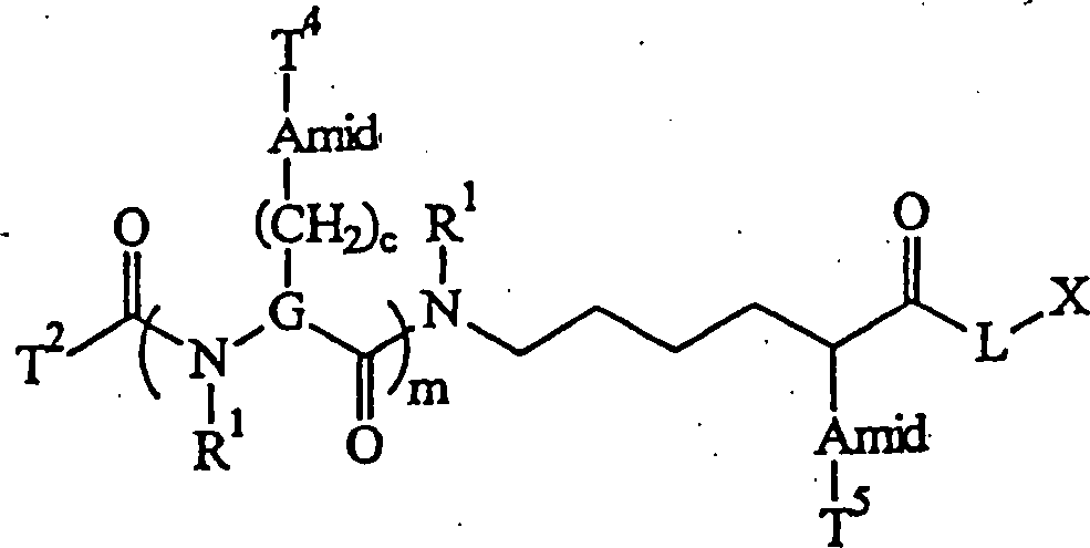


Applications Claiming Priority (7)
| Application Number | Priority Date | Filing Date | Title |
|---|---|---|---|
| US898180 | 1997-07-22 | ||
| US08/898,501 US6027890A (en) | 1996-01-23 | 1997-07-22 | Methods and compositions for enhancing sensitivity in the analysis of biological-based assays |
| US898564 | 1997-07-22 | ||
| US08/898,180 US6312893B1 (en) | 1996-01-23 | 1997-07-22 | Methods and compositions for determining the sequence of nucleic acid molecules |
| US08/898,564 US6613508B1 (en) | 1996-01-23 | 1997-07-22 | Methods and compositions for analyzing nucleic acid molecules utilizing sizing techniques |
| PCT/US1998/015008 WO1999005319A2 (en) | 1997-07-22 | 1998-07-22 | Methods and compounds for analyzing nucleic acids by mass spectrometry |
| US898501 | 2004-07-22 |
Publications (2)
| Publication Number | Publication Date |
|---|---|
| DE69814629D1 DE69814629D1 (en) | 2003-06-18 |
| DE69814629T2 true DE69814629T2 (en) | 2004-03-25 |
Family
ID=27420575
Family Applications (1)
| Application Number | Title | Priority Date | Filing Date |
|---|---|---|---|
| DE69814629T Expired - Lifetime DE69814629T2 (en) | 1997-07-22 | 1998-07-22 | METHOD AND CONNECTIONS FOR ANALYZING NUCLEIC ACIDS BY MASS SPECTROMETRY |
Country Status (12)
| Country | Link |
|---|---|
| EP (1) | EP0990047B1 (en) |
| JP (2) | JP4414086B2 (en) |
| CN (1) | CN1270598A (en) |
| AT (1) | ATE240408T1 (en) |
| AU (1) | AU738237B2 (en) |
| CA (1) | CA2297158C (en) |
| DE (1) | DE69814629T2 (en) |
| DK (1) | DK0990047T3 (en) |
| ES (1) | ES2200355T3 (en) |
| NZ (1) | NZ501919A (en) |
| PT (1) | PT990047E (en) |
| WO (1) | WO1999005319A2 (en) |
Cited By (1)
| Publication number | Priority date | Publication date | Assignee | Title |
|---|---|---|---|---|
| DE10350484A1 (en) * | 2003-12-20 | 2005-07-28 | Ehrfeld Mikrotechnik Bts Gmbh | Method for labeling proteins |
Families Citing this family (88)
| Publication number | Priority date | Publication date | Assignee | Title |
|---|---|---|---|---|
| WO1999022839A1 (en) * | 1997-10-30 | 1999-05-14 | Transgenomic, Inc. | Method for high resolution liquid chromatographic separation of polynucleotides |
| GB9815164D0 (en) * | 1998-07-13 | 1998-09-09 | Brax Genomics Ltd | Compounds for mass spectrometry |
| US7225079B2 (en) | 1998-08-04 | 2007-05-29 | Transgenomic, Inc. | System and method for automated matched ion polynucleotide chromatography |
| CN1371356A (en) * | 1999-08-31 | 2002-09-25 | 佳根基因组学公司 | Enatiomerically-enriched compounds having photocleavable bond(s) and methods related thereto |
| WO2001046687A2 (en) * | 1999-12-22 | 2001-06-28 | Transgenomic, Inc. | System and method for automated matched ion polynucleotide chromatography |
| EP1385869A2 (en) * | 2000-02-08 | 2004-02-04 | The Regents Of The University Of Michigan | Protein mapping |
| JP2001218598A (en) * | 2000-02-09 | 2001-08-14 | Asahi Breweries Ltd | Method for specifically and quantitatively detecting bacterium belonging to pectinatus |
| WO2001073119A2 (en) | 2000-03-29 | 2001-10-04 | Cambia | Methods for genotyping by hybridization analysis |
| US7255999B2 (en) | 2001-05-21 | 2007-08-14 | Monogram Biosciences, Inc. | Methods and compositions for analyzing proteins |
| US7771929B2 (en) | 2000-04-28 | 2010-08-10 | Monogram Biosciences, Inc. | Tag library compounds, compositions, kits and methods of use |
| US7160735B2 (en) | 2000-04-28 | 2007-01-09 | Monogram Biosciences, Inc. | Tagged microparticle compositions and methods |
| CA2419159A1 (en) * | 2000-08-11 | 2002-02-21 | Agilix Corporation | Ultra-sensitive detection systems |
| US7666588B2 (en) | 2001-03-02 | 2010-02-23 | Ibis Biosciences, Inc. | Methods for rapid forensic analysis of mitochondrial DNA and characterization of mitochondrial DNA heteroplasmy |
| US20040121313A1 (en) | 2002-12-06 | 2004-06-24 | Ecker David J. | Methods for rapid detection and identification of bioagents in organs for transplantation |
| US7226739B2 (en) | 2001-03-02 | 2007-06-05 | Isis Pharmaceuticals, Inc | Methods for rapid detection and identification of bioagents in epidemiological and forensic investigations |
| US20030027135A1 (en) | 2001-03-02 | 2003-02-06 | Ecker David J. | Method for rapid detection and identification of bioagents |
| EP1950305A1 (en) | 2001-05-09 | 2008-07-30 | Monsanto Technology, LLC | Tyr a genes and uses thereof |
| HUP0501021A3 (en) | 2001-05-21 | 2006-06-28 | Aclara Biosciences Inc Mountai | Methods and compositions for analyzing proteins |
| WO2002097112A2 (en) | 2001-05-26 | 2002-12-05 | Aclara Biosciences, Inc. | Catalytic amplification of multiplexed assay signals |
| US7217510B2 (en) | 2001-06-26 | 2007-05-15 | Isis Pharmaceuticals, Inc. | Methods for providing bacterial bioagent characterizing information |
| US8073627B2 (en) | 2001-06-26 | 2011-12-06 | Ibis Biosciences, Inc. | System for indentification of pathogens |
| WO2003033741A1 (en) * | 2001-10-16 | 2003-04-24 | Aclara Biosciences, Inc. | Universal e-tag primer and probe compositions and methods |
| GB0205455D0 (en) | 2002-03-07 | 2002-04-24 | Molecular Sensing Plc | Nucleic acid probes, their synthesis and use |
| US20040229294A1 (en) | 2002-05-21 | 2004-11-18 | Po-Ying Chan-Hui | ErbB surface receptor complexes as biomarkers |
| JP2006516193A (en) | 2002-12-06 | 2006-06-29 | アイシス・ファーマシューティカルス・インコーポレーテッド | Rapid identification of pathogens in humans and animals |
| CA2488584C (en) | 2003-01-30 | 2011-10-11 | Applera Corporation | Methods, mixtures, kits and compositions pertaining to analyte determination |
| US6997031B2 (en) | 2003-05-01 | 2006-02-14 | Waters Corporation | Fraction collector for composition analysis |
| US7964343B2 (en) | 2003-05-13 | 2011-06-21 | Ibis Biosciences, Inc. | Method for rapid purification of nucleic acids for subsequent analysis by mass spectrometry by solution capture |
| US8158354B2 (en) | 2003-05-13 | 2012-04-17 | Ibis Biosciences, Inc. | Methods for rapid purification of nucleic acids for subsequent analysis by mass spectrometry by solution capture |
| US8546082B2 (en) | 2003-09-11 | 2013-10-01 | Ibis Biosciences, Inc. | Methods for identification of sepsis-causing bacteria |
| US7956175B2 (en) | 2003-09-11 | 2011-06-07 | Ibis Biosciences, Inc. | Compositions for use in identification of bacteria |
| US8097416B2 (en) | 2003-09-11 | 2012-01-17 | Ibis Biosciences, Inc. | Methods for identification of sepsis-causing bacteria |
| CA2542941C (en) * | 2003-11-26 | 2013-02-12 | Applera Corporation | Analysis of mass spectral data in the quiet zones and label selection therefor |
| US20050148087A1 (en) | 2004-01-05 | 2005-07-07 | Applera Corporation | Isobarically labeled analytes and fragment ions derived therefrom |
| US20050148771A1 (en) | 2004-01-05 | 2005-07-07 | Applera Corporation. | Active esters of N-substituted piperazine acetic acids, including isotopically enriched versions thereof |
| US7355045B2 (en) | 2004-01-05 | 2008-04-08 | Applera Corporation | Isotopically enriched N-substituted piperazine acetic acids and methods for the preparation thereof |
| US7666592B2 (en) | 2004-02-18 | 2010-02-23 | Ibis Biosciences, Inc. | Methods for concurrent identification and quantification of an unknown bioagent |
| CA2567839C (en) | 2004-05-24 | 2011-06-28 | Isis Pharmaceuticals, Inc. | Mass spectrometry with selective ion filtration by digital thresholding |
| US20050266411A1 (en) | 2004-05-25 | 2005-12-01 | Hofstadler Steven A | Methods for rapid forensic analysis of mitochondrial DNA |
| US7811753B2 (en) | 2004-07-14 | 2010-10-12 | Ibis Biosciences, Inc. | Methods for repairing degraded DNA |
| WO2006135400A2 (en) | 2004-08-24 | 2006-12-21 | Isis Pharmaceuticals, Inc. | Methods for rapid identification of recombinant organisms |
| US8084207B2 (en) | 2005-03-03 | 2011-12-27 | Ibis Bioscience, Inc. | Compositions for use in identification of papillomavirus |
| US8182992B2 (en) | 2005-03-03 | 2012-05-22 | Ibis Biosciences, Inc. | Compositions for use in identification of adventitious viruses |
| AU2006272776B2 (en) | 2005-07-21 | 2012-01-19 | Ibis Biosciences, Inc. | Methods for rapid identification and quantitation of nucleic acid variants |
| US8088582B2 (en) | 2006-04-06 | 2012-01-03 | Ibis Biosciences, Inc. | Compositions for the use in identification of fungi |
| EP2064332B1 (en) | 2006-09-14 | 2012-07-18 | Ibis Biosciences, Inc. | Targeted whole genome amplification method for identification of pathogens |
| WO2008104002A2 (en) | 2007-02-23 | 2008-08-28 | Ibis Biosciences, Inc. | Methods for rapid forensic dna analysis |
| US9598724B2 (en) | 2007-06-01 | 2017-03-21 | Ibis Biosciences, Inc. | Methods and compositions for multiple displacement amplification of nucleic acids |
| KR101322624B1 (en) * | 2007-06-13 | 2013-10-29 | 삼성전자주식회사 | Field effect transistor-based detection device for biomolecules containing adsorptive media and detection method using the same |
| US7888127B2 (en) | 2008-01-15 | 2011-02-15 | Sequenom, Inc. | Methods for reducing adduct formation for mass spectrometry analysis |
| CN101251511B (en) * | 2008-03-14 | 2012-06-06 | 毅新兴业(北京)科技有限公司 | Method for testing SNP using restriction enzymes double zyme cutting |
| CN101251510B (en) * | 2008-03-14 | 2012-06-06 | 毅新兴业(北京)科技有限公司 | Method for testing SNP combining restrictive zyme cutting method and mass spectrometry |
| CN101246142B (en) * | 2008-04-03 | 2012-06-20 | 毅新兴业(北京)科技有限公司 | Method for detecting mononucleotide polymorphism |
| EP2344893B1 (en) | 2008-09-16 | 2014-10-15 | Ibis Biosciences, Inc. | Microplate handling systems and methods |
| EP2349549B1 (en) | 2008-09-16 | 2012-07-18 | Ibis Biosciences, Inc. | Mixing cartridges, mixing stations, and related kits, and system |
| EP2347254A2 (en) | 2008-09-16 | 2011-07-27 | Ibis Biosciences, Inc. | Sample processing units, systems, and related methods |
| EP2396803A4 (en) | 2009-02-12 | 2016-10-26 | Ibis Biosciences Inc | Ionization probe assemblies |
| US9719083B2 (en) | 2009-03-08 | 2017-08-01 | Ibis Biosciences, Inc. | Bioagent detection methods |
| US9393564B2 (en) | 2009-03-30 | 2016-07-19 | Ibis Biosciences, Inc. | Bioagent detection systems, devices, and methods |
| WO2011008972A1 (en) | 2009-07-17 | 2011-01-20 | Ibis Biosciences, Inc. | Systems for bioagent identification |
| US8950604B2 (en) | 2009-07-17 | 2015-02-10 | Ibis Biosciences, Inc. | Lift and mount apparatus |
| WO2011014811A1 (en) | 2009-07-31 | 2011-02-03 | Ibis Biosciences, Inc. | Capture primers and capture sequence linked solid supports for molecular diagnostic tests |
| EP3098325A1 (en) | 2009-08-06 | 2016-11-30 | Ibis Biosciences, Inc. | Non-mass determined base compositions for nucleic acid detection |
| EP2473630B1 (en) | 2009-09-04 | 2017-11-08 | QIAGEN GmbH | Optimized probes and primers and methods of using same for the detection, screening, isolation and sequencing of vancomycin resistance genes and vancomycin resistant enterococci |
| EP3225695A1 (en) | 2009-10-15 | 2017-10-04 | Ibis Biosciences, Inc. | Multiple displacement amplification |
| WO2011115840A2 (en) | 2010-03-14 | 2011-09-22 | Ibis Biosciences, Inc. | Parasite detection via endosymbiont detection |
| TWI510781B (en) | 2010-10-29 | 2015-12-01 | Scinopharm Taiwan Ltd | Real-time monitor solid phase peptide synthesis by mass spectrometry |
| WO2012138470A2 (en) | 2011-04-04 | 2012-10-11 | Intelligent Medical Devices, Inc. | Optimized oligonucleotides and methods of using same for the detection, isolation, amplification, quantitation, monitoring, screening, and sequencing of group b streptococcus |
| US9891193B2 (en) | 2012-07-24 | 2018-02-13 | Massachusetts Institute Of Technology | Reagent impregnated swipe for chemical detection |
| US9588095B2 (en) | 2012-07-24 | 2017-03-07 | Massachusetts Institute Of Technology | Reagents for oxidizer-based chemical detection |
| WO2014058508A2 (en) * | 2012-07-24 | 2014-04-17 | Massachusetts Institute Of Technology | Reagents for oxidizer-based chemical detection |
| US20140127674A1 (en) | 2012-10-25 | 2014-05-08 | Intelligent Medical Devices, Inc. | Optimized probes and primers and methods of using same for the binding, detection, differentiation, isolation and sequencing of influenza a; influenza b and respiratory syncytial virus |
| WO2014137906A1 (en) | 2013-03-05 | 2014-09-12 | Intelligent Medical Devices, Inc. | Optimized probes and primers and methods of using same for the detection, screening, isolation and sequencing of mrsa, mssa, staphylococcus markers, and the antibiotic resistance gene mec a |
| US9305756B2 (en) | 2013-03-13 | 2016-04-05 | Agena Bioscience, Inc. | Preparation enhancements and methods of use for MALDI mass spectrometry |
| US10816530B2 (en) | 2013-07-23 | 2020-10-27 | Massachusetts Institute Of Technology | Substrate containing latent vaporization reagents |
| WO2015154087A1 (en) | 2014-04-04 | 2015-10-08 | Massachusetts Institute Of Technology | Reagents for enhanced detection of low volatility analytes |
| US10370673B2 (en) | 2014-09-26 | 2019-08-06 | Purecircle Sdn Bhd | Single nucleotide polymorphism (SNP) markers for stevia |
| KR101701998B1 (en) * | 2014-10-23 | 2017-02-03 | 한국과학기술연구원 | Gas chromatography-electrospray mass spectrometry with high resolution and a method ionizing organic compounds of gas phase using it |
| CN107208162B (en) * | 2015-02-26 | 2020-12-29 | 株式会社日立高新技术 | Method for constructing nucleic acid molecule |
| WO2018170436A1 (en) | 2017-03-16 | 2018-09-20 | Jacobs Farm Del Cabo | Basil with high tolerance to downy mildew |
| CN106979942B (en) * | 2017-05-04 | 2020-07-07 | 华中师范大学 | Raman spectrum analysis method for individual quantification of solid-phase synthesized compound combinatorial library and application thereof |
| US20210198734A1 (en) * | 2018-05-25 | 2021-07-01 | New York Institute Of Technology | Direct nucleic acid sequencing method |
| CN111208300B (en) * | 2018-11-21 | 2021-10-29 | 中国科学院大连化学物理研究所 | Method for enriching and identifying lysine nitrogen-linked phosphorylation post-translational modification |
| CN111239402B (en) * | 2018-11-29 | 2021-07-09 | 北京大学 | Mass spectrum immunoassay method for detecting disease marker and application |
| CN113075114B (en) * | 2019-12-17 | 2022-07-01 | 北京大学 | Organic mass spectrometry flow analysis method for single cell analysis |
| AU2021276656A1 (en) | 2020-05-19 | 2022-11-24 | Cybin Irl Limited | Deuterated tryptamine derivatives and methods of use |
| CN114984032B (en) * | 2022-06-27 | 2023-07-07 | 四川大学 | DNA tetrahedron framework nucleic acid-chlorogenic acid complex and application thereof in preparation of medicines for treating hepatic fibrosis |
| CN115356411B (en) * | 2022-08-09 | 2023-07-28 | 河北一品生物医药有限公司 | Method for detecting related substances of L-piperidine hydrochloride by high performance liquid chromatography |
Family Cites Families (12)
| Publication number | Priority date | Publication date | Assignee | Title |
|---|---|---|---|---|
| US5118605A (en) * | 1984-10-16 | 1992-06-02 | Chiron Corporation | Polynucleotide determination with selectable cleavage sites |
| US5565324A (en) * | 1992-10-01 | 1996-10-15 | The Trustees Of Columbia University In The City Of New York | Complex combinatorial chemical libraries encoded with tags |
| EP0679196B1 (en) * | 1993-01-07 | 2004-05-26 | Sequenom, Inc. | Dna sequencing by mass spectrometry |
| GB9315847D0 (en) * | 1993-07-30 | 1993-09-15 | Isis Innovation | Tag reagent and assay method |
| US5849542A (en) * | 1993-11-17 | 1998-12-15 | Amersham Pharmacia Biotech Uk Limited | Primer extension mass spectroscopy nucleic acid sequencing method |
| EP0751950B1 (en) * | 1994-03-23 | 2000-10-25 | The Penn State Research Foundation | Method for identifying members of combinatorial libraries |
| CZ228598A3 (en) * | 1996-01-23 | 1998-12-16 | Rapigene, Inc. | Processes and compositions for analyzing molecules of nucleic acids by making use of classification procedure |
| DE69701671T3 (en) * | 1996-01-23 | 2006-08-17 | Qiagen Genomics, Inc., Bothell | METHOD AND COMPOUNDS FOR THE SEQUENCE DETERMINATION OF NUCLEIC ACID MOLECULES |
| CA2243989A1 (en) * | 1996-01-23 | 1997-07-31 | Rapigene, Inc. | Methods and compositions for detecting binding of ligand pair using non-fluorescent label |
| EP0963443B1 (en) * | 1996-12-10 | 2006-03-08 | Sequenom, Inc. | Releasable nonvolatile mass-label molecules |
| ATE374836T1 (en) * | 1997-01-15 | 2007-10-15 | Xzillion Gmbh & Co Kg | MASS MARKED HYBRIDIZATION PROBE |
| WO1999002728A1 (en) * | 1997-07-11 | 1999-01-21 | Brax Group Limited | Characterising nucleic acids |
-
1998
- 1998-07-22 WO PCT/US1998/015008 patent/WO1999005319A2/en active IP Right Grant
- 1998-07-22 CN CN98809047A patent/CN1270598A/en active Pending
- 1998-07-22 EP EP98936928A patent/EP0990047B1/en not_active Expired - Lifetime
- 1998-07-22 AU AU85765/98A patent/AU738237B2/en not_active Ceased
- 1998-07-22 PT PT98936928T patent/PT990047E/en unknown
- 1998-07-22 AT AT98936928T patent/ATE240408T1/en not_active IP Right Cessation
- 1998-07-22 CA CA002297158A patent/CA2297158C/en not_active Expired - Fee Related
- 1998-07-22 DE DE69814629T patent/DE69814629T2/en not_active Expired - Lifetime
- 1998-07-22 DK DK98936928T patent/DK0990047T3/en active
- 1998-07-22 NZ NZ501919A patent/NZ501919A/en unknown
- 1998-07-22 JP JP2000504286A patent/JP4414086B2/en not_active Expired - Lifetime
- 1998-07-22 ES ES98936928T patent/ES2200355T3/en not_active Expired - Lifetime
-
2009
- 2009-04-14 JP JP2009098483A patent/JP4870791B2/en not_active Expired - Lifetime
Cited By (2)
| Publication number | Priority date | Publication date | Assignee | Title |
|---|---|---|---|---|
| DE10350484A1 (en) * | 2003-12-20 | 2005-07-28 | Ehrfeld Mikrotechnik Bts Gmbh | Method for labeling proteins |
| DE10350484B4 (en) * | 2003-12-20 | 2006-02-02 | Ehrfeld Mikrotechnik Bts Gmbh | Method and device for labeling proteins |
Also Published As
| Publication number | Publication date |
|---|---|
| JP4870791B2 (en) | 2012-02-08 |
| DK0990047T3 (en) | 2003-09-15 |
| EP0990047B1 (en) | 2003-05-14 |
| AU738237B2 (en) | 2001-09-13 |
| CN1270598A (en) | 2000-10-18 |
| CA2297158C (en) | 2009-12-22 |
| PT990047E (en) | 2003-10-31 |
| AU8576598A (en) | 1999-02-16 |
| NZ501919A (en) | 2001-11-30 |
| WO1999005319A2 (en) | 1999-02-04 |
| CA2297158A1 (en) | 1999-02-04 |
| WO1999005319A9 (en) | 1999-06-17 |
| EP0990047A2 (en) | 2000-04-05 |
| ES2200355T3 (en) | 2004-03-01 |
| JP4414086B2 (en) | 2010-02-10 |
| DE69814629D1 (en) | 2003-06-18 |
| JP2009159985A (en) | 2009-07-23 |
| WO1999005319A3 (en) | 1999-05-14 |
| JP2001511359A (en) | 2001-08-14 |
| ATE240408T1 (en) | 2003-05-15 |
Similar Documents
| Publication | Publication Date | Title |
|---|---|---|
| DE69814629T2 (en) | METHOD AND CONNECTIONS FOR ANALYZING NUCLEIC ACIDS BY MASS SPECTROMETRY | |
| DE69701671T3 (en) | METHOD AND COMPOUNDS FOR THE SEQUENCE DETERMINATION OF NUCLEIC ACID MOLECULES | |
| US6613508B1 (en) | Methods and compositions for analyzing nucleic acid molecules utilizing sizing techniques | |
| US6027890A (en) | Methods and compositions for enhancing sensitivity in the analysis of biological-based assays | |
| AU717330B2 (en) | Methods and compositions for detecting binding of ligand pair using non-fluorescent label | |
| AU717945B2 (en) | Methods and compositions for analyzing nucleic acid molecules utilizing sizing techniques | |
| US6623928B2 (en) | Methods and compositions for determining the sequence of nucleic acid molecules | |
| WO1997027331A9 (en) | Methods and compositions for determining the sequence of nucleic acid molecules | |
| EP0992511B1 (en) | Methods and compositions for determining the sequence of nucleic acid molecules | |
| EP0962464A2 (en) | Methods and compositions for detecting binding of ligand pair using non-fluorescent label |
Legal Events
| Date | Code | Title | Description |
|---|---|---|---|
| 8364 | No opposition during term of opposition | ||
| 8327 | Change in the person/name/address of the patent owner |
Owner name: AGILENT TECHNOLOGIES, INC. (N.D.GES.D. STAATES, US |
|
| 8328 | Change in the person/name/address of the agent |
Representative=s name: DERZEIT KEIN VERTRETER BESTELLT |