Download
1 / 35
460 likes | 1.1k Views
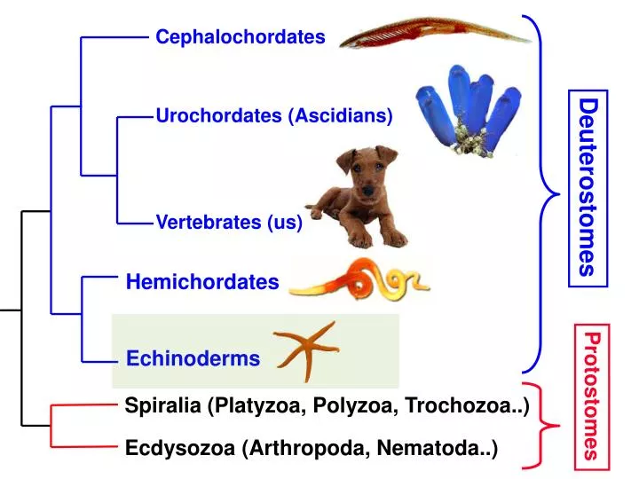
Cephalochordates Urochordates (Ascidians) Vertebrates (us). Deuterostomes. Hemichordates Echinoderms. Protostomes. Spiralia (Platyzoa, Polyzoa, Trochozoa..) Ecdysozoa (Arthropoda, Nematoda..). Embryonic Development: Early Cleavage. Zygote. Protostomes. Deuterostomes. Radial,

E N D
Cephalochordates Urochordates (Ascidians) Vertebrates (us) Deuterostomes Hemichordates Echinoderms Protostomes Spiralia (Platyzoa, Polyzoa, Trochozoa..) Ecdysozoa (Arthropoda, Nematoda..)
Embryonic Development: Early Cleavage Zygote Protostomes Deuterostomes • Radial, • Indeterminate • Cleavage • each cell can • potentially • develop into a • complete embryo • Spiral, • Determinate • Cleavage • fate of cells is • set early in • development 2 cells 4 cells 8 cells 8 cells, top view
Spiral Cleavage in Protostomes 32 cell stage - labeled with Wilson’s coding system Annelid cross Rosette cross Molluscan cross
From Blastula to Gastrula ectoderm blasto- coel Invagination endoderm archenteron blastopore Gastrulation = formation of embryonicgerm layers - separates cells fated to interact with the environment (movement, sensory, protection) from those that will process food (= the gut)
Protostome Deuterostome GASTRULA STAGE Fate of Blastopore Coelom Formation
Coelom formation Schizocoely (protostomes) Enterocoely (deuterostomes) Ectoderm Blastocoel future mesoderm Archenteron (larval gut) Endoderm Mesoderm formsout of ball of cells descended from 4d micromere cell Mesoderm formsfrom cells that pinch off from blastopore
Coelom formation via schizocoely Solid mass of mesoderm, derived from 4d micromere cell; gives rise to paired coelomic spaces - associated with segmentation in annelids, arthropods
Coelom formation via enterocoely coelom Pouching of archenteron production of mesoderm-lined coelomic spaces - may results in a 3-part coelom in deuterostome embryos (and some lophophorates) 3-part coelom
Evolution of the Bilateria Ancestor probably had deuterostome features as an embryo Protostomes Deuterostomes - spiral cleavage - blastopore becomes mouth - mesoderm derived from 4d cell - ventral nerve chord - coelom formed by schizocoely, hollowing out of mesodermal mass from 4d micromere cell - radial cleavage - blastopore becomes anus - mesoderm from archenteron - dorsal nerve chord - coelom forms by enterocoely, pinching out of gut (with a 3-part arrangement in body)
~7,000 spp. Phylum Echinodermata • Deuterostomes with penta-radialsymmetry as adults, • bodies organized along oral-aboralaxis (mouth anus) • - Calcified endoskeleton derived from mesoderm; bony • ossicles or plates filled with living tissue (stroma) • Water vascular system, derived from coelom, powers the • podia (=tube feet) and serves as circulatory system • - Decentralized nerve ring + radial nerves • - Mutable connective tissue that can harden, soften • - Mostly dioecious (separate sexes) • Bilaterally symmetriclarvae, such as bipinnaria (starfish) • and pluteus larvae (urchins, brittlestars)
Aboral surface Oral surface Madreporite: opening to water vascular system ambulacral (open on grooves asteroids) mouth central disc body rays = ambulacra podia(tube feet) line open grooves
Body Orientation in Echinoderms Ophiuroids Crinoids Asteroids (stars) Echinoids (urchins) Holothurians (cucumbers) mouth ambulacral surface, w/ podia
Endoskeleton Echinoderms have an epidermis covering a mesoderm-derived dermis layer containing the calcified endoskeleton - skeleton is composed of ossicles, porous plates of CaCO3 filled with living dermal cells called stroma stroma tissue fills these spaces Under the dermis layer (i.e., beneath the skeleton): - layer of muscle - coelom (i.e., components of the water vascular system)
Endoskeleton ossicles may be spread throughout the body, embedded in connective tissue as in sea stars & sea cucumbers Pisaster ossicles ossicles may fuse together to form the solid test (inner shell) & spines of sea urchins, or the arm vertebrae of brittle stars
Pedicellariae Pincer-like structures on aboral surface, formed from ossicles - have their own muscles, nerves + reflex arcs - may be hollow & inject toxins Function in both physical and chemicaldefense by pinching and poisoning predators - also anti-fouling: keep surface clean by pinching any larvae or algal spores that try to settle on body surface
Phylum Echinodermata Class Asteroidea - sea stars Class Ophiuroidea - brittle stars & basket stars Class Crinoidea - crinoids Class Echinoidea - sea urchins & sand dollars Class Holothuroidea - sea cucumbers
Phylum Echinodermata Class Asteroidea - sea stars
Class Asteroidea – sea stars 1,500 species in 5 orders - 5 or more arms, not distinct from central disc - open ambulacral grooves lined with podia that have internal ampullae (fluid-filled bulbs) gills found in grooves & across body surface
surface of Astrometis spines gills pedicellariae podia
Water Vascular System each lateral canal ends in a tube foot - extends around central disc regulate internal pressure
Water Vascular System • Seawater enters through madreporite, mixes with coelomic fluid • and is is pumped through the system by cilia • When an ampulla contracts, it pushes fluid into the tube foot • (1) sucker is pushed against substrate; adheres via secretions • (2) muscles in tube foot contract, pushing fluid back out • (3) muscles pull up sucker of foot, creating vacuum– this • creates the tremendous suction that holds foot to substrate • (4) suction is released when ampulla again contracts, sending • fluid into foot sucker and relieving vacuum
paired digestive glands run down each arm everted for feeding
Sea stars use their tube feet to pull open shells of bivalves such as mussels, clams - then turntheir stomach inside-out, into the shell - digest the soft bivalve tissue inside its own shell sea star hunched over a mussel, ready to start pulling its shell open Stomach of the bat star Asterina pushed out against glass
Asteroids as keystone predators Pisaster ochraceous and P. giganteus function as keystone predatorsin rocky intertidal habitats, preserving biodiversity - mussels are competitive dominantsfor space, so they crush everything else off the rocks if their populations grow unchecked loss of biodiversity - by consuming mussels, sea stars create free space for other organisms promotes biodiversity in rocky intertidal zone
Asteroids as keystone predators maximum foraging distance - sea stars consume any mussel they can crawl to, open, and eat during a single high tide verticalzonation, with mussels restricted to the tops of rocks above foraging reach of sea stars, which hang out near the base where it’s wet and cool
Crown-of-Thorns starfish, Acanthaster plancki • eats live coral polyps • in last few decades, major outbreaks have resulted in massive • coral mortality in Australia; human influences suspected
Sea stars are famous for regenerating lost arms - some (Linkia) re-grow whole body from a dropped arm ! Asteroid larvae can also bud off asexually, the only known larvae capable of reproduction Which is healing? which is asexual reproduction?...
Phylum Echinodermata Class Asteroidea - sea stars Class Ophiuroidea - brittle stars & basket stars Class Crinoidea - crinoids Class Echinoidea - sea urchins & sand dollars Class Holothuroidea - sea cucumbers
Class Ophiuroidea – brittle stars • 2,000 species in 3 orders • Body with 5 or more arms, distinctfrom central disc • Tube feet have no suckers; • no anus is present • Coelom in arms is reduced, • due to large vertebral ossicles • Ambulacral grooves closed • - Madreporite on oral surface • (underneath)
Ophiuroid Cross-section Central discArm Digestive system confined to central disc
Ophiuroid External Anatomy oral surface aboral surface
Ophiuroid Oral Surface bursal slits, openings that draw in water for gas exchange with coelomic fluid-filled sinuses mouth ventral arm shield arm spines
Ophiuroids are predominantly deposit or suspension feeders - food particles are trapped in mucous or passed podia-to-podia to the mouth Basket stars use multi-branched arms to suspension feed or catch larger prey; incomplete gut