
Recommended
More Related Content
What's hot
What's hot (20)
Similar to Lysosomes
Similar to Lysosomes (20)
More from Kanchan Rawat
Recently uploaded
Recently uploaded (20)
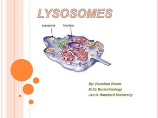
Lysosomes
- 2. Historical background Introduction Lysosomal enzymes Structure Functions Lysosomes and human disease Chaperone-mediated autophagy: roles in disease and aging References
- 4. Christian de Duve, discovered lysosomes in 1955. In 1949 he was studying insulin action, wanted to find location of glucose-6- phosphatase on liver cells. de Duve and colleagues ruptured the rat liver cells and then fractionated the samples using differential centrifugation. They found sacs containing large amount of acid phosphatase, later de Duve coined the term lysosomes.
- 5. The name “lysosomes” is derived from the Latin word “Lyein” (means to dissolve /to release/ to loosen) and “Soma” (means body). These structures are termed as the “digestive system of the cell” or “suicide bags”. Are present in animal cells, while in yeast and plants same roles are performed by lytic vacuoles. These are membrane bound vesicles (0.1–1.2 μm). They vary greatly in size and shape.
- 6. lysosomes are formed by the fusion of transport vesicles budded from the trans Golgi network with endosomes, which contain molecules taken up by endocytosis at the plasma membrane.
- 7. - Primary lysosomes ( do not contain particle or membrane for digestion) - Secondary lysosomes ( contain particles or membranes for digestion)
- 8. Contain over 50 hydrolytic enzymes which breaks down - proteins, nucleic acids, carbohydrates, and lipids. Enzymes include proteases, nucleases, lipases, glycosidases, phosphatases, phospholipases and sulfatases. All enzymes are acid hydrolases because for optimal activity they require an acid environment and the lysosome provides a pH of about 5.0 in its interior. synthesized in the cytosol and the endoplasmic reticulum, where they receive a mannose-6-phosphate tag that targets them for transport to the lysosome.
- 9. A membrane seals off the lysosome's acidic environment, preventing its enzymes from harming the rest of the cell. A H+ pump in the lysosomal membrane uses the energy of ATP hydrolysis to pump H+ into the lysosome, thereby maintaining the acidic pH of lumen.
- 11. Generally spherical, but they can vary in and shape as a result of differences in materials that have been taken up for digestion Enzyme-filled sacs Surrounded by single membrane Hydrolytic enzymes and lysosomal membrane are made by RER and then transferred to the Golgi apparatus for processing
- 12. It is the lysosomal digestion of extracellular materials by the process of endocytosis. Heterophagy are of two types namely Phagocytosis and Pinocytosis. cellular process of ingestion, in which the plasma membrane engulfs substances and pinches off to form a particle containing vacuole.
- 14. pinocytosis is the process of engulfment of fluid particles through the plasma membrane. Autolysis refers to the digestion of own cells by the lysosomes It is also otherwise known as programmed cell death or apoptopic lysis Autolysis occurs during amphibian metamorphosis, insect metamorphosis, mensuration etc.
- 16. During fertilization process, acrosome (giant lysosome) of sperm head ruptures and releases enzymes on the surface of the egg. These enzymes digest the egg membrane and provide way for the entry of sperm nucleus into the egg. This action also activates the egg for the developmental processes.
- 17. Due to the presence of DNase enzyme, lysosome had an ability to attacks chromosome and cause chromosomal breakages. These breakages can leads to diseases like cancer etc.
- 18. - Caused by the deficiency of an enzyme N-acetyglucosamine phosphotransferase. Symptoms: A rare inherited metabolic disorder characterized by coarse facial features, skeletal abnormalities and mental retardation. Characterized by the deficiency of a single lysosomal enzyme and corresponding accumulation of undegraded substance. Eg: Result from the absence of a lysosomal enzyme, α-glucosidase. Symptoms: accumulation of glycogen in all organs, cardiorespiratory failure and death , usually before age 2. Treatment: Enzyme replacement therapy.
- 19. Deficiency of the enzyme β-N-hexosaminidase. Symptoms: brain impairment by accumulation of lipids. Treatment: preventing problems with the lungs and airways ,relieving any feeding or swallowing problems (dysphagia), medication to control fits and muscle stiffness. It is caused by a hereditary deficiency of the enzyme glucocerebrosidase. Symptoms: bruising, fatigue, anemia, low blood platelets, and enlargement of the liver and spleen. Treatment: Enzyme replacement treatment with intravenous recombinant glucocerebrosidase.
- 20. Cytosolic Proteins are targeted to the lysosomal membrane and then gain access to the lumen by directly crossing its membrane This process is chaperone-mediated autophagy. Most of the lysosomal delivery involves vesicles but CMA don’t. Proteins that undergo degradation are selected individually through a recognition motif in their amino acid sequences. This allows for the removal of specific proteins without disturbance of neighboring ones and makes CMA an efficient system for degradation of damaged or abnormal proteins. This selectivity permits CMA to play a regulatory role in multiple cellular processes by contributing to modulate intracellular levels of enzymes, transcription factors and cell maintenance proteins.
- 21. CMA involves: substrate recognition and lysosomal targeting; substrate binding and unfolding; substrate translocation substrate degradation in the lysosomal lumen Recognition takes place in cytosol through binding of constitutive chaperone hsc70 to a pentapeptide motif present in the amino acid sequences of all CMA substrates. Once bound to the chaperone, the substrate is targeted to the surface of the lysosomes where it interacts with the cytosolic tail of the single-span membrane protein LAMP-2A. Substrate unfolding is mediated by hsc70 and some of its cochaperones present at lysosomal membrane, and is completed before the LAMP-2A complex is fully assembled.
- 22. As inserting LAMP-2A in proteoliposomes in the presence of hsc70 mediate substrate translocation. After substrate translocation into the lysosomal lumen, LAMP- 2A is rapidly dissembled from the translocation complex into monomers where substrates can bind again. Although both CMA and endosomal microautophagy share hsc70 as the targeting chaperone, the dependence on LAMP- 2A is exclusive for CMA. The best way to confirm that a protein is a CMA substrate is, to reproduce its binding and translocation across the membrane of isolated lysosomes.
- 23. amino acid recycling during prolonged starvation: - CMA is gradually activated after 8-10 h of starvation and persists at maximal activity for up to three days -this form degradation allows for changes in the proteome aimed at adapting the cell to the new conditions. For example, degradation of regulatory metabolic enzymes by CMA has been shown to contribute to the metabolic changes that allow for cancer cells to adapt to low nutrient conditions.
- 24. quality control- remove single proteins from the cytosol CMA is upregulated during oxidative stress, where it contributes to the degradation of oxidized proteins. CMA is also upregulated in other conditions that lead to protein damage such as exposure to denaturing toxic compounds. Activation of CMA has also been shown to support survival of retinal cells upon activation of a pro-apoptotic program in those cells. degradation of the transcription factor Pax2 by CMA in kidney is important to control tubular cell growth.
- 25. STEPS AND PHYSIOLOGICAL FUNCTIONS OF CMA
- 26. Parkinson’s disease (PD): Caused by loss of dopaminergic neurons subsequent motor deficits Dysfunction in CMA has been described in both familial and sporadic PD. In the case of familial PD, sequence analysis reveals the presence of CMA-targeting motifs in the majority of PD-related proteins, supporting an important role for CMA in the control of their intracellular levels. the 2 most commonly mutated proteins in patients with familial PD, α-synuclein and LRRK2, degrade in lysosomes via CMA using various experimental systems such as isolated lysosomes, primary mouse neuronal cultures, mouse models of PD and even neuronal-differentiated induced pluripotent stem cells and brains from familial, and sporadic PD patients.
- 27. These protein fail to reach the lysosomal lumen to be degraded by CMA It is caused due to aberrant interactions of these toxic proteins with LAMP-2A toxic interactions of α-synuclein and LRRK2 mutants with the CMA transporter preclude not only their own degradation, but also inhibit the degradation of other CMA substrates
- 29. Abnormally enhanced CMA and cancer: upregulation of CMA has been linked to the survival and proliferation of cancer cells. Activation of CMA is mostly due to an increase in the LAMP- 2A levels in these cancer cells and tumors. Genetic knock-down of LAMP-2A in cancer cells helped to establish that CMA is required for cancer cell proliferation, optimal tumor growth and metastasis. Selective blockage of CMA in cancer cells results in transcriptional attenuation of several rate-limiting glycolytic enzymes, and the subsequent reduction in glycolysis and ATP production. the inactive forms of PKM2, one of the rate-limiting glycolytic enzymes, are eliminated via CMA. CMA takes on an anti-oncogenic role in non proliferating tumor cells by reducing the cellular levels of mutant p53 through CMA degradation.
- 30. blockage of CMA in human tumor explants in mice through knock-down of LAMP-2A has proven effective in not only reducing tumor growth and metastasis but also in inducing tumor shrinkage through cancer cell necrotic death.
- 31. How does aging affect CMA? Reduced CMA activity has been observed in many cell types and tissues of old rodents, as well as in cells derived from aged individuals. Age-dependent decay in CMA appears to be caused by age- related changes in the lipid constituents of lysosomal membrane that alter the dynamics and stability of LAMP-2A in the lysosomes of old organisms. Experimental blockage of CMA activity in cultured cells suggests that a direct consequence of the age dependent failure in CMA is the loss of CMA-mediated homeostasis such as the removal of oxidatively damaged proteins and the ability to respond to stressors. Genetic manipulation to preserve CMA function in old rodents by expressing an exogenous copy of LAMP-2A in mouse liver has proven effective in improving the health span of aged animals.
- 32. http://www.newworldencyclopedia.org/entry/Lysosome# http://www.ncbi.nlm.nih.gov/books/NBK9953/ http://www.sivabio.50webs.com/lysosomes.htm http://www.nature.com/scitable/topicpage/the-discovery-of- lysosomes-and-autophagy-14199828 http://www.ncbi.nlm.nih.gov/pmc/articles/PMC3746853/ https://books.google.co.in/books https://en.wikipedia.org/wiki/Gaucher%27s_disease http://www.mayoclinic.org/diseases-conditions/tay-sachs- disease/care-at-mayo-clinic/treatment/con-20036799 http://www.ncbi.nlm.nih.gov/books/NBK9953/figure/A1524/?report =objectonly http://centennial.rucares.org/index.php?page=Exploring_Cells_Ce ntriguge http://www.ivyroses.com/Biology/Organelles/Lysosomes.php Cell Research (2014) Ana Maria Cuervo1, Esther Wong2 24:92- 104 © 2014 IBCB, SIBS, CAS, http://www.nature.com/cr