Structure and functions of Mitochondria
•Download as PPTX, PDF•
33 likes•12,028 views

This Power Point Presentation (PPT) entitled “Structure and Functions of Mitochondria” consists of 118 slides with following sub-heads INTRODUCTION HISTORY ORIGIN AND EVOLUTION OF MITOCHONDRIA SYNTHESIS OF MITOCHONDRIA ISOLATION OF MITOCHNDRIA SHAPE , SIZE AND NUMBER OF MITOCHONDRIA STRUCTURE OF MITOCHONDRIA CHEMICAL COMPOSITION OF MITOCHONDRIA FUNCTIONS OF MITOCHONDRIA MITOCHONDRIA –POWER HOUSE OF CELL MITOCHONDRIAL DNA/ GENOME TRANSPORT OF PROTEINS INTO MITOCHONDRIA MITOCHONDRIAL INHERITANCE MITOCHONDRIAL DISEASES IN HUMAN SUMMARY QUESTIONS BOOKS CONSULTED REFERENCES

Recommended
More Related Content
What's hot
What's hot (20)
Similar to Structure and functions of Mitochondria
Similar to Structure and functions of Mitochondria (20)
More from ICHHA PURAK
More from ICHHA PURAK (20)
Recently uploaded
Recently uploaded (20)
Structure and functions of Mitochondria
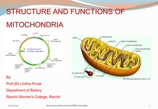
- 1. STRUCTURE AND FUNCTIONS OF MITOCHONDRIA By Prof (Dr.) Ichha Purak Department of Botany Ranchi Women’s College, Ranchi 11/21/2020 Structure and function of Mitochondria 1
- 2. 11/21/2020 2 CONTENTS INTRODUCTION HISTORY ORIGIN AND EVOLUTION OF MITOCHONDRIA SYNTHESIS OF MITOCHONDRIA ISOLATION OF MITOCHNDRIA SHAPE , SIZE AND NUMBER OF MITOCHONDRIA STRUCTURE OF MITOCHONDRIA OUTER MEMBRANE INNER MEMBRANE INTERMEMBRANOUS SPACE MATRIX CHEMICAL COMPOSITION OF MITOCHONDRIA FUNCTIONS OF MITOCHONDRIA MITOCHONDRIA –POWER HOUSE OF CELL MITOCHONDRIAL DNA/ GENOME TRANSPORT OF PROTEINS INTO MITOCHONDRIA MITOCHONDRIAL INHERITANCE MITOCHONDRIAL DISEASES IN HUMAN SUMMARY QUESTIONS BOOKS CONSULTED REFERENCES Structure and function of Mitochondria
- 3. Mitochondrion , a membrane bound organelle found in the cytoplasm of almost all eukaryotic cells (cells with clearly defined nuclei), the primary function of which is to generate large quantities of energy in the form of adenosine triphosphate (ATP). Mitochondria are oxygen consuming rod shaped cellular organelles of immense importance floating free throughout the cell. They are known as the “powerhouse of the cell” since these organelles supply all the necessary biological energy to the cell by oxidizing the food substrates available. Mitochondria are abundantly found on those sites where more energy is required such as sperm tail, muscle cell, liver cell (up to 1600 mitochondria per cell ), microvilli etc. Human oocyte contains 100,000 to 300,000 mitochondria per cell but each mitochondrion contains only one copy of mtDNA. Typically, there are about 2000 mitochondria per cell, representing around 25% of the cell volume. INTRODUCTION 11/21/2020 Structure and function of Mitochondria 3
- 4. Rudolf Albert von Kölliker (1817-1905) C Benda (1897) Peter Mitchell (1961) HISTORY OF MITOCHONDRIA 11/21/2020 Structure and function of Mitochondria 4
- 5. HISTORY Rudolf Albert von Kolliker (1857), first observed cytoplasmic granules in striped muscles of insects. He termed them as sarcosomes, now known as mitochondrion. He was studying human muscle cells when he noted strange granules in them. Richard Altman (1890) employed a dye technique to identify granules, terming them as “bioblasts”.He hypothesized these organelles as basic units of cell activity in his book "Die Elementarorganismen" ("The Elementary Organism") Benda (1897) coined the term mitochondria (mitos-thread,chondrion-grannules) for these cell inclusions. Michaelis (1898) demonstrated that mitochondria play a significant role in respiration. All these earlier informations about mitochondria were obtained from animal cells. The first evidence for the presence of mitochondria in plant cells (Nymphaea) was given by Meves (1904). Since then mitochondria have been shown in all kinds of plant and animal cells. Bensley and Hoerr (1934) for the first time isolated mitochondria from liver cells. Eugene Kennedy and Albert Lehninger (1950) were able to prove that mitochondria contain the respiratory assembly, the enzymes of the citric acid cycle, and of fatty acid oxidation. 11/21/2020 Structure and function of Mitochondria 5
- 6. All mitochondria present in a cell are collectively called chondriosome In 1957, Philip Siekevitz termed mitochondria as the “powerhouse of the cell.” Efraim Racker and co-workers (1960) isolated, from mitochondria, the enzyme "F1- F0 ATPase" now called ATP synthase Peter D. Mitchell (1978) was awarded the Nobel Prize in Chemistry "for his contribution in formulation of the chemiosmotic theory (1961) for synthesis of ATP in Mitochondria. Wallace (1992) identified degenerative diseases caused by mtDNA mutations. Professor Paul Boyer, John E Walker and Skou J (1997) won the Nobel Prize for discovering the role of mitochondria in the combination of adenosine diphosphate and inorganic phosphate to produce ATP (a high energy compound) 11/21/2020 Structure and function of Mitochondria 6
- 7. ORIGIN AND EVOLUTION OF MITOCHONDRIA About 1.45 billion years ago , some independent prokaryotic bacteria like organisms which accidently entered a host (Eukaryotic) cell and established a successful symbiotic union with it as cell organelle mitochondria Mitochondria are similar to bacteria in certain features, which support endosymbiotic theory of origin of mitochondria from bacteria.(Table :- 1) Similarities are as follows : 1) Mitochondrial DNA is circular (like bacteria) 2) Mitochondrial ribosomes are like bacteria (55S & 70S) 3) Protein synthesis of bacteria and mitochondria are sensitive to different antibiotics (e g Chloramphenicol and streptomycin) while cytoplasmic protein synthesis is inhibited by antibiotics like cycloheximide. 4) New mitochondria arise by growth and fisson (as in bacteria) , not de novo. 5) Special adhesion sites are present in between outer and inner membrane of mitochondria. These sites facilitate the transport of many proteins (synthesized in the cytoplasm) from cytoplasm to matrix . In bacteria (E coli) similar sites are present in the membrane which play important role in transport of proteins from cytoplasm to outside of cell. 11/21/2020 Structure and function of Mitochondria 7
- 8. Character Mitochondria Bacteria 1. size-width length 0.2μm -1.0μm 1.0-4 μm-10 μm 0.2μm -2.0 μm 0.3- 5 μm -10 μm 2. Lipoprotein membrane 6nm -7 nm 7nm -8 nm 3.Invagination of membrane Cristae Mesosome 4. Respiratory control Marked Low or absent 5.Inhibitors of phosphorylation like CN-CO azide , antimycin, DNP and oligomycin Effective Less effective or ineffective 6. DNA shape Closed circle Closed circle 7. Inhibitor of protein synthesis Chloramphenicol Chloramphenicol 8. Ribosomes 55S/ 70S (variable) 70S 9.Growth & Reproduction By fission as bacteria By Binary fission 10. Adhesion sites Between outer and Inner membrane help in transport of proteins from cytoplasm to matrix In membrane,help in transport of proteins from cytoplasm to outside cell Table: - Comparision between mammalian mitochondria and bacteria 11/21/2020 Structure and function of Mitochondria 8
- 9. Ivan Wallin(1926) an American Biologist made first experimental work on endosymbiotic theory. Lynn Margulis (1967) later on proposed her Serial Endosymbiotic theory stating that eukaryotic cells (cells with true nuclei) evolved from the symbiotic merger of non-nucleated bacteria that had previously existed independently Lynn Margulis (1938-2011) Figure :- 1 Serial Endosymbiosis Theory (SET) proposed by Lynn Margulis (1967) for Evolutionary origin of Mitochondria 11/21/2020 Structure and function of Mitochondria 9
- 10. Monophyletic origin of mitochondrion from a free living proteobacterium (rather than polyphyletic origin) Woese et al (1985) analyzed sequence of rRNA of small subunit (16S) of bacterial and mitochondrial ribosome mentioning similar structure. Complete sequences of numerous mitochondrial, many prokaryotic, and several nuclear genomes are now available. These data confirm that the mitochondrial genome originated from some bacteria , Rickettsia l subdivision of the α –proteobacteria. During 1997-99, complete DNA sequence of bacteria like mitochondrial genome (eg Reclinomonas americana) and mitochondria like eubacterial genome sequence (eg Rickettsia prowazekii) have been worked out, suggesting strongly that all extant mitochondrial DNAs had their origin in a single ancestral promitochondrial genome, which in turn must have originated from a eubacterial genome. 11/21/2020 Structure and function of Mitochondria 10
- 11. A serial endosymbiosis model has also been proposed for the origin of nuclear genome and mitochondria In this model, it is assumed that in the first step, the nuclear genome of the host itself resulted from fusion of eubacterial and archaebacterial partners and that only in the second step, the mitochondrion was acquired as a symbiont A group of eukaryotes known as Archezoa, do lack mitochondria, suggesting that perhaps eukaryotes without mitochondria originated first as a primitive form and that the mitochondria entered the cells later. The above view, however, has been challenged (1997-99) and it is suggested that the eukaryotes acquired the nucleus and mitochondria simultaneously, and that Archezoa (lacking mitochondria) must have resulted due to loss of mitochondria. This view was supported by the observation that genes for several mitochondrial proteins of bacterial origin are found in the genome of a mitochondriate Archezoa and also in the hydrogenome, recently discovered in protists lacking mitochondria. 11/21/2020 Structure and function of Mitochondria 11
- 12. This view is described as ‘ hydrogen hypothesis’ since it assumes chimeric origin of eukaryotic nucleus, due to symbiotic association between a eubacterium ( a protobacterium, the symbiont), producing H2 as the end product of anaerobic metabolism and a hydrogen requiring autotrophic archaebacterium (the host) . This hypothesis thus allows the possibility of simultaneous origin of the ancestor of eukaryotic cell and its mitochondrion. (Figure:- 2) 11/21/2020 Structure and function of Mitochondria 12
- 13. Figure : 2 Alternative hypotheses describing the origin of eukaryotic cell and its mitochondria Simultaneous creation of the eukaryotic nucleus (gray) and mitochondrion (orange) by fusion of a hydrogen-requiring, methanogenic Archaebacterium (host) with a hydrogen-producing α- Proteobacterium (symbiont). Initially involving formation of an amitochondriate eukaryote by fusion of an Archaebacterium and a Proteobacterium followed by acquisition of the mitochondrion through endosymbiosis with an α- Proteobacterium 11/21/2020 Structure and function of Mitochondria 13
- 14. In concern with origin of mitochondria, it can be summarized that all mitochondria are descendants from a single, alpha proteobacterial ancestor. The acquisition of the mitochondrial endosymbiont triggered eukaryogenesis. The host of the mitochondrial, alpha proteobacterial endosymbiont was a prokaryote. Figure :-3 Endosymbiotic origin of Mitochondria 11/21/2020 Structure and function of Mitochondria 14
- 15. SYNTHESIS OF MITOCHONDRIA The DNA in the cell nucleus does not code for the construction of mitochondria. Mitochondria are not formed de novo, existing mitochondria divide by binary fission as that of bacteria. Mitochondria are generated by existing mitochondria by inward furrowing like bacterial binary fission. Mitochondrial Fission and Mitochondrial Fusion (when two separate mitochondria join) are almost balanced. Figure : -4 Mitochondrial fission and fusion These processes involve both outer and inner mitochondrial membranes. (A) An electron micrograph of a dividing mitochondrion in a liver cell. (B) Fusion (courtesy of Daniel S. Friend.) From : Molecular Biology of the Cell. 4th edition. Alberts et al. (2002). The Genetic Systems of Mitochondria and Plastids As mitochondria are enclosed by a double membrane, fisson and fusion are complex processes. Mitochondria require dynamin on the cytosolic face for fission and for outer membrane fusion GTPase and Mitofusin 1 & 2 and OPA1 for inner membrane fusion. 11/21/2020 Structure and function of Mitochondria 15
- 16. ISOLATION OF MITOCHONDRIA. Mitochondria can be isolated from plant tissue for example potato tuber or roots of pea seedling or animal tissue as mouse liver or skeletal tissue. Isolation of mitochondria involves cell disruption (breaking open of cell to spill out contents within cell) and differential centrifugation. The cell disruption step should be gentle enough not as to mutilate structure of the organelles The fresh tissue is gently homogenized to disrupt cells and release contents. Mitochondria are pelleted by differential centrifugation, which separates cell components based on differences in rate at which they may sediment by using specific solvents and buffers. Further purification is carried out by sucrose gradient centrifugation. 11/21/2020 Structure and function of Mitochondria 16
- 17. SHAPE , SIZE AND NUMBER OF MITOCHONDRIA Mitochondria can be seen under high power of light microscope by using specific vital stain Janus Green ,which was used by Michaelis (1900) . Commonly mitochondria appear rod shaped (2 μm -3 μm ) in higher plant cell. Shape of mitochondria under goes considerable change, as they move around by cytoplasmic streaming . They are some time globular, cylindrical or branched and sometimes split into portions or fuse with one another ,when globular have diameter (0.5 μm -1.5μm) and when cylindrical reach 6 μm-8 μm in length . In some cases oval mitochondria range in size from 0.5μm to 10 μm. The number of mitochondria in a cell depends on the type and functional state of the cell. It varies from cell to cell and from species to species. Cells with higher cellular activity and high energy requirements, contain large number of mitochondria. Less active cells possess fewer mitochondria. The number of mitochondria for a given cell is fixed per unit volume of cytoplasm (1/5th ) . 11/21/2020 Structure and function of Mitochondria 17
- 18. Mitochondria are often concentrated in more active regions of the cell where energy requiring processes take place ( e g at base of cilia) . Number of mitochondria in Amoeba (Chaos chaos) are about 50,000 mitochondria. In rat liver cells, about 1000 to 1600. Sea urchin eggs contain about 14,000 to 15,000 mitochondria per cell . Some oocytes contain as many as 300,000 or even more mitochondria. The green plant cells possess less number of mitochondria in comparison to animal cells, since chloroplast in mesophyll cells can also synthesize ATP by photophosphorylation. Some algal cells and Trypanosoma contain only one mitochondrion. The number of mitochondria per cell varies widely, in humans, erythrocytes (red blood cells) do not contain any mitochondria, whereas liver and muscle cells may contain hundreds or even thousands. The only eukaryotic organism known to lack mitochondria is Oxymonad Monocercomonoides species. (Karnkowska et al., 2016) (Encyclopaedia Britannica) 11/21/2020 Structure and function of Mitochondria 18
- 19. STRUCTURE OF MITOCHONDRIA The fine structure of a mitochondrion can change in different cells of a tissue at different stages of development or in different physiological conditions. Mitochondria are surrounded by double membranous envelope of phospholipid bilayer and proteins. (Figure:-5) If outer membrane of mitochondria is removed then structure is called mitoplast. Two mitochondrial membranes have different permeability properties. The outer membrane is smooth, freely permeable to low molecular weight compounds and number of proteins, whereas inner membrane is impermeable to many ions, low molecular weight compounds and proteins and possesses specific trans-membranous transport system. Both mitochondrial membranes are very rich in proteins Outer membrane has more phospholipids (phosphatidylcholine and cholesterol) as compared to inner membrane. Phospholipid in inner membrane is mainly diphosphatidyl glycerol (Cardiolipin) The outer membrane has 3 times higher lipid content than inner membrane and has a totally different components of enzymes, some of which are identical to Endoplasmic Reticulum. 11/21/2020 Structure and function of Mitochondria 19
- 20. Each membrane is 60-75 A° thick and separated by 80-100 A° thick peri - mitochondrial space having enzymes required for oxidation of fats and pyruvic acid. Outer surface of inner membrane is called C-face while inner surface called M-face. Figure: - 5 Showing components of mitochondria (From Encyclopaedia Britanica) 11/21/2020 Structure and function of Mitochondria 20
- 21. 11/21/2020 Structure and function of Mitochondria 21 Mitochondrion has an outer membrane and an inner membrane. The inner membrane contains folds, called cristae, which increase its surface area. The space between the two membranes is called the intermembrane space, and the space inside the inner membrane is called the mitochondrial matrix.( Figure:-6) ATP synthesis takes place on the inner membrane. Outer mitochondrion membrane is quite smooth (6nm -7.5 nm thick) having some pores. It has protein: phospholipid ratio 1:1 as that of plasma membrane. It contains large numbers of integral membrane proteins called porins. Porins allow small molecules (less than 5000 dalton ) to be exchanged between the cytoplasm and the intermembrane space ULTRASTRUCTURE OF MITOCHONDRION
- 22. ULTRASTRUCTURE OF MITOCHONDRION Figure :6 This electron micrograph shows a mitochondrion as viewed with a transmission electron microscope. (credit: modification of work by Matthew Britton; scale-bar data from Matt Russell) Intermembrane space 11/21/2020 Structure and function of Mitochondria 22
- 23. .Some sessile particles , attached to outer membrane are known as “subunits of parson” Outer membrane of mitochondria is similar in structure to gram-negative bacterial membrane. Larger proteins can enter the mitochondria if a signaling sequence at their N-terminus binds to translocase (a large multisubunit protein ) present in outer membrane, which then actively moves them across the membrane. Outer membrane contains certain important enzymes as monoamine oxidase, rotenone-insensitive NADH-cytochrome-C-reductase, kynurenine hydroxyalase, and fatty acid CoA ligase. (Novikoff and Holtzman,1970). The mitochondrial outer membrane can associate with the endoplasmic reticulum (ER) membrane, in a structure called MAM (mitochondria-associated ER-membrane). This is important in the ER-mitochondria calcium signaling and is involved in the transfer of lipids between the ER and mitochondria. 11/21/2020 Structure and function of Mitochondria 23
- 24. Intermembrane space The outer mitochondrial membrane is separated from Inner mitochondrial membrane by Inter membranous (perimitochondrial) space which is 6nm-10 nm wide. It has same composition as that of cell’s cytoplasm. Intermembranous space continues with the intracristae space. It contains Cytochrome c , adenylate kinase and nucleoside diphosphokinase enzymes . Mitochondrial Inner membrane It contains proteins with three types of functions Specific transport proteins that regulate passage of metabolites in and out of mitochondrial matrix ATP synthase enzyme complex which generates ATP in the matrix. Proteins of Electron transport chain for redox reactions. Inner membrane is rich in phospholipid cardiolipin but does not have cholesterol. The inner mitochondrial membrane is rich in many enzymes, coenzymes, and other components of electron transport chain. 11/21/2020 Structure and function of Mitochondria 24
- 25. Inner membrane is completely impermeable to all molecules even small molecules (With the exception of O2, CO2, and H2O) . It also contains proton pumps and many permease proteins for the transport of various molecules such as citrates, ADP, phosphate, and ATP. Numerous transporters in the inner membrane ensure import and export of important metabolites. Proteins are transported into the matrix via Translocase of Inner Membrane (TIM) complex. Cristae The inner mitochondrial membrane gives out finger or plate like outgrowths crista(S),cristae (P) or crests towards the lumen (matrix) of the mitochondrion . Palade (1952) coined the term cristae for these infoldings of inner membrane. The cristae may be unbranched or branched forming a complex network which provides access to the respiratory enzymes. The cristae are tubular in shape, having a narrow neck extending into a globular or plate like expansion. 11/21/2020 Structure and function of Mitochondria 25
- 26. The inner membrane is folded differently. These foldings are tubular in plants, are known as tubuli or microvilli, crest like in animals. In some mitochondria intermediary types of foldings are seen. In Fungi crista are plate like while in Euglena cristae are vesicle shaped. Mitochondria from cells that have a greater demand for ATP, such as muscle cells, contain more cristae. Regarding the location of Electron transport chain and ATP synthase enzymes there are different views and concepts. According to old concept The cristae bears stalked particles (respiratory particles) or oxysomes extending into matrix. The particles are present at constant distance having a spherical head, stalk and base . These particles carry out the electron transport chains as complexes (I,II,III & IV). The spherical head of particles contain ATPase or coupling factor or F1 particle which again has a stalk and head, transports protons for creating proton gradient which facilitates ATP synthesis. 11/21/2020 Structure and function of Mitochondria 26
- 27. According to Modern concept The components of respiratory electron transport chain are directly built into inner membrane or infoldings of inner membrane (cristae) as structural units and occupy about 10% area of inner membrane in the form of complexes( I,II,II &IV). It contains NADH dehydrogenase, Flavoproteins ,Succinic dehydrogenase , Ubiquinone, Cytochrome b & c , Cytochrome a & a3 (cytochrome oxidase) as final electron acceptors. Inner mitochondrial membrane as well cristae contain along with components of ETC , ATPase (coupling factor) or F1-F0 stalked particles ,involved in synthesizing ATP (Figure:- 7& 8 ) The proteins of ETC are embedded in the inner mitochondrial membrane or cristae membrane, actually traversing the lipid bilayer and protruding from the inner and outer surfaces 11/21/2020 Structure and function of Mitochondria 27
- 28. Figure : 8 Electron Transport Chain (ETC) and ATPsynthase located on inner membrane of Mitochondria Figure : 7 Structure of inner membrane of mitochondria 11/21/2020 Structure and function of Mitochondria 28
- 29. Intracristal Space is the space enclosed by cristae. It is continuous with intermembranous space. Matrix Inner mitochondrial membrane encloses a finally granular, dense, highly proteinaceous matrix. The matrix is generally homogenous, but contains some fibrillar (Nucleoid) and filamentous structures. Matrix contains two types of electron dense particles, ribosomes and calcium containing granules ( consisting of calcium phosphate ) The mitochondrial ribosomes (70S) are smaller than cytoplasmic ribosomes having 2 subunits 30S and 50S. 30S contains only one rRNA 16S and 50S contains 2 rRNAs 5S and 23S . All rRNAs are coded by mitochondrial DNA. Both subunits of mitoribosome contain considerable number of proteins, majority of which are coded by nuclear genes. Mitochondria of mammals have 55S ribosomes . The matrix contains a highly concentrated mixture of hundreds of enzymes, mitoribosomes, tRNAs, and several copies of the mitochondrial DNA genome. 11/21/2020 Structure and function of Mitochondria 29
- 30. The mitochondrial matrix contains all the enzymes for Citric Acid Cycle and for oxidation of fatty acids, except flavoproteins and succinic dehydrogenase which are bound to inner surface of inner membrane or cristae. Colloidal or gel like matrix of mitochondria contains soluble enzymes of Krebs cycle which completely oxidize acetyl-CoA to produce CO2, H2O and hydrogen ions, which reduce NAD and FAD, both of which pass on hydrogen ions to respiratory or electron transport chain where oxidative phosphorylation takes place to generate energy-rich ATP molecules. Mitochondria contains several copies (2-6) of circular histone free DNA ,similar to bacterial DNA in shape ,but smaller molecules having about 15000-75000 base pairs. Mitochondria have their own genetic material, and machinery to manufacture their own RNAs and proteins. About 100 or more proteins are present in mitochondria, majority of which are coded by nuclear genes. Since mitochondria can synthesize 10 percent of their proteins by their own protein-synthetic machinery, they are considered as semi-autonomous organelles 11/21/2020 Structure and function of Mitochondria 30
- 31. Enzyme DNA polymerase is found in matrix, it shows that mitochondrial DNA is independently synthesized in mitochondria Mitochondrial DNA carries information for synthesis of only 30 proteins required in mitochondria, but this is not enough to build all the proteins required to make a new mitochondria, therefore, for rest of the proteins, it has to depend upon nuclear DNA, cytoplasmic enzymes and other molecules supplied by cell. Kreb’s cycle enzymes, electron carriers and outer membrane elements are synthesized by the cytoplasmic nuclear protein synthesizing system The proteins of matrix and inner membrane are synthesized by both nuclear and mitochondrial genomes. Human mitochondrial DNA has been sequenced, which reveals 16569 base pairs encoding 37 genes, 22tRNA, 2rRNA and 13 peptide . Mt-DNA is rich in G-C content. 11/21/2020 Structure and function of Mitochondria 31
- 32. CHEMICAL COMPOSITION OF MITOCHONDIA Mitochondria contains 65-70% protein, 25-30% lipids, 0.5% RNA and small amount of DNA in matrix. Proteins and lipids make about 80-99% of the organelle. The important lipids present are lecithins, cephalins, cholesterol, and cardiolipins. The inner membrane possess more of unsaturated fatty acids in the lipids. Proteins are also abundant in inner membrane and act as electron carriers. In matrix of Mitochondria, ribosomes are present which are diverse in size having 2 subunits (SSU and LSU). Mitoribosomes consist of several specific proteins and less rRNAs Mammalian mitochondrial ribosomes (mitoribosome ) are 55S particles having two subunits 28S and 39S (Greber et al,2015) Mitochondria also has a number of enzymes in outer membrane, perimitochondrial space, Inner membrane and matrix. Enzymes for DNA replication (DNA polymerase), RNA polymerase, Amino acid activating enzymes are also present. (Table No- 2 ) The mitochondrial DNA differs from nuclear DNA and resembles bacterial DNA. Mitochondrial DNA is extrachromosomal DNA . Mitochondrial genes cause cytoplasmic or maternal inheritance . 11/21/2020 Structure and function of Mitochondria 32
- 33. Table :-2 Enzyme distribution in mitochondria Enzymes of outer membrane Enzyme of peri - mitochondrial space Enzymes of inner membrane Enzymes of matrix Monoamine oxidase NADH-cytochrome C reductase Kynurenin hydroxylase Fatty acid CoA ligase Adenylate kinase Nucleoside diphosphokinase NADH dehydrogenase complex(NAD) Flavin adenine dinucleotide (FAD) Diphosphopyridine nucleotide (DPN) Cytochrome b-c complex Cytochrome oxidase complex Ubiquinone or Coenzyme Q ATP synthase /F1F0ATPase Succinate dehydrogenase Β-hydroy butyrate dehydrogenase Carnitine fatty acid transferase Pyruvate dehydrogenase complex Malate dehydrogenase Isocitrate dehydrogenase Fumarase Aconitase Citrate synthethase Keto glutaric acid dehydrogenase 11/21/2020 Structure and function of Mitochondria 33
- 34. Although the best-known role of mitochondria is in aerobic respiration (ATP synthesis for production of energy) , they carry out other important tasks as well. 1. ATP Synthesis/ Production of energy The mitochondrion is the site of ATP synthesis for the cell. (Figure:--9) ATP is produced in mitochondria through a series of reactions ( Krebs cycle) which takes place on the folds or cristae of the inner membrane resulting in formation of chemicals as NADH2 and FADH2 which are oxidized through electron transport chain to produce ATP (oxidative phosphorylation ) . In molecules of ATP, energy is stored in the form of chemical bonds, when these chemical bonds are broken, the energy released can be used. The most primary and important function of mitochondria is to produce energy through aerobic cellular respiration which is dependent on presence of oxygen so Seikevitz (1957) proposed mitochondria as “power house” of the cell . FUNCTIONS OF MITOCHONDRIA 11/21/2020 Structure and function of Mitochondria 34
- 35. The 3 main stages in the overall process of aerobic respiration are 1. Glycolysis : spilting sugar ( Glucose ) molecules to pyruvic acid in cytoplasm 2. TCA cycle (in mitochondria) : Pyruvic acid to Acetyl CoA 3. Electron Transport (in mitochondria) and oxidative phosphorylation (ATP synthesis) Figure: 9 Role of Mitochondria in Aerobic Respiration Note : Visit Appendix Role of Mitochondria in Aerobic Respiration for details 11/21/2020 Structure and function of Mitochondria 35
- 36. 2. Production of Heat: non-shivering thermogenesis Under certain conditions, protons can re-enter the mitochondrial matrix without contributing to ATP synthesis. This process is known as proton leak or mitochondrial uncoupling and is due to the facilitated diffusion of protons into the matrix. The process results in the unharnessed potential energy of the proton electrochemical gradient being released as heat. The process is mediated by a proton channel found in brown adipose tissue or brown fat and is responsible for non-shivering thermogenesis. In humans, brown adipose tissue is present at birth and decreases with age. In brown adipose tissue mitochondria have an alternative function of heat production using the electron transport chain. 11/21/2020 Structure and function of Mitochondria 36
- 37. 3. Mitochondria plays various roles as independent unit within eukaryotic cells by virtue of having its own mitochondrial DNA (mt-DNA) and protein synthesis machinery (ribosomes and RNAs) , which can replicate itself and synthesize some proteins and RNAs. Mitochondria contain their own genetic material which is independent of the cell in which they are located. Mitochondrial genome can code for a number of proteins acting as enzymes for its metabolic activities . Each mitochondria has 2-6 circular DNAs . The human mitochondrial genome contains 37 genes that encode 13 proteins, 22 tRNAs, and 2 rRNAs. Mitochondrial DNA (mtDNA) is maternally inherited. Mutations of mitochondrial genes can result in certain mitochondrial diseases. 11/21/2020 Structure and function of Mitochondria 37
- 38. 4. Role in the process of apoptosis (Programmed cell death ) Cell death, also called apoptosis, is an essential part of life. As cells become old or broken, they are cleared away and destroyed. Mitochondria help decide which cells are to be destroyed. Mitochondria release cytochrome C, which activates caspase, one of the chief enzymes involved in destroying cells during apoptosis. Cytochrome emerges from the mitochondrion into the cytosol or the intracellular fluid of the eukaryotic cell which stimulates a cascade of biochemical changes that lead to apoptotic death of the cell. Because certain diseases, such as cancer, involve a breakdown in normal apoptosis, mitochondria are thought to play a role in the disease. 11/21/2020 Structure and function of Mitochondria 38
- 39. 5. Mitochondria can store calcium, which maintains homeostasis of calcium levels in the cell. Mitochondria help the cells to maintain proper concentration of calcium ions within the compartments of the cell. Storage of Calcium (Ca2+ ) ions has many important functions in the physiology and biochemistry of cells as Signal transduction pathway Neurotransmitter release from neurons Contraction of muscle cells Many enzymes need Ca2+ as a cofactor. e. g. in blood clotting Fertilization Mitochondria play a part in this by quickly absorbing calcium ions and holding them until they are needed. In case of mammals, including humans, bone tissue is the main mineral storage site. Release and re-absorption of Ca2+ from and into bone is regulated by hormones . At a cellular level , Ca2+ is held within two types of organelles, the endoplasmic reticulum and mitochondria. 11/21/2020 Structure and function of Mitochondria 39
- 40. 6. Some mitochondrial functions are performed only in specific types of cells. For example, mitochondria in liver cells contain enzymes that allow them to detoxify ammonia, a waste product of protein metabolism. 7. An increase in Reactive Oxygen Species (ROS) generated by mitochondria is associated with a decrease in regeneration potential of human bone marrow stem cells that are important for making and repairing skeletal tissues, such as cartilage and bone. 8.Regulation of Innate Immunity- Mitochondrial antiviral signalling protein (MAVS) plays a key role in the innate response to viral infections, helping to induce antiviral and anti-inflammatory pathways. 9.Enzymes required for the synthesis of lipids ,amino acids ,cholesterol and hormones as testosterone and Oestrogen are present in the mitochondria . 11/21/2020 Structure and function of Mitochondria 40
- 41. 10. In plants Mitochondria play additional role in a process known as Photorespiration which takes place in mesophyll cells in leaves of C3 Plants Photorespiration involves three cell organelles chloroplast, peroxisome and Mitochondria. Photorespiration is enhanced respiration during day time under high oxygen levels Ribulose 1,5,di phosphate reacts with oxygen instead of CO2 by the enzyme RUBISCO forming 3PGA and 2C compound Phosphoglycolic acid ,which diffuses from chloroplast to Peroxisome where it is converted to glycine which passes to mitochondria . 2 molecules of Glycine in mitochondria are converted to one molecule of serine ,releasing one molecule of CO2 (respiration ) and NH3 , Serine passes back to peroxisome and then to chloroplast as 3PGA. 11/21/2020 Structure and function of Mitochondria 41
- 42. MITOCHONDRIA –POWER HOUSE OF CELL /ROLE IN ATP PRODUCTION Mitochondria are commonly known as power house of the cell as they are involved in synthesis of ATP (energy rich compound ) by a process oxidative phosphorylation with the help of components of Electron Transport Chain (ETC) embedded in cristae (in foldings of inner membrane of mitochondria) along with the Enzyme ATPase (F1-F0) particle. Mitochondria are tiny organelles inside cells that are involved in releasing energy from food. This process is known as cellular respiration. It is for this reason that mitochondria are often referred to as the powerhouses of the cell. The popular term “powerhouse of the cell” was coined by Philip Siekevitz in 1957. Philip Siekevitz (1957) in his paper “Powerhouse of the cell” mentioned that it is the mitochondrion, a small body which appears to play a central role in the oxidation of foodstuff. Its structure, as revealed by the electron microscope, mirrors its function. The energy is released in the form of adenosine triphosphate (ATP) . It is an energy currency of the cell. 11/21/2020 Structure and function of Mitochondria 42
- 43. MITOCHONDRIAL DNA/ GENOME Mitochondria contain their own genetic system, which is separate and distinct from the nuclear genome of the cell. Mitochondrial genomes are usually circular DNA molecules, like those of bacteria but are much smaller, which are present in multiple copies (2-6) per organelle. The similarity supports the hypothesis that mitochondria arose from intracellular bacterial symbiosis (endosymbiotic theory). In some organisms mtDNA may be linear as for example ciliates, protozoa, algae, slime molds and oomycetous fungi. Entire nucleotide sequence of mitochondrial DNA of many plant and animal species has been worked out , which throws light on origin of mitochondria from bacteria. Mitochondrial RNA – is different from RNA of nuclear origin as it is resistant to ribonuclease enzyme. Mitochondrial ribosomal proteins (MRPs), are encoded by the nuclear genome, translated in the cytoplasm and then imported into the mitochondria. 11/21/2020 Structure and function of Mitochondria 43
- 44. Figure : -10 The human mitochondrial genome Courtsey : The Cell: A Molecular Approach. 2nd edition. Cooper GM.Sunderland (MA): Sinauer Associates; 2000 The genome contains 13 protein coding sequences, which are designated as components of respiratory complexes I, III, IV, or V. In addition, the genome contains genes for 12SrRNA(for SSU of ribosome )and 16SrRNA (for LSU of ribosome) and for 22 tRNAs, which are designated by the one-letter code for the corresponding amino acid. The region of the genome designated “D loop” contains an origin of DNA replication and transcriptional promoter sequences. The human mitochondrial genome (mt DNA) is a double stranded, circular molecule of 16,569 bp (representing a small fraction of the total DNA in cells), contains 37 genes of which 13 encodes proteins involved in electron transport and oxidative phosphorylation . In addition, human mitochondrial DNA encodes 12S and 16S rRNAs and 22 tRNAs, which are required for translation of the proteins encoded by the organelle genome. (Figure :-10) 11/21/2020 Structure and function of Mitochondria 44
- 45. Mitochondrial DNA molecules range in size from less than 6kb in Plasmodium to more than 300kb in some land plants In malaria parasites of human, Plasmodium falciparum mt genome is of particular relevance, due to its small size (~6 kb), containing only 3 protein-coding genes viz. cytochrome c oxidase I (cox1), cytochrome c oxidase III (coxIII) and cytochrome b (cytb ),these proteins are required for heme and coenzyme Q biosynthesis, and oxidative phosphorylation. Plants contain large mitochondrial genomes, which are several times as complex as those in animals, fungi or algae. However, genome size is not correlated with information content. Mitochondrial genome (mtDNA) shows large range of variation in size, from 15-19 kb in diptera, mammals, amphibians and fishes; from 15-176 kb in protists and fungi. 11/21/2020 Structure and function of Mitochondria 45 The complete DNA sequences for more than 200 mitochondrial genomes have been determined. The lengths of a few of these mitochondrial DNAs are shown to scale as circles for circular genomes and lines for linear genomes. The largest circle represents the genome of Rickettsia prowazekii, a small pathogenic bacterium whose genome most closely resembles that of mitochondria. (Figure:- 11)
- 46. The mitochondrial DNA(mtDNA) of Saccharomyces cerevisiae (yeast) is 85.8 kb in size and codes for about 9 proteins , small (rns) and (rnl) rRNA subunits and approximately 24 tRNA genes. It also encodes subunits I,II and III of cytochrome c oxidase (cox 1, cox 2 and cox 3), apocytochrome b (cob), subunits 6, 8 and 9 of ATPase and a ribosome-associated protein (VAR1). The size of mitochondrial genomes does not correlate well with the number of proteins encoded in them: while human mitochondrial DNA encodes 13 proteins, the 22-fold larger mitochondrial DNA of Arabidopsis encodes only 32 proteins. The extra DNA that is found in Arabidopsis, Marchantia, and other plant mitochondria may be “junk DNA”. 11/21/2020 Structure and function of Mitochondria 46 The mitochondrial genome (mtDNA) of Arabidopsis specifies only 57/58 genes in 367/372 kb, whereas the 184 kb mtDNA in the liverwort Marchantia polymorpha codes for 66 genes, 58 kb genome in the green alga Prototheca wickerhamii encodes 63 genes and 67kb genome of Chara vulgaris encodes 68 genes( Unseld et al 1997.Turmel et al 2003, Wolff et. al 1994 ). (Figure:-12)
- 47. Figure:-11 Various sizes of mitochondrial genomes From :- The Genetic Systems of Mitochondria and Plastids Molecular Biology of the Cell. 4th edition.Alberts et al (2002) New York Figure:-12 Scale comparison of mitochondrial genome size between different organisms. The two outermost circles represent the size of two plant mitochondrial genomes, Nicotiana tabacum and Arabidopsis thaliana. Most animal mitochondrial genomes are 16.5 kbp in length. Great size disparity can be seen between plants and organisms from different phyla as yeast,insects and mammals 11/21/2020 Structure and function of Mitochondria 47
- 48. The largest number of mitochondrial genes has been found in mitochondrial DNA of the protozoan Reclinomonas americana, which is 69 kb and contains 97 genes. (Gray et al ,1999). The mitochondrial genome of Reclinomonas closely resemble the bacterial genome from which mitochondria evolved than most present-day mitochondrial genomes, which encode only a small number of proteins that are essential components of the oxidative phosphorylation system. Mitochondrial genomes encode all of the ribosomal RNAs and most of the transfer RNAs needed for translation of these protein-coding sequences within mitochondria. Other mitochondrial proteins are encoded by nuclear genes, which are thought to have been transferred to the nucleus from the ancestral mitochondrial genome. The DNAs of many other animal mitochondrial genomes have also been completely sequenced. Most of these animal mitochondrial DNAs encode precisely the same genes as humans, with the gene order being identical for animals that range from mammals to fish. The genomes of human and most other animal mitochondria are only about 16 kb, but substantially larger mitochondrial genomes are found in yeasts (approximately 80 kb) and plants (more than 200 kb). 11/21/2020 Structure and function of Mitochondria 48
- 49. The number of genes in mtDNA varies from 5 to 94. The four genes that are common in all mtDNAs are cob (cytochrome b) ,cox1(cytochrome c oxidase 1) and two genes for ribosomal RNAs (rns and rnl) The gene content in mtDNA is relatively low, when compared with that of chloroplast DNA in green plants. Number of genes in chloroplast DNA (cpDNA) is generally more than 100. Generally Mitochondrial DNA is about less than 1% of cellular DNA ,but yeast mtDNA represents as high as 18% of cellular DNA. Mitochondria are not self-supporting entities but rely heavily for their functions on imported nuclear gene products. Mitochondria contain one or more molecules of DNA which are circular in shape , highly twisted and measure about 5 μm in length having about 15000-75000 base pairs. Mt- DNA behaves like a chromosome and duplicates in usual manner into several circles. Due to DNA, mitochondria are capable to undergo self reproduction and thus may exhibit cytoplasmic inheritance. 11/21/2020 Structure and function of Mitochondria 49
- 50. Mitochondrial DNA differs from nuclear DNA in several aspects such as Mitochondrial DNA contains more (GC) contents than nuclear DNA,having higher density. Denaturation or melting temperature of mitochondrial DNA is higher than nuclear DNA Mitochondrial DNA is generally circular in shape as in bacteria. But in some organisms mtDNA may be linear Rate of renaturation of Mt-DNA is more The Mt-DNA is shorter than nuclear DNA. The genome contains little non-coding DNA ( Junk DNA or introns). Mt-DNA can undergo replication and duplication. Some mitochondrial codons resemble those of purple non-sulfur bacteria. Different stop codons are present in the mitochondrial DNA : AGA AAG but not UGA. DNA polymerase of Mitochondria is also different from nuclear DNA polymerase . In human mitochondrial genome is built of 16,569 DNA base pairs, whereas the nuclear genome is made of 3.3 billion DNA base pairs Some bases are part of two different genes, as the last base of one gene and as the first base of the next gene 11/21/2020 Structure and function of Mitochondria 50
- 51. Transport of proteins into Mitochondria Mitochondria contains a number of proteins of which only few are encoded by mitochondrial genome. Mitochondrial genes code and form some subunits of protein complexes meant for inner membrane of mitochondria. All other proteins are encoded by nuclear genes, so are synthesized by free ribosomes present in cytoplasm and later transported into mitochondria. New mitochondria are produced by the growth of preexisting mitochondria, it mainly depends on the import of proteins from the cytosol . Some important mitochondrial proteins imported from cytosol are alcohol dehydrogenase (yeast),Citrate synthase and other citric acid cycle enzymes, DNA polymerase, Ribosomal proteins, RNA polymerase,F1ATPase subunits, CoenzymeQ, Cytochrome c reductase, Cytochrome c peroxidase and Mitochondrial porins. 11/21/2020 Structure and function of Mitochondria 51
- 52. These proteins may belong to either outer membrane or inner membrane or intermembranous space or matrix, the mechanism of transport may differ in each case. Transport of protein to outer membrane or matrix requires a single signal peptide whereas transport of protein to inner membrane and inter membranous space requires second signal peptide also. Mitochondrial proteins are first fully synthesized as precursor proteins in the cytosol and then translocated into mitochondria by a post translational mechanism. Most of the mitochondrial precursor proteins have a signal sequence at their N terminus that is rapidly removed after import by a protease (the signal peptidase) present in the mitochondrial matrix 11/21/2020 Structure and function of Mitochondria 52
- 53. Figure:- 13 A signal sequence for mitochondrial protein import in cytochrome oxidase Cytochrome oxidase is a large multiprotein complex located in the inner mitochondrial membrane acts as terminal enzyme in the ETC. The first 18 amino acids of the precursor of subunit IV of this enzyme serve as signal sequence for import of the subunit into the mitochondrion. The signal sequence becomes folded as an amphipathic α helix by clustering of positively charged residues on one side and negatively charged residues on opposite face, which is recognized by specific receptor proteins on the mitochondrial surface. (Figure:-13 ) 11/21/2020 Structure and function of Mitochondria 53 Courtsey: Alberts et al 1994.Molecular Biology of the cell)
- 54. Figure:- 14 Three protein translocators (TOM,TIM and OXA) in the mitochondrial membranes (Courtsey: Alberts et al 1994.Molecular Biology of the cell) Protein translocation across mitochondrial membranes is mediated by multi-subunit protein complexes that function as protein translocators as one TOM (translocase of outer membrane) and two TIM (Translocase of Inner membrane) TIM 23 and TIM 22 complexes and one OXA complex (Figure :- 14 ) TOM is required for import of all nucleus encoded mitochondrial proteins . TIM23 transports some of these proteins into matrix space . TIM22 complex mediates insertion of a subclass of inner membrane proteins including carrier proteins. A third protein translocator in the inner mitochondrial membrane, the OXA complex, mediates the insertion of inner membrane proteins that are synthesized within the mitochondria.11/21/2020 Structure and function of Mitochondria 54
- 55. Mitochondrial precursor proteins are imported as unfolded polypeptide chains Mitochondrial precursor proteins remain unfolded through interaction with other proteins (chaperones belonging to hsp70 family ),bind directly to their signal sequence. In the import process, as a first step, the mitochondrial precursor proteins bind to import receptor proteins of the TOM complex, which recognize the mitochondrial signal sequence. The interacting proteins are then stripped off, with the help of signal sequence , the unfolded polypeptide chain is fed into the translocation channel. Transport of proteins to mitochondrial matrix Translocation into the mitochondrial Matrix depends on a signal sequence and protein translocators Proteins imported into the matrix of mitochondria are usually taken up from cytosol within seconds or minutes of their release from ribosomes. 11/21/2020 Structure and function of Mitochondria 55
- 56. Mitochondrial precursor proteins are imported into the matrix at contact sites that join the inner and outer membranes Electron microscopic studies of single mitochondrion reveal several contact sites at which the inner and outer mitochondrial membranes are closely apposed, and it seems likely that translocation occurs at or near these sites. At these contact sites, there appear to be separate protein translocators in outer and inner membranes, whose functions are coupled to allow translocation across both the membranes at the same time. It is thought that the TOM complex first transports the mitochondrial targeting signal across the outer membrane. Once it reaches in the intermembrane space, the targeting signal binds to a TIM complex, opening the channel in the complex through which the polypeptide chain either enters the matrix or inserts into the inner membrane. (Figure:- 15) 11/21/2020 Structure and function of Mitochondria 56
- 57. Figure:- 15 Protein import by mitochondria utilizing signal peptide in the protein and a receptor in the mitochondrial membrane (redrawn from Alberts et al 1994.Molecular Biology of the cell) The N-terminal signal sequence of the precursor protein is recognized by receptors of the TOM complex. The protein is thought to be translocated across both mitochondrial membranes at or near special contact sites. The signal sequence is cleaved off after import by a signal peptidase in the matrix to form the mature protein. The free signal sequence is then rapidly degraded. Although the functions of the TOM and TIM complexes are usually coupled to allow protein transport across both membranes at the same time, both protein types of translocator can work independently 11/21/2020 Structure and function of Mitochondria 57
- 58. Thus transport of a protein from the cytosol to mitochondrial matrix requires i) a signal peptide attached to NH2 terminus of protein to be transported ii) a receptor protein at the outer membrane iii) ATP hydrolysis to supply energy and iv) In addition, another energy source is required: an electrochemical H+ gradient across the inner mitochondrial membrane. (Figure:-16) It has also been shown that cytosolic chaperone proteins ( chaperonins) belonging to the hsp70 family also help in import of mitochondrial and ER proteins by binding this protein in its unfolded state during translocation. These chapronins also help in correct folding of cytosolic proteins. 11/21/2020 Structure and function of Mitochondria 58 Despite the independent functional roles of the TOM and TIM translocators, the two mitochondrial membranes at contact sites may be permanently held together by the TIM23 complex, which spans both membranes
- 59. Figure: 16 The role of energy in protein import into the mitochondrial matrix (Alberts et al 1994,Molecular Biology of the cell) 1) Bound cytosolic hsp70 is released from the protein in a step that depends on ATP hydrolysis. After initial insertion of the signal sequence and of adjacent portions of the polypeptide chain into the TOM complex, the signal sequence interacts with a TIM complex. (2) The signal sequence is then translocated into the matrix in a process that requires an electrochemical H+ gradient across the inner membrane, positioning the unfolded polypeptide chain so that it transiently spans both membranes. (3) Mitochondrial hsp70 binds to regions of the polypeptide chain as they become exposed in the matrix, there by pulling the protein into the matrix. ATP hydrolysis then removes the mitochondrial hsp70, allowing the imported protein to fold. 11/21/2020 Structure and function of Mitochondria 59
- 60. Transport of proteins into inner membrane and intermembrane space Protein transport into inner mitochondrial membrane and intermembrane space requires two signal sequences Proteins that are integrated into the inner mitochondrial membrane or the intermembrane space are initially transported from the cytosol by the same mechanism that transports proteins into the matrix. In some cases they are first transferred into the matrix . A hydrophobic amino acid sequence, however, is strategically placed after the N-terminal signal sequence that guides import into the matrix. Once the N-terminal signal sequence has been removed by the matrix signal peptidase, the hydrophobic sequence functions as a new N-terminal signal sequence to translocate the protein from the matrix into or across the inner membrane, using the OXA complex as the translocator . The OXA complex is also used to insert proteins encoded in the mitochondrion into the inner membrane (Figure :-17 A). 11/21/202017A17 Structure and function of Mitochondria 60
- 61. These proteins may follow one of the two pathways i) they are prevented from completing journey to the matrix and get localized to intermembrane space/inner membrane ii) they may reach the matrix and then transported back across inner membrane to reach their final destination. In the first direct pathway, a translocator in the inner membrane binds the hydrophobic sequence following the amino-terminal end of the signal peptide. The signal peptide is then cleaved off and disintegrates in the matrix. The translocators in the outer and inner membrane become uncoupled , so that the protein is pulled into inter membrane space and may remain attached to the inner membrane with the help of stop-transfer peptide. In case of proteins destined for the intermembranous space , the protein is cleaved off by a signal peptidase to release the mature soluble protein in the space. They may later become attached to the outer surface of inner membrane. (Figure:17 ) 11/21/2020 Structure and function of Mitochondria 61
- 62. Figure:-17(A-D) Protein import from the cytosol into the inner mitochondrial membrane or intermembrane space 11/21/2020 Structure and function of Mitochondria 62
- 63. A)Pathway that requires two signal sequences and two translocation events is thought to be used to move some proteins from the cytosol to the inner membrane. The precursor protein is first imported into the matrix space . Cleavage of the signal sequence (red) used for the initial translocation, however, unmasks an adjacent hydrophobic signal sequence (orange) at the new N terminus. This signal then directs the protein into the inner membrane, presumably by the same OXA-dependent pathway that is used to insert proteins encoded by the mitochondrial genome. B) In some cases, the hydrophobic sequence that follows the matrix-targeting signal binds to the TIM23 translocator in the inner membrane and stops translocation. The remainder of the protein is then pulled into the intermembrane space through the TOM translocator in the outer membrane, and the hydrophobic sequence is released into the inner membrane. (C) Some soluble proteins of the intermembrane space may also use the pathways shown in (A) and (B) before they are released into the intermembrane space by a second signal peptidase, which has its active site in the intermembrane space and removes the hydrophobic signal sequence. D) The import pathway used to insert metabolite carrier proteins into the inner mitochondrial membrane utilizes the TIM22 complex, which is specialized for the translocation of multipass membrane proteins. Explanation of Figure : 17 - A,B,C & D 11/21/2020 Structure and function of Mitochondria 63
- 64. MITOCHONDRIAL INHERITANCE The genes controlling cytoplasmic inheritance are present outside nucleus, in the cytoplasm, they are known as plasma genes, cytoplasmic genes, cytogenes, extra nuclear genes or extra chromosomal genes. Nass and co-workers (1963) proved the existance of DNA in mitochondria The sum total of the genes present in cytoplasm of a cell are known as Plasmon. All genes present in a mitochondria are known as chondrioms and all genes present in a plastid are known as plastoms. During reproduction (Sexual) nuclear DNA is inherited in equal proportions from both parents, but mtDNA has a unique inheritance pattern. MtDNA is inherited from maternal parent (mother ) over generations . It is passed down strictly along the direct maternal line from a mother to all of her children. Males will carry mtDNA of their mother, but when they have children, their children will carry the mtDNA of their own mother, not their father. Thus, only daughters pass the mtDNA on to future generations. 11/21/2020 Structure and function of Mitochondria 64
- 65. During fertilization (oogamous) which takes place in higher plants, many animals and human, ovum is very large and non motile ,contains cytoplasm along with cell organelles like mitochondria ,where as the sperm is very small and motile, has very little cytoplasm, only nucleus of sperm enters the ovum. (Figure: -18) The male and female nuclei fuse to form diploid zygote nucleus.(Figure:-19) The zygote contains cytoplasm contributed mostly by ovum (Figure :-20) Further development of embryo takes place by mitotic division of zygote. The mitochondria are transmitted into daughter cells during cell division Since mtDNA is found only in the cytoplasm, all of our mtDNA comes from our mother, not our father. As the embryo continues to develop into a full grown human, all of the cells in the resulting human contain strictly the cytoplasm and mtDNA of the mother, not the father. 11/21/2020 Structure and function of Mitochondria 65
- 66. Figure :-18 The steps in fertilization and formation of zygote Figure:-19 Haploid sperm fuses with haploid egg to form diploid zygote Figure:- 20 zygote/embryo receives most of cytoplasm from egg, so mitochondria are contributed by mother MITOCHONDRIAL INHERITANCE 11/21/2020 Structure and function of Mitochondria 66
- 67. Mitochondria originate from pre-existing mitochondria only. Mitochondria contain DNA, RNA and ribosomes. Many plasmagenes are located in the mitochondrial DNA (mt-DNA). The available evidences show that, generally mitochondria are transmitted to the progeny from only maternal parent. Therefore characters governed by the genes located in mt-DNA show cytoplasmic inheritance (Maternal inheritance) . Examples of maternal inheritance: Petites mutant (very small yeast) Poky mutant of Neurospora Cytoplasmic male sterility in some crops 11/21/2020 Structure and function of Mitochondria 67
- 68. Extra-Nuclear Inheritance by Mitochondria of Yeast In Yeast (Saccharomyces cerevisiae)- unicellular ascomycetes fungus, sexual reproduction takes place by fusion of two somatic cells to form diploid zygote nucleus which divides by two successive divisions (meiosis) forming four haploid daughter nuclei ,all of which take part in ascospore formation in Zygote (ascus) cell . Respiration in yeast cell takes place both aerobically and anaerobically (Fermentation). Certain mutant yeast cells are comparatively small sized and are unable to utilize oxygen for aerobic respiration . These mutant strains are known as petites. Petite strains lack cytochrome b and c and also enzyme cytochrome oxidase a and a3 for aerobic terminal respiration. These components are present in the cell of normal strain where they are associated with the inner membrane of mitochondria. 11/21/2020 Structure and function of Mitochondria 68
- 69. Petite strain can be maintained indefinitely in the vegetative state and can be mated with normal yeast cells. When such matings are carried out, three petite varieties can be classified Figure: 21 Results of crosses between different kinds of petites and Normal strain of yeast 11/21/2020 Structure and function of Mitochondria 69
- 70. i)Nuclear (segregational) Petites:When a normal haploid strain of yeast is crossed with a haploid petite strain, a normal diploid zygote is produced. The haploid ascospores produced from zygote by sporulation are segregated in the ratio 1 : 1 (petite : normal), following ordinary nuclear mendelian inheritance ii) Neutral Petites: In this type, only normal wild ascospores are produced from mattings between petite and normal strain of yeast. The petite characteristic is absent in product of segregation. It shows the non-Mendelian inheritance,caused by extra nuclear inheritance, not by nuclear genes iii) Suppressive Petites: In this type, all ascospores produced from mating between normal and petite strain are petite type. Such petites seem to suppress normal respiratory behaviour and the suppressive petite factor acts as a dominant.(Figure :-21) 11/21/2020 Structure and function of Mitochondria 70 It has becomes clear that there are two different genetic causes for respiratorty deficiency in yeast, one is nuclear and other is extra nuclear.
- 71. Extra-Nuclear Inheritance by Mitochondria of Poky Strain of Neurospora crassa Neurospora crassa (Ascomycetes fungi) has a slow growing mutant called poky. This mutant strain exhibits poorly differentiated mitochondria which are deficient in Cytochrome b and enzymes cytochrome oxidase a and a3 of respiratory ETC When poky female parent is crossed with a normal strain as a male parent, the progeny are found to be poky. In reciprocal cross, the progeny are normal. The only difference between reciprocal crosses is the contribution of cytoplasm. Thus nuclear genotype has no effect on this particular phenotype. The trait is inherited via female parent in non-mendelian fashion indicate its extra chromosomal nature 11/21/2020 Structure and function of Mitochondria 71
- 72. Cytoplasmic male sterility in some crops is controlled by mitochondrial DNA Male sterility is the failure of plants to produce functional anthers, pollen grains , or male gametes. Male sterility in plants can be controlled by nuclear genes or cytoplasmic genes or by both. Therefore at least three types of male sterility are seen in plants. Genetic male sterility, Cytoplasmic male sterility and Cytoplasmic-genetic male sterility. Cytoplasmic male sterility (CMS) is produced by plasmagenes located in mt- DNA In maize, wheat, sugar-beets, onions etc. male sterility is controlled by cytoplasmic factors. This type of male sterility is referred to as cytoplasmic male sterility. Nuclear genes does not control this type of male sterility It is transmitted over generations through egg cytoplasm (mitochondria ) . In other plants however ,male sterility is controlled entirely by nuclear genes. 11/21/2020 Structure and function of Mitochondria 72
- 73. Rhodes (1933) discovered and analyzed cytoplasmic male sterility in maize In this plant pollens are aborted in the anther. He demonstrated that male sterility was contributed by female parent. This was shown by crossing male sterile plants with wide range of fertile males and by observing that in subsequent generations all progenies were male sterile . The male sterile plant is produced when an egg cell containing cytoplasmic male sterility factor is fertilized by pollen from normal male fertile plants. In such cases if female parent is male sterile, F1 progeny would always be male sterile (Fig. -22), because cytoplasm is mainly derived from egg obtained from male sterile female parent In maize, three cytoplasmic male sterile sources (CMS) are known, which are T(Texas), C (Charnua) and S(USDA) . Some dominant nuclear genes, called restorer genes, nullify the effects of CMS cytoplasm on male fertility. 11/21/2020 Structure and function of Mitochondria 73
- 74. Figure :-22 Maternal inheritance of cytoplasmic male sterility in plants. 11/21/2020 Structure and function of Mitochondria 74
- 75. Three types of cytoplasm, T, C and S show differential response to the restorer gene . The normal male fertile cytoplasm is called N cytoplasm. CMS is extensively used in hybrid seed production in crops like maize, jowar,bajara,etc. Each of the three CMS cytoplasms exhibits strict maternal inheritance and even when all chromosomes were replaced by a male fertile source through repeated backcrosses, male sterility could not be overcome Recent studies have shown that cytoplasmic male sterility is controlled by either some unique polypeptide produced by mitochondria or by some plasmid like elements which are not found in the mitochondria of normal cytoplasm It indicates that male sterility is contributed by the cytoplasm of female parent. It is also observed that when a male sterile female plant is crossed with wide range of fertile males, all progenies are male sterile in the subsequent generations. Therefore, it is confirmed that male sterility is not controlled by nuclear gene. In rare cases, male sterile plants produce a few fertile pollen grains. 11/21/2020 Structure and function of Mitochondria 75
- 76. MITOCHONDRIAL DISEASES IN HUMAN Human Mitochondrial DNA contains 37 genes which all are essential for normal function of the mitochondria. Mitochondrial DNA (mtDNA) is highly susceptible to mutations, largely because it does not possess DNA repair mechanisms found in nuclear DNA. Many genetic conditions are related to changes (mutation) in particular mitochondrial genes. Mitochondria are energy factories in cells of human body. Mitochondrial diseases occur when mitochondria fail to produce enough energy required for the body to function properly. Mitochondrial diseases can be present at birth, but can also occur at any age and are often inherited. One in 5000 individuals has a genetic mitochondrial disease. Symptoms might include poor growth, developmental delays and muscle weakness. 11/21/2020 Structure and function of Mitochondria 76
- 77. Mitochondria produce 90% energy our body needs for various metabolic activities and support organ function. When mitochondria malfunction, organs start to fail, people get sick and even die. Mitochondrial diseases can affect almost any part of the body, including the cells of the brain, nerves, muscles, kidneys, heart, liver, eyes, ears, pancreas, skeletal muscles and digestive tract . Mitochondrial diseases are not curable but treatment and physiotherapy can reduce symptoms and slow decline in health. Mutations in at least three mitochondrial genes can cause cytochrome c oxidase deficiency. The mitochondrial genes associated with cytochrome c oxidase deficiency provide instructions for making proteins that are part of a large enzyme group (complex) called cytochrome c oxidase Cytochrome c oxidase is responsible for the last step in oxidative phosphorylation before the generation of ATP. 11/21/2020 Structure and function of Mitochondria 77
- 78. A mitochondrial disease that causes prominent muscular problems is called a mitochondrial myopathy (myo means muscle, and pathos means disease), while a mitochondrial disease that causes both prominent muscular and neurological problems is called a mitochondrial encephalomyopathy (encephalo refers to the brain). The symptoms of mitochondrial myopathies include muscle weakness or exercise intolerance, heart failure or rhythm disturbances, dementia, movement disorders, stroke-like episodes, deafness, blindness, droopy eyelids, limited mobility of the eyes, seizures. diabetes, and stunted growth. 11/21/2020 Structure and function of Mitochondria 78
- 79. Some examples of Mitochondrial diseases : Leigh Syndrome MELAS : Mitochondrial Encephalomyopathy Lactic Acidosis and Stroke-like episodes DAD- Diabetes mellitus and deafness KSS- Kearns Sayre syndrome Pearson syndrome PD-Parkinson Disease Leigh syndrome and MELAS are the most common mitochondrial myopathies Leigh syndrome causes brain abnormalities that can result in ataxia (impaired coordination), dystonia (involuntary muscle movement), external ophthalmoplegia (paralysis of eye muscles), progressive neurodegeneration seizures, lactic acidosis (buildup of lactate in the body), vomiting, weakness, hypotonia ,developmental delays, and altered control over breathing. This condition is characterized by progressive loss of mental and movement abilities (psychomotor regression) and typically results in death within two to three years, usually due to respiratory failure. 11/21/2020 Structure and function of Mitochondria 79
- 80. MELAS (Mitochondrial Encephalopathy, Lactic Acidosis, and Stroke-like episodes) syndrome is a rare disorder that begins in childhood, usually between two and fifteen years of age, and mostly affects the nervous system and muscles. The most common early symptoms are seizures, recurrent headaches, loss of appetite and recurrent vomiting. Stroke-like episodes with temporary muscle weakness on one side of the body may also lead to vision and hearing loss, loss of motor skills and intellectual disability. DAD (Diabetes mellitus and deafness ) is a subtype of diabetes which is caused by point mutation in human mitochondrial DNA which affects the gene encoding tRNAleu . It is characterized by hearing loss Maternal DD is characterized by both a defect in insulin secretion and hearing loss. Other associated abnormalities are mascular dystrophy, myopathy, cardiac disorders, renal and gastrointestinal disorders. 11/21/2020 Structure and function of Mitochondria 80
- 81. KSS (People with Kearns Sayre Syndrome ) develop paralysis of eye muscles that affects eye movements and cause drooping eyelids. The features of KSS usually appear before age of 20. This disorder is defined by chronic progressive external ophthalmoplegia (CPEO), which consists in slowly progressive weakness of the muscles that control the eye movement plus pigmentary retinopathy, that can affect vision. Other common symptoms are deafness, diabetes, anemia, and ataxia ( lack of coordination of voluntary movements ) Pearson syndrome- This syndrome causes severe anemia, low level of immune cells (neutrophils) ,low blood platelet count and malfunction of pancreas. PD (Parkinson's disease) is a progressive disorder that affects nerve cells in the brain responsible for body movement. Parkinson’s disease (PD) is a complex neurodegenerative disorder ,evidences indicate that mitochondrial dysfunction is a central factor behind this disease 11/21/2020 Structure and function of Mitochondria 81
- 82. SUMMARY Mitochondrion, a membrane-bound organelle found in the cytoplasm of almost all eukaryotic cells, the primary function of which is to generate large quantities of energy in the form of adenosine triphosphate (ATP). Mitochondria are oxygen consuming rod shaped or spherical cellular organelles. Mitochondria are known as the power house of the cell since these organelles supply all the necessary biological energy to the cell by oxidizing the food substrates available. Normally mitochondria vary in size from 0.5 µm to 2.0 µm to 10µm. Mitochondria can be seen under high power of light microscope by using specific vital stain Janus Green ,which was used by Michaelis (1900) . Mitochondria are surrounded by double membranous envelope of phospholipid bilayer and proteins. Mitochondrion has following distinct parts as Outer mitochondrial membrane, intermembrane space ,inner mitochondrial membrane, cristae (infoldings of inner membrane ) , matrix(space within the inner membrane) and intracristal space (space inside cristae) . 11/21/2020 Structure and function of Mitochondria 82
- 83. Mitochondria contain 65-70% protein, 25-30% lipids, 0.5% RNA and small amount of DNA is present in its matrix. The important lipids present are lecithins, cephalins, cholesterol, and cardiolipins. The inner membrane possess more of unsaturated fatty acids in the lipids. Proteins are also abundant in inner membrane and act as electron carriers. The mitochondria are organelles that reproduce themselves semi-autonomously within eukaryotic cells. Mitochondria contains a number of proteins of which only few are encoded by mitochondrial genome. All other proteins are encoded by nuclear genes, so are synthesized by free ribosomes present in cytoplasm and later transported into mitochondria. Mitochondrial precursor proteins are imported into the matrix at contact sites that join the inner and outer membranes. Mitochondria have their own genome .Mitochondria contain multiple copies of circular DNAs. The human mitochondrial genome (mt DNA) is a double stranded, circular molecule having 16569 bp, consisting of 37 genes which encode 13 proteins,16S and 12S rRNAS and 22tRNAs 11/21/2020 Structure and function of Mitochondria 83
- 84. Maternal inheritance is expression of some characters which are governed by genes located in mt- DNA.Examples of maternal inheritance are Petites mutant (very small yeast),Poky mutant of Neurospora and cytoplasmic male sterility in some crops . Mitochondrial DNA (mtDNA) is highly susceptible to mutations, largely because it does not possess the DNA repair mechanisms found in nuclear DNA. Mitochondrial diseases can affect almost any part of the body, including the cells of the brain, nerves, muscles, kidneys, heart, liver, eyes, ears, pancreas, skeletal muscles and digestive tract . 11/21/2020 Structure and function of Mitochondria 84
- 85. MCQs (Multiple Choice Questions) on Mitochondria 1. Who introduced the name Mitochondria first? a) Jams Long b) Benda c) L Pasture d) Harvard 2. Following in which Mitochondria is absent? a) Muscle fibers b) RBC c) Renal tubular cells d) Liver cells 3. On which factor number of mitochondria depends? a) Energy requirement of the cell b) Size of the plant or animal. c) Condition of plasma membrane d) Requirement of secretion 4. Life span of mitochondria:- a) 1 to 2 years b) 2 to 4 year c) 4 to 10 years d) 10 to 14 years. 5. Percentage of RNA in a mitochondria? a) 1% b) 5% c) 1.5% d) 0.5% 6. Which of the following is Mitochondria’s job a) To make energy b) To protect the cell c) To make proteins d) To hold genetic information 7. How many mitochondria are in a cell ? a) 1 b) 2 c) 1000 d) Depends on the complexity of the cell 8. What molecule do mitochondria use to make energy? a) DNA b) RNA c) ATP d) ADP 9. The mitochondria take food particles and combine them with a) Carbon dioxide ) Nitrogen c) water d) Oxygen 10. Which of the following parts of the mitochondria is wrinkled with lots of folds? a) Outer Membrane b) Inner Membrane c) Cristae d) Matrix 11/21/2020 Structure and function of Mitochondria 85 QUESTIONS
- 86. 11. What is the purpose of the cristae? a) To protect the mitochondria b) To increase the surface area of the inner membrane c) To hold ribosomes and DNA d) To make proteins 12. Which of the following is the space inside the inner membrane? a) Outer Membrane b) Inner Membrane c) Cristae d) Matrix 13) The inner membrane of the mitochondria is usually, highly convoluted forming a series of infoldings known as a) Thylakoids b) Lamellae c) Cristae d) grana 14) Oxysomes of F0-F1 particles occur on a) Thylakoid b) Mitochondrial surface c) chloroplast surface d) Inner Mitochonfrial membrane 15) Select the wrong statement from the following . a) Both chloroplast and mitochondria contain an inner and outer membrane. b) Both chloroplast and mitochondria have an internal compartment, the thylakoid space bound by the thylakoid membrane c) The chloroplast are generally much larger than mitochondria d) Both chloroplast and mitochondria contain DNA 16) Which of the following statements regarding mitochondrial membrane is not correct? a) The outer membrane resembles a sieve b) The enzymes of the Electron Transport Chain are embedded in the outer membrane. c) The outer membrane is permeable to all kinds of molecules d) The outer membrane is permeable to all kinds of molecules ? 17) In mitochondria, cristae act as sites for a) Protein synthesis b) Phosphorylation of flavoproteins c) breakdown of macromolecules d) Oxidation reduction reaction 11/21/2020 Structure and function of Mitochondria 86
- 87. 18) Mitochondrial inner membrane is rich in phospholipid a) Cardiolipin b) Phosphatidyl inositol c) Phosphatidyl serine d) Phosphatidyl choline 19) All the statements are true except a) Mitochondria are called as the “power house “ of the cell b) mitochondrial DNA is called mt DNA c) Mitochondria is the site of oxidative phosphorylation and Krebs cycle d) Mitochondria is the site of Calvin cycle 20) Which of the following is not a function of mitochondria a) Electron transport chain and associated ATP production b) Glycolysis and associated ATP synthesis c) Fatty acid breakdown d) Non shivering thermogenesis 21) mt DNA is a) Simple,double stranded linear DNA molecule b) Simple single stranded DNA molecule c) Simple , single stranded circular DNA molecule d) Simple double stranded circular DNA molecule 22) The inner mitochondrial membrane has a) NADH dehydrogenase complex b) Cytochrome b-c1 complex c) Cytochrome oxidase complex d) All of these 23) Which of the following cell organelle can be viewed by a light microscope a) Ribosome b) Endoplasmic Reticulum c) Golgi Apparatus d) Mitochondria 24) Which of the following statement about mitochondria is not true ? a) Size and shape of mitochondria varies in a cell . b) Mitochondria in the cell can fuse with one another . c) large mitochondria in the cell can split into two d) In all cells, one mitochondria will be exceptionally larger than others. 11/21/2020 Structure and function of Mitochondria 87
- 88. 25) Nebenkern of Insect sperm cells is a modified ------------------------ a) Nucleus b) Golgi Apparatus c) Mitochondria d) Centrosome 26) Mitochondria in the human sperm cell are occupied at --------------------- a) Sperm head b) Mid piece c) Sperm tail d) No mitochondria in the sperm 27) Which of the following statement is true regarding the membrane system of mitochondria ? a) Chemical composition of both inner and outer membrane of mitochondria are same b) Outer membrane contain more protein/lipid ratio (by weight) than inner membrane c) Inner membrane contain more proteins/lipid ratio (by weight) than outer membrane d) Phospholipids are completely absent on the outer membrane of mitochondria. 28) Which of the following membrane lipid constituent can be considered as the lipid marker of inner mitochondrial membrane ? a) Lecithin b) Cardiolipin c) Ceramide d) Sphongo-ceramide 29) The only other place where cardiolipin can be found naturally other than mitochondrial inner membrane is ----------------------- a) Inner membrane of Golgi apparatus b) Apoptosome c) Bacterial plasma membrane d) Membrane of glyoxysome 30) Which of the following statement regarding the distribution of cholesterol is true in the membrane system of mitochondria ? a) Cholesterol is completely absent in mitochondrial membrane. b) Cholestrol is absent in the inner membrane c) Cholesterol is present only in the inner membrane d) Cholesterol is present in both inner and outer membrane 11/21/2020 Structure and function of Mitochondria 88
- 89. 31) Mitochondrial membrane system processes a variety of porin proteins which allow the passage of selective molecules.Which of the following statement is true regarding the porin protein in the mitochondrial membrane ? a) Mitochondrial outer membrane possess porins but they lack the usual beta barrel structure of the usual porins. b) Both inner and outer mitochondrial membrane possess porin proteins. c) Only the inner mitochondrial membrane possess porin proteins d) Many porin proteins which are structurally similar to the porins of bacterial cell are present in the outer membrane of mitochondria 32) ATP,NAD and CoA can be ----------------------. a) Freely transported through both outer and inner membrane of mitochondria. b) Freely transported through the outer membrane of mitochondrion. c) Freely transported through the inner membrane of mitochondria. d) Cannot be transported freely through outer and inner membrane of mitochondria. 33) Cellular organelles (s) involved in the regulation of Ca2+ level in the cell -------------- a) Mitochondria b) Endoplasmic Reticulum c) Endoplasmic Reticulum and Mitochondria d) Mitochondria and vesicles 34) The only enzyme in citric acid cycle which is attached to the inner mitochondrial membrane is a) Succinic dehydrogenase b) NADPH dehydrogenase c) Isocitric dehydrogenase d) Malate dehydrogenase 11/21/2020 Structure and function of Mitochondria 89
- 90. 35) Which of the following can be used as an enzyme marker for inner mitochondrial membrane ? a) Succinic dehydrogenase b) ATP synthase c) Succinyl Co-A synthase d) Cardiolipin 36) Which of the following is a mobile electron carrier in the mitochondrial electron transport system ? a) NADH dehydrogenase b) FADH dehydrogenase c) Ubiquinone d) Succinic dehydrogenase 37) Which of the following cellular event is NOT directly involved with mitochondria ? a) Apoptosis b) ATP synthesis c) Controlling cell cycle d) protein degradation 38) Which of the following statement is NOT true about mitochondria ? a) Hepatocytes have more than 2000 mitochondria per cell. b) RBC completely lack mitochondria. c) Sperm cells of insects completely lack mitochondria. d) Mitochondrial number may change in each cell of a species . 39) Examples of autonomous organelle (s) a) Mitochondria b) Chloroplast c) Mitochondria and Chloroplast d) Chloroplast, Mitochondria and Golgi Apparatus 40) Pick out the odd one : a) Kearne-Sayre Syndrome b) Tay-sach Syndrome c) MELAS Syndrome d) Leber’s hereditary optic neuropathy 11/21/2020 Structure and function of Mitochondria 90
- 91. 41) Mitochondrial DNA (MtDNA) is considered as one of the best marker tool for population biologists and evolutionary biologists . The reason for this is : a) Mitochondrial DNA undergoes spontaneous mutation b) Mitochondrial DNA can be easily isolated c) Mitochondrial genes are specific to mtDNA d) Absence of genetic recombination in mt DNA 42) Cyanide is a mitochondrial toxin. The mechanism of action of cyanide is by inhibiting : a) NADH dehydrogenase b) Succinate dehydrogenase c) Cytochrome c oxidase d) ATP synthase 43) Who observed the mitochondria first a) Kolliker b) Robert Brown c) Robert Hook d) Altman 44) The mitochondrial DNA differes from the nuclear DNA because of a) Being linear b) Having A=T and C-G c) Lacking binding histones d) Being highly twisted 45) ATP is formed in a) Mitochondria b) Nucleus c) Nucleolous d) Ribosomes 46) F1 particles are also called a) Electron transport particles b) Elementary particles c) Cytochrome d) Cristae 47) Prokaryotic origin of mitochondria was proposed by a) Rabinowitch b) Altman and Schimper c) Salton d) Morrison 11/21/2020 Structure and function of Mitochondria 91
- 92. 48) Mitochondria are related to a) Prokaryotes b) Plasmids c) plastids d) Viruses 49) F1particles/oxysome/elementary particles are present in a) Endoplasmic Reticulum b) Chloroplast c) Mitochondria d) Golgi complex 50) The number of mitochondria increases in cells of a) Dormant seeds b) Germinating seeds c) Dry seeds d) Dead seeds 51) Who first introduced the term mitochondrion a) Kolliker b) Robert Brown c) Benda d) Altman 52) Glycogen occurs in – a) Mitochondria b) Kreb’s cycle c) Cytoplasm d) None of them 53) The proteins forming the membranes of mitochondria are called a) Mitochondrial proteins b) Structural proteins c) Skeletal proteins d) All the above 54) Mitochondria are non-existent in a) Red algae b) Some bacteria c) Green algae d) Brown algae 55) Mitochondria supply most of the necessary biological energy by a) Breaking down of sugar b) Oxidizing substrates of TCA cycle c) Reducing NADP d) Breaking down of protein 56) Mitochondria are usually found in a) Reproductive cells b) Vegetative cells c) Both reproductive and vegetative cells d) Non of these 57) Which organelle has Electron Transport System a) Ribosomes b) Sphaerosomes c) Mitochondria d) Lysosome 11/21/2020 Structure and function of Mitochondria 92
- 93. 58) Respiratory enzymes are present in a) Mitochondria b) Chloroplast c) Golgi bodies d) Lysosome 59) Autonomic genome system is present in a) Ribosome and chloroplast b) Mitochondria and Ribosome c) Mitochondria and Chloroplast d) Golgi bodies and Mitochondria 60) First plant cell in which mitochondria was observed a) Lily b) Nymphea c) Nelumbium d) Nerium 61) In which part of mitochondria, ATP is generated a) Matrix b) Cristae c) Outer membrane d) F1 particle 62) Function of mitochondria is a) To provide CoA b) To synthesize PGA c) To release energy during respiration d) All of the above 63) Single mitochondria is found in a) Microsteria b) Rhizopus c) Nostoc d) Ulothrix 64) Oxidative enzymes occur mostly in a) Lysosome b) Golgi bodies c) Mitochondria d) Ribosome 65) The presence of DNA in Mitochondria and Chloroplast supports the hypothesis that a) Mitochondria and Chloroplast both originated as independent free living organisms b) Glycolysis occurs in mitochondria and chloroplast c) ATP is produced in Mitochondria and chloroplast d) Mitochondria and chloroplast undergo meiosis and mitosis independent of nucleus 66) Percentage of mitochondrial DNA in the cell is a) 10 % of the total cellular DNA b) 1 % of total cellular DNA c) 2.5 % of the total cellular DNA d) None of the above 67) mitochondria are numerous and densely packed in a) inactive tissue b) Less active tissue c) very active tissue d) Damaged tissue 11/21/2020 Structure and function of Mitochondria 93
- 94. 68) In prokaryotes, the mitochondria are absent. Even then Krebs cycle takes place. What is the site of krebs cycle in bacteria. a) Ribosome b) Nucleoid c) Cytoplasm d) Mesosome 69) The site for cellular respiration is a) Nucleus b) Ribosome c) Mitochondria d) Endoplasmic Reticulum 70) Which cell organelle participates in photo-respiration a) Chloroplast b) Peroxisome c) mitochondria d) All of the above 71) Mitochondria arises a) By growth and division of pre-existing mitochondria b) From non-mitochondrial membranes c) From pre-cursors of the cytoplasm d) None of the above 72) Chondriospheres are formed due to a) Fusion of mitochondria b) Division of mitochondria c) DNA replication d) transcription 73) Mitochondria are store house of a) Fats b) ATP c) Glucose d) Glycogen 74) Which of the following is correct pair a) DNA synthesis-Ribosomes b) Protein synthesis-Smooth E R c) Aerobic respiration-Cristae d) Suicidal sacs –Dictyosome 75) Mesosome were taken as a) Golgi bodies b) Plastids c) Mitochondria d) Endoplasmic Reticulum 76) In which of the following parts of mitochondria succinic dehydrogenase enzyme is located a) outer membrane b) Inner membrane c) Perimitochondrial space d) Matrix 77) Which of the following is present in mitochondria a) Polysome b) Mesosome c) Quantasome d) Oxysome 11/21/2020 Structure and function of Mitochondria 94
- 95. 78) Mitochondria are site for a) phosphorylation b) Oxidative phosphorylation c) Transpiration d) Carboxylation 79) Which of the following organelle is considered to be rich in catalytic enzymes a) E R b) Lysosome c) Golgi Body d) Mitochondria 80) The size of mitochondria is a) 5-10u b) 50-100u c) 0.5-1.0u d) 150-300u 81) DNA is present in a) Carboxysome b) Ribosome c) Lysosome d) Mitochondria and Chloroplast 82)Rackers particles are found in a) chromosome b) Mitochondria c) Nucleus d) Golgi body 83) What is the typical size of a mitochondrion ? a) 0.75-3 centimeters b) 0.75-3 micrometer c) 0.75-3 nanometers d)0.75-3 meters 84) What is the main function of the mitochondria ? a) Site of glucose synthesis b) Site of ATP synthesis c) Site of protein synthesis d) Site of lipid synthesis 85) Which of the following stored by the mitochondria, help in triggering apoptosis? a) Caspases b) Lipases c) Endonucleases d) Peptidases 86) What alternate function can mitochondria carry out in brown adipose tissue ? a) Heat production b) Glucose production c) Protein production d) lipid production 87) What is the lineage of mitochondria ? a) Maternal b) Paternal c) Maternal and Paternal d)none 11/21/2020 Structure and function of Mitochondria 95
- 96. 88) _______is called powerhouse of the cell a) Mitochondria b) Endoplasmic reticulum c) Nucleus d) Golgi complex 89) The site of aerobic respiration in eukaryotic cells is___________ a) Peroxisome b) Plastid c) Mitochondria d) Cilia 90) How do the small molecules pass through the outer membrane of mitochondria? a) ATP pump b) Carrier protein c) Channels d) Porins 91) Which of the following division technique is similar in mitochondria and bacteria? a) Binary fission b) Budding c) Binary fusion d) Meiosis 92) Name the organelle which is used for aerobic respiration and ATP synthesis in Entamoeba histolytica. a) Hydrogenosome b) Mitochondria c) Mitosome d) Peroxisome 93) Which of the following is INCORRECT evidence to support the endosymbiotic theory? a) Mitochondria are self-repeating bodies like microscopic organisms b) Mitochondria have their own particular DNA c) Mitochondrial ribosomes, and enzymes are similar to the bacteria d) Mitochondria and bacteria differ in size Answers : Note : Options in red colour are correct choice for MCQs 11/21/2020 Structure and function of Mitochondria 96
- 97. 11/21/2020 Structure and function of Mitochondria 97 Short Answer Questions Write short notes on the following a) Outer membrane of mitochondria b) Inner membrane of mitochondria c) Cristae d) Matrix of mitochondria e) Mitochondrial DNA /genome f) ATP Synthase in mitochondria g) F1-F0 particle h) Cytochrome c Oxidase i) Electron Transport Chain (ETC) of Mitochondria j) Mitochondrial fusion k) Biogenesis of mitochondria l) Intermembranous space of mitochondria m) Chemical composition of mitochondria n) Oxysome o) Mitochondrial ribosome p) Mitochondrial inheritance q) D loop in mitochondrial DNA
- 98. Explain the following in brief a) Symbiotic origin of mitochondria b) Enzyme distribution in mitochondria c) Succinate dehydrogenase d) Oxidative phosphorylation e) Chemiosmotic hypothesis as proposed by Mitschell (1961) f) Role of Mitochondria in heat production g) Mitochondrial inheritance h) Cytoplasmic male sterility i) Mitochondrial diseases J) Role of mitochondria in Apoptosis K) Protein transport into matrix of mitochondria L) Protein transport into inner mitochondrial membrane m) TOM complex n) TIM complex 11/21/2020 Structure and function of Mitochondria 98
- 99. o) OXA complex p) MITOCHONDRIAL DISEASES q) MELAS r) Leigh Syndrome s) Mitochondrial myopathy t) Chondrioms 11/21/2020 Structure and function of Mitochondria 99
- 100. 11/21/2020 Structure and function of Mitochondria 100 Long Answer Questions 1.Why is mitochondria called powerhouse of cell ? 2. Describe briefly main functions of mitochondria. 3.How many mitochondria are in a cell ? 4. Why mitochondria is called endosymbiont ? 5. Mitochondria are called semi autonomous cell organelle –Discuss 6. How did mitochondria get into eukaryotic cells ? 7. Can eukaryotic cells survive without mitochondria? 8. Is mitochondria inherited from the mother? 9. Why mitochondrial DNA is passed from mothers only ? 10. Do all humans have the same mitochondrial DNA? 11. What is mitochondrial inheritance?
- 101. 12.Describe the various components of mitochondria and their functions. 13. What Is the Purpose of Mitochondrial Membranes? 14. Briefly describe the structure of mitochondria 15. What are cristae? 16. What difference exist in structure and function between the inner and outer membranes of mitochondria ? 17. Describe the biogenesis of the mitochondria 18. Give a short account of symbiotic origin of mitochondria. 19. What is the special function of brown fat mitochondria and how is it carried out ? 20. Describe with the help of suitable diagram mitochondrial genome. 11/21/2020 Structure and function of Mitochondria 101
- 102. Very short Answer Questions -2 marks each 1. Do mitochondria have circular DNA? 2. What kind of eukaryotic cells have mitochondria? 3. Do prokaryotic cells have mitochondria? 4. Where are mitochondria located in the cell ? 5. Do sperm have mitochondria? 6. Do eggs have mitochondria? 7. What Is the Origin of Mitochondria? 8. During evolution, When did mitochondria appear? 9. Why red blood cells do not have mitochondria? 10. Are mitochondrial diseases curable? 11. What is the most common mitochondrial disease? 11/21/2020 Structure and function of Mitochondria 102
- 103. Molecular Biology of the Cell. 4th edition. Alberts B, Johnson A, Lewis J, et al.New York: Garland Science; 2002. Chapter 12 Transport of proteins into Mitochondria and Chloroplast and Chapter 14 The Mitochondrion and The Genetic Systems of Mitochondria and Plastids The Cell: A Molecular Approach. 2nd edition. Cooper GM. Sunderland (MA): Sinauer Associates; 2000. ,Chapter 10: Mitochondria Cell and molecular biology Concepts and Experiments Gerald Karp 5th Edition 2007 John Willey and Sons, Inc Chapter 5 The Mitochondrion and Bioenergetics Cell and Molecular biology By P K Gupta: Rastogi Publications Cell Biology (Cytology,Biomolecules and Molecular Biology) By Verma P S and Agarwal V K S Chand & Company Pvt Ltd. Cell Biology by Dr S P Singh and Dr B S Tomar Rastogi Publication,Meerut,U P ,India Cell Biology 1997 Dr SC Roy and Dr K K De New Central Book Agency (P ) Ltd. Kolkatta , India BOOKS CONSULTED 11/21/2020 Structure and function of Mitochondria 103
- 104. Molecular Cell Biology. 4th edition.2000 H Lodish, A Berk and S L Zipursky W. H. Freeman; New York A B Novikoff and E Holtzman ( 1970) Cells and Organelles Published by Holt, Rinchart and Winston New York 337 pages Lehninger Principles of Biochemistry 2001 Fourth Edition David L Nelson and Michael M Cox Chapter 19: Oxidative Phosphorylation and Photophosphorylation Biocyclopaedia Is online for students and researchers in Biology Biocyclopedia.com 11/21/2020 Structure and function of Mitochondria 104
- 105. REFERENCES Altmann, Richard. (1890) Die Elementarorganismen und ihre Beziehungen zu den Zellen. Zweite vermehrte Auflage (The Elementary Organisms and Their Relationships to the Cells. Second Extended Edition). Leipzig: Verlag Von Veit and Company, Oxford Univ. Press, New York Benda C (1898). "Ueber die Spermatogenese der Vertebraten und höherer Evertebraten. II. Theil: Die Histiogenese der Spermien". Arch. Anal. Physiol.: 393–398. Bensley RR and Hoerr N.l (1934) Studies on cell structure by freezing -drying method VI ; The preparation and properties of mitochondria. The Anatomical Records : 60:449–455 Boyer P., Walker B J and Skou J (1997) The Nobel Prize in Chemistry for their elucidation of the enzymatic mechanism underlying the synthesis of adenosine triphosphate (ATP) and for the first discovery of an ion-transporting enzyme, Na+, K+ -ATPase. Gray M W (2017) Lynn Margulis and the endosymbiont hypothesis: 50 years later Molecular Biology of the Cell 28:1285-1287 Gray M W,Burger G and Lang B F (1999) Mitochondrial Evolution . Science :05 (283): 1476-1481 11/21/2020 Structure and function of Mitochondria 105 Greber et al (2015) Ribosome. The complete structure of the 55S mammalian mitochondrial ribosome.Science: 348 (6232) : 303-308
- 106. Karnkowska et al., 2016, A Eukaryote without a Mitochondrial Organelle oxymonad Monocercomonoides species : Current Biology 26, 1274–1284 Kennedy, E. P., and Lehninger, A. L. (1949) Oxidation of Fatty Acids and Tricarboxylic Acid Cycle Intermediates by Isolated Rat Liver Mitochondria J. Biol. Chem. 179(2): 957–972 Margulis L (1967) On the Origin of Mitosing Cells. Journal of Theoretical Biology 14: 225–274. Margulis,L (1970) Origin of Eukaryotic Cells (Yale Univ. Press, New Haven, CT) Meves F. (1904). "Über das Vorkommen von Mitochondrien bezw. Chondromiten in Pflanzenzellen". Ber. Dtsch. Bot. Ges. 22: 284–286. Michaelis, L. (1900). Die vitale Farbung, eine Darstellungsmethode der Zellgranula. Archiv für mikroskopische Anatomie 55:558-575 Mitchell, P. (1961). "Coupling of Phosphorylation to Electron and Hydrogen Transfer by a Chemi- Osmotic type of Mechanism" Nature. 191 (4784): 144–148. Mitchell,P (1978) was awarded the 1978 Nobel Prize for Chemistry for his discovery of the chemiosmotic mechanism of ATP synthesis 11/21/2020 Structure and function of Mitochondria 106
- 107. Nass, M. K. and Nass. S(1963) Intramitochondrial Fibers with DNA Characteristics. I. Fixation and Electron Staining Reactions." Journal of Cell Biology 19: 593–611. Palade G. (1952). The fine structure of mitochondria. Anatomical Record 114, 427–451 Racker E and Pullman M E ,Penefsky H S and Datta (1960) Partial resolution of the enzymes catalyzing oxidative phosphorylation I. Purification and properties of soluble dinitrophenol-stimulated adenosine triphosphatase. J. Biol. Chem. 235:3322-3329. Rhoades, M. (1933). The cytoplasmic inheritance of male sterility in Zea mays. J. Genet. 27:71–93. Sagan L (1967) :On the origin of mitosing cells; Journal of Theoretical Biology Siekevitz P (1957) Powerhouse of the cell . Scientific American Vol 197(1) : 131-144 Turmel M,Otis C and Lemieux C(2003) The mitochondrial genome of Chara vulgaris: insights into the mitochondrial DNA architecture of the last common ancestor of green algae and land plants. Plant Cell : 15(8): 1888-1903 Unseld M, Marienfeld JR, Brandt P, Brennicke A.(1997) The mitochondrial genome of Arabidopsis thaliana contains 57 genes in 366,924 nucleotides Nat Genet 15(1) : 57-61 11/21/2020 Structure and function of Mitochondria 107
- 108. Wallin,Ivan E (1925) On the nature of mitochondria IX . Demonstration of the bacterial nature of mitochondria . American Journal of Anatomy 36 (1) : 131-149. Wallace . DC (1992 ) Mitochondrial genetics: a paradigm for aging and degenerative diseases? Science 256 : 628-632 Wallace DC. (1992) Diseases of the mitochondrial DNA. Annu Rev Biochem. 1992;61:1175–1212 Woese et al (1985) Sequence of the 16S rRNA gene from the archaebacterium Sulfoiobus solfataricus and its evolutionary implications Journal of molecular evolution 22(4):m301-307 Wolff G, Plante I, Lang BF, Kück U and , Burger G.(1994) Complete sequence of the mitochondrial DNA of the chlorophyte alga Prototheca wickerhamii. Gene content and genome organization. J Mol Biol. 18:237(1):75-86. 11/21/2020 Structure and function of Mitochondria 108
- 109. THE END 11/21/2020 Structure and function of Mitochondria 109
- 110. ROLE OF MITOCHONDRIA IN AEROBIC RESPIRATION ATP Synthesis/ Production of energy The mitochondrion is the site of ATP synthesis for the cell.(Figure:-1) ATP is produced in mitochondria through a series of reactions ( Krebs cycle) which takes place on the folds or cristae of the inner membrane resulting in formation of chemicals as NADH2 and FADH2 which are oxldized through electron transport chain to produce ATP (oxidative phosphorylation ) . In molecules of ATP, energy is stored in the form of chemical bonds, when these chemical bonds are broken, the energy released can be used. The most primary and important function of mitochondria is to produce energy through aerobic cellular respiration which is dependent on presence of oxygen so Seikevitz (1957) proposed mitochondria as “power house” of the cell . 11/21/2020 Structure and function of Mitochondria 110 APPENDIX-1
- 111. The 3 main stages in the overall process of aerobic respiration are 1. Glycolysis : spilting sugar ( Glucose ) molecules to pyruvic acid in cytoplasm(Figure:-2) 2. TCA cycle (in mitochondria) : Pyruvic acid to Acetyl CoA 3. Electron Transport (in mitochondria) and oxidative phosphorylation (ATP synthesis) Figure: 1 Role of Mitochondria in Aerobic Respiration 11/21/2020 Structure and function of Mitochondria 111
- 112. In Glycolysis which takes place in cytoplasm Glucose through a series of reactions changes into 2 molecules of Pyruvic acid. 2ATP molecules are synthesized by Substrate level phosphorylation 2NAD+ is reduced to 2 NADH2 Figure: - 2 Flowchart diagram of Glycolysis 11/21/2020 Structure and function of Mitochondria 112