. The origin of floral structures : through insect and other agencies. Plants; Flowers; Flowers. THE RECEPTACULAE TUBE. 97 an outer epidermis, and an intermediate ground tissue, apparently nearly uniform in character, from one epidermis to the other (as in Fig. 14, a to e, p. 68). A definite number of fibro-vascalar cords penetrates this ground tissue. Theo- retically, if this structure consist of two parts, viz. the interior carpels and the exterior "tube," some line of demarcation might be expected to be traceable; but in the majority of cases it would seem that, as neither the inn
Image details
Contributor:
The Book Worm / Alamy Stock PhotoImage ID:
RDC0DWFile size:
7.1 MB (143.7 KB Compressed download)Releases:
Model - no | Property - noDo I need a release?Dimensions:
1405 x 1778 px | 23.8 x 30.1 cm | 9.4 x 11.9 inches | 150dpiMore information:
This image is a public domain image, which means either that copyright has expired in the image or the copyright holder has waived their copyright. Alamy charges you a fee for access to the high resolution copy of the image.
This image could have imperfections as it’s either historical or reportage.
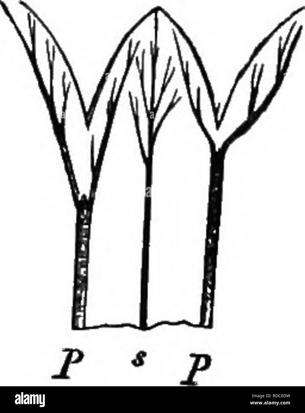
. The origin of floral structures : through insect and other agencies. Plants; Flowers; Flowers. THE RECEPTACULAE TUBE. 97 an outer epidermis, and an intermediate ground tissue, apparently nearly uniform in character, from one epidermis to the other (as in Fig. 14, a to e, p. 68). A definite number of fibro-vascalar cords penetrates this ground tissue. Theo- retically, if this structure consist of two parts, viz. the interior carpels and the exterior "tube, " some line of demarcation might be expected to be traceable; but in the majority of cases it would seem that, as neither the inner epidermis of the tube nor the outer one of the carpels are required, they are not developed at all; and so the internal tissues of the two organs become confluent and uniform, and this accounts for the fact that the dorsal cords at least are simply embedded in this common tissue. Nevertheless, in some cases there actually is a certain differentiation in the tissue, as Van Tieghem has shown in the case of Alstroemeria versicolor (Fig. 30), where a yellow band of cells marks the. Please note that these images are extracted from scanned page images that may have been digitally enhanced for readability - coloration and appearance of these illustrations may not perfectly resemble the original work.. Henslow, George, 1835-1925. New York : Appleton